. 1998 Aug;18(8):4833-43.
doi: 10.1128/MCB.18.8.4833.

Cytoplasmic tail regulates the intercellular adhesion function of the epithelial cell adhesion molecule

Affiliations

Cytoplasmic tail regulates the intercellular adhesion function of the epithelial cell adhesion molecule

M Balzar et al. Mol Cell Biol. 1998 Aug.

Abstract

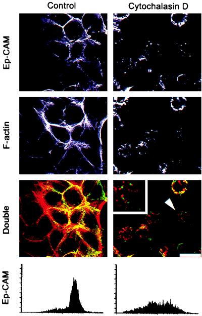
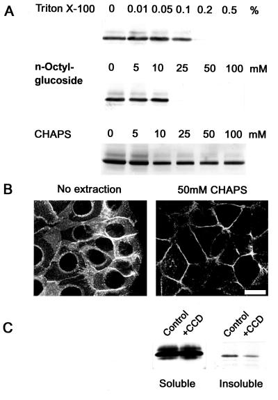
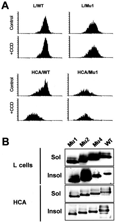
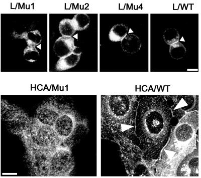
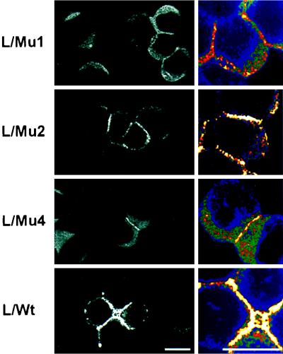
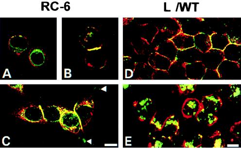
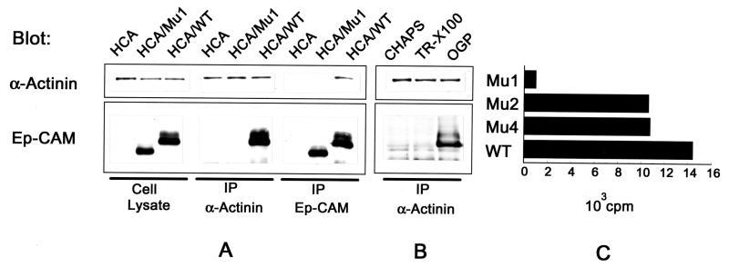
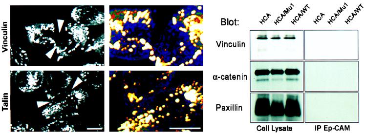
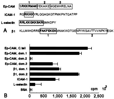
Ep-CAM, an epithelium-specific cell-cell adhesion molecule (CAM) not structurally related to the major families of CAMs, contains a cytoplasmic domain of 26 amino acids. The chemical disruption of the actin microfilaments, but not of the microtubuli or intermediate filaments, affected the localization of Ep-CAM at the cell-cell boundaries, suggesting that the molecule interacts with the actin-based cytoskeleton. Mutated forms of Ep-CAM were generated with the cytoplasmic domain truncated at various lengths. All of the mutants were transported to the cell surface in the transfectants; however, the mutant lacking the complete cytoplasmic domain was not able to localize to the cell-cell boundaries, in contrast to mutants with partial deletions. Both the disruption of the actin microfilaments and a complete truncation of the cytoplasmic tail strongly affected the ability of Ep-CAM to mediate aggregation of L cells. The capability of direct aggregation was reduced for the partially truncated mutants but remained cytochalasin D sensitive. The tail truncation did not affect the ability of the transfectants to adhere to solid-phase-adsorbed Ep-CAM, suggesting that the ability to form stable adhesions and not the ligand specificity of the molecule was affected by the truncation. The formation of intercellular adhesions mediated by Ep-CAM induced a redistribution to the cell-cell boundaries of alpha-actinin, but not of vinculin, talin, filamin, spectrin, or catenins. Coprecipitation demonstrated direct association of Ep-CAM with alpha-actinin. Binding of alpha-actinin to purified mutated and wild-type Ep-CAMs and to peptides representing different domains of the cytoplasmic tail of Ep-CAM demonstrates two binding sites for alpha-actinin at positions 289 to 296 and 304 to 314 of the amino acid sequence. The results demonstrate that the cytoplasmic domain of Ep-CAM regulates the adhesion function of the molecule through interaction with the actin cytoskeleton via alpha-actinin.


Figures

FIG. 1
FIG. 1
Effect of cytochalasin D on the actin cytoskeleton and the subcellular localization of Ep-CAMs in human epithelial RC-6 cells. Control cells and cells treated for 2 h with 10 μg of cytochalasin D per ml were stained for either Ep-CAM with MAb 323/A3 (green fluorescence) or polymerized actin filaments with phalloidin-TRITC (red fluorescence). Note the substantial disappearance of the Ep-CAMs from the cell-cell boundaries after the treatment. Internalized Ep-CAM does not colocalize with actin patches in the treated cells in the cytochalasin D-treated, double-stained cells (note the cell marked by an arrowhead and presented at larger magnification in the upper left corner). The internalization of Ep-CAM was verified by staining with MAb 323/A3 of living RC-6 cells (flow cytometry histograms below). Bar, 25 μm.
FIG. 2
FIG. 2
Detergent-soluble and -insoluble fractions of cellular Ep-CAM. (A) Monolayers of RC-6 cells (106 cells/sample) were extracted with various concentrations of Triton X-100, n-octylglucoside, or CHAPS. The presence of Ep-CAM in detergent-insoluble pellets was detected by immunoblotting with MAb 323/A3. Note that CHAPS at concentrations above 10 mM discriminates between the detergent-soluble and -insoluble fractions of Ep-CAM. (B) The CHAPS-insoluble fraction of Ep-CAM represents the molecules that are localized at the cell-cell boundaries, as was revealed by immunofluorescent staining of the nonextracted and extracted (50 mM CHAPS) cells with MAb 323/A3. Note the disappearance of the intracellular fraction of Ep-CAM from the extracted cells. Bar, 20 μm. (C) Decrease of insoluble Ep-CAM in cells pretreated with cytochalasin D (+CCD [10 μg/ml, 2 h]) prior to extraction with 50 mM CHAPS.
FIG. 3
FIG. 3
Ep-CAM mutants with a deletion in the cytoplasmic domain. (A) Amino acid sequences of the intracellular domains for the wild-type (Wt) and mutant Ep-CAMs. (B) Expression of the wild-type and mutant Ep-CAMs in transfected human epithelial HCA cells, as detected by immunoblotting with MAb 323/A3 in lysates of individual cell lines transfected with the respective form of Ep-CAM.
FIG. 4
FIG. 4
Internalization and extractability of mutant and wild-type (WT) Ep-CAMs in transfected cells. (A) Decrease of surface wild-type Ep-CAM after cytochalasin D (+CCD) treatment as observed in HCA cells and not in L cells. The CCD treatment had no effect on the presence of the tailless Mu1 molecules at the cell surface in both cell lines. Cells were pretreated with 10 μg of cytochalasin D per ml for 2 h, detached, and stained with MAb 323/A3, and the immunofluorescence was analyzed by flow cytometry. The results are presented as flow cytometry histograms. (B) Extractability of mutant Ep-CAMs from HCA and L cells. The cells were lysed in 50 mM CHAPS, and the soluble (Sol) and insoluble (Insol) fractions were analyzed by immunoblotting with MAb 323/A3.
FIG. 5
FIG. 5
Subcellular localization of the wild-type and mutant Ep-CAMs in transfected fibroblast L cells and human epithelial HCA cells. The cells were fixed and stained with the anti-Ep-CAM MAb 323/A3, followed by a secondary FITC-labeled antimouse antibody. Note the absence of Mu1 molecules and the presence of Mu2, Mu4, and wild-type (WT) molecules at the cell-cell boundaries between L cell transfectants (marked by arrowheads). Similarly, in HCA cells, Mu1 molecules were located on the entire cell surface, in contrast to the wild-type Ep-CAM, which was present at the cell-cell boundaries (marked by arrowheads). Mu2 and Mu4 molecules were localized in HCA cells similar to the wild type (not shown). Bar, 10 μm.
FIG. 6
FIG. 6
Effect of truncations in the cytoplasmic domain on cell adhesion properties of the Ep-CAM in L cells. (A) Aggregation in suspension (2 h in the absence of Ca2+) of the L929 transfectants, expressing various forms of Ep-CAM, is presented as the degree of aggregation. The relative levels of Ep-CAM expression at the surface of the transfectant cells used for the assay are presented (here and in panels B and D) above the bars as mean cell fluorescence, determined by flow cytometry with the 323/A3 MAb. LMC, mock transfectants of L cells. (B) After overnight culture in suspension, the cell aggregates were dispersed, and the cells were allowed to aggregate for 4 h in either the absence (solid bars) or presence (open bars) of 10 μg of cytochalasin D per ml in the aggregation media. For aggregation assays (A and B), the data presented were obtained from 12 independent measurements, and the standard deviation did not exceed 10%. (C) Micrographs of aggregates formed by L cells transfected with wild-type Ep-CAM in the presence and absence of cytochalasin D. (D) Adhesion of wild-type, Mu1, and blank vector (mock)-transfected L cells to the solid-phase-adsorbed purified Ep-CAM. The assay was performed as described in Materials and Methods, and the results are presented as percentages of cells attached to the substrate after a 2-h assay. The data presented were obtained from 12 independent measurements. The standard deviation did not exceed 10%.
FIG. 7
FIG. 7
Distribution of α-actinin in L cell transfectants expressing different forms of Ep-CAM, as detected by immunofluorescent staining of fixed cells with an α-actinin MAb, CB-11. Note the concentration of α-actinin along the areas of the intercellular contacts in Mu2, Mu4, and wild-type (WT) transfectants, in contrast to Mu1 transfectants. On the right are shown enlarged areas of cell-cell contact; the intensity of the fluorescent signal is presented as a pseudocolor increasing from blue to white. Note that the concentration of α-actinin at the cell-cell boundaries of Mu1 transfectants does not differ from the average density in cytoplasm, in contrast to all other cell types. Bar, 10 μm.
FIG. 8
FIG. 8
Colocalization of Ep-CAM and α-actinin in RC-6 (A, B, and C) and L (D and E) cells. Double-immunofluorescence staining was performed with fixed RC-6 and L cells with MAb 323/A3 for Ep-CAM (red fluorescence) and MAb CB-11 for α-actinin (green fluorescence). The L cells shown in panel E were cultured for 2 h in the presence of 10 μg of cytochalasin D per ml prior to fixation. Note the absence of colocalization between the two molecules in single cells and also in the areas of adhesion plaques (arrows), as well as in L cells treated with cytochalasin D. Bars, 10 μm (A, B, and C) and 5 μm (D and E).
FIG. 9
FIG. 9
Interaction of Ep-CAM and α-actinin. (A) Detection of Ep-CAM and α-actinin by immunoblotting with the respective antibodies (323/A3 and CB-11) in cell lysates and immunoprecipitates (IP) obtained with antibodies to Ep-CAM (MAb 323/A3) and α-actinin (polyclonal serum) from 50 mM n-octylglucoside extracts of the parental HCA cells, Mu1, or wild-type (WT) Ep-CAM transfectants. (B) Detection of Ep-CAM and α-actinin in immunoprecipitates with anti-α-actinin antibody from lysates of HCA/WT transfectants prepared with either CHAPS (50 mM), Triton X-100 (0.2%), or n-octylglucoside (OGP [50 mM]). Note that Ep-CAM is coprecipitated only from the lysates obtained with n-octylglucoside. (C) Binding of α-actinin to a solid-phase immobilized wild-type and mutant Ep-CAM molecules. Mu1, -2, and -4 and wild-type Ep-CAM from 1% Triton X-100 (TR-X100) lysates of the respective HCA cell transfectants were immobilized on Sac-Cel beads precoated with anti-Ep-CAM MAb. Binding of 125I-α-actinin to the immobilized wild-type or mutant Ep-CAM molecules is presented after subtraction of the background binding of α-actinin to the beads precoated with nontransfected HCA cell lysate.
FIG. 10
FIG. 10
Vinculin, talin, and α-catenin are not involved in the Ep-CAM adhesion complex. (Left) Immunofluorescent staining of interacting wild-type Ep-CAM-transfected L (L/WT) cells with antibodies to vinculin and talin. Note the absence of both proteins in the areas of cell-cell contact, as marked by arrows, and their presence in cell-substrate adhesions. The marked areas are also presented at a larger magnification as pseudocolor pictures demonstrating that both molecules are present at the cell-cell boundaries at their average concentration in other areas not involved in adhesion. (Right) Immunoblotting of total lysates and immunoprecipitates (IP) from the n-octylglucoside lysates with anti-Ep-CAM MAb from HCA cell transfectants (similar to Fig. 9, the same precipitates are involved). Note the absence of all three molecules in immunoprecipitates. Bar, 10 μm.
FIG. 11
FIG. 11
Binding of α-actinin to peptides representing the fragments of cytoplasmic domains of various CAMs. (A) Sequences of the cytoplasmic domains of Ep-CAM, ICAM-1, L-selectin, and β1 integrin. Boxes indicate various peptides used for the assay. Numbers indicate the respective domains of the cytoplasmic tail of Ep-CAM or β1 integrin. Boldface letters indicate the previously identified α-actinin binding sequences within the cytoplasmic domains of ICAM-1, L-selectin, and β1 integrin molecules and the sequence of a similar amino acid composition in the cytoplasmic domain of Ep-CAM. (B) Binding of purified 125I-labelled α-actinin to various cytoplasmic domain peptides (tested in parallel assays [n = 10]; the error bars represent ± 2ς). The whole cytoplasmic domain of Ep-CAM (C. tail) or its separate fragments (domains [dom.] 1, 2, and 3) were used. Peptide sequences used for other molecules are indicated in panel A by the fragments in boxes.

References

    1. Ben-Ze’ev A. Cytoskeletal and adhesion proteins as tumor suppressors. Curr Opin Cell Biol. 1997;9:99–108. - PubMed
    1. Bergsagel P L, Victor-Kobrin C, Timblin C R, Trepel J, Kuehl W L. A murine cDNA encodes a pan-epithelial glycoprotein that is also expressed on plasma cells. J Immunol. 1992;148:590–596. - PubMed
    1. Berndorff D, Gessner R, Kreft B, Schnoy N, Lajous-Petter A-M, Loch N, Reutter W, Hortsch M, Tauber R. Liver-intestine cadherin: molecular cloning and characterization of a novel Ca2+-dependent cell adhesion molecule expressed in liver and intestine. J Cell Biol. 1994;125:1353–1369. - PMC - PubMed
    1. Carpén O, Pallai P, Staunton D E, Springer T A. Association of intercellular adhesion molecule-1 (I-CAM1) with actin-containing cytoskeleton and α-actinin. J Cell Biol. 1992;118:1223–1234. - PMC - PubMed
    1. Cheung P H, Culic O, Qui Y, Early K, Thompson N, Hixson D C, Lin S H. The cytoplasmic domain of C-CAM is required for C-CAM mediated adhesion function: studies of a C-CAM transcript containing an unspliced intron. Biochem J. 1993;295:427–435. - PMC - PubMed

Publication types

MeSH terms

LinkOut - more resources