. 1998 Aug;18(8):4863-71.
doi: 10.1128/MCB.18.8.4863.

Structure-function analysis of Qk1: a lethal point mutation in mouse quaking prevents homodimerization

Affiliations

Structure-function analysis of Qk1: a lethal point mutation in mouse quaking prevents homodimerization

T Chen et al. Mol Cell Biol. 1998 Aug.

Abstract

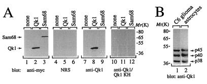
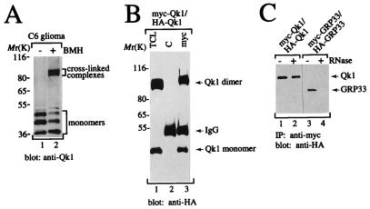
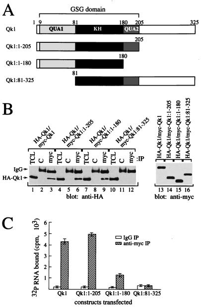
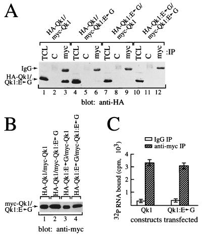
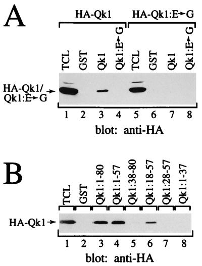
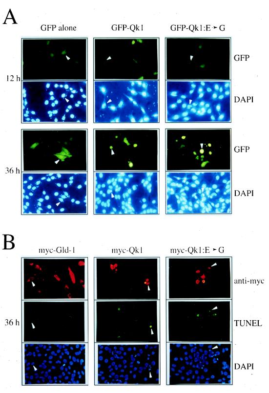
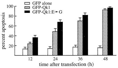
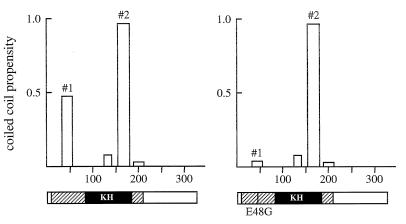
Qk1 is a member of the KH domain family of proteins that includes Sam68, GRP33, GLD-1, SF1, and Who/How. These family members are RNA binding proteins that contain an extended KH domain embedded in a larger domain called the GSG (for GRP33-Sam68-GLD-1) domain. An ethylnitrosourea-induced point mutation in the Qk1 GSG domain alters glutamic acid 48 to a glycine and is known to be embryonically lethal in mice. The function of Qk1 and the GSG domain as well as the reason for the lethality are unknown. Here we demonstrate that the Qk1 GSG domain mediates RNA binding and Qk1 self-association. By using in situ chemical cross-linking studies, we showed that the Qk1 proteins exist as homodimers in vivo. The Qk1 self-association region was mapped to amino acids 18 to 57, a region predicted to form coiled coils. Alteration of glutamic acid 48 to glycine (EG) in the Qk1 GSG domain (producing protein Qk1:EG) abolishes self-association but has no effect on the RNA binding activity. The expression of Qk1 or Qk1:EG in NIH 3T3 cells induces cell death by apoptosis. Approximately 90% of the remaining transfected cells are apoptotic 48 h after transfection. Qk1:EG was consistently more potent at inducing apoptosis than was wild-type Qk1. These results suggest that the mouse quaking lethality (EG) occurs due to the absence of Qk1 self-association mediated by the GSG domain.


Figures

FIG. 1
FIG. 1
Characterization of the anti-Qk1 antibody. (A) Total cell lysates from HeLa cells transfected with vector (none), myc-Qk1 (Qk1), or myc-Sam68 (Sam68) were separated by SDS-PAGE. The proteins were transferred to nitrocellulose and immunoblotted with anti-myc, normal rabbit serum (NRS), anti-Qk1, or anti-Qk1 antibodies preabsorbed with 1 μg of GST-Qk1KH antigen/ml (anti-Qk1/Qk1 KH). The positions of Qk1 and Sam68 are shown on the left, and those of molecular mass markers (in kilodaltons) are on the right. (B) Total cell extracts from rat C6 glioma cells and rat astrocytes were immunoblotted with anti-Qk1. The presence of three Qk1-immunoreactive proteins with approximate molecular masses of 45, 40, and 38 kDa is shown.
FIG. 2
FIG. 2
Qk1 self-associates into homodimers in the absence of cellular RNA. (A) C6 glioma cells were treated in situ with (+) or without (−) BMH. The cells were lysed in sample buffer, and the proteins were separated by SDS-PAGE. The proteins were transferred to nitrocellulose and immunoblotted with rabbit anti-Qk1 antibody. The bands representing the three Qk1 isoforms are shown as monomers, and the cross-linked complexes are indicated. The positions of molecular mass markers (in kilodaltons) are indicated on the left. (B) HeLa cells cotransfected with myc- and HA-Qk1 were treated in situ with BMH. The cells were lysed, and an aliquot of the total cell lysate (TCL) as well as anti-myc (myc) and IgG (C) immunoprecipitates were separated by SDS-PAGE. The proteins were transferred to nitrocellulose and immunoblotted with anti-HA antibodies. (C) Qk1, unlike GRP33, does not require cellular RNA for self-association. HeLa cells expressing myc-Qk1, HA-Qk1, myc-GRP33, or HA-GRP33 were lysed separately, and each cell lysate was divided into two portions, either treated with RNase A (+) or not treated (−), mixed, and incubated with anti-myc antibodies. The anti-myc immunoprecipitates (IP) of the mixed lysates were separated by SDS-PAGE and immunoblotted with anti-HA antibodies.
FIG. 3
FIG. 3
Mapping of the Qk1 self-association and RNA binding regions within the GSG domain. (A) Schematic diagrams of the Qk1 constructs utilized are shown. The black box denotes the KH domain, whereas the gray boxes represent the QUA1 and QUA2 regions as indicated. The entire region encompassing QUA1, KH, and QUA2 is the GSG domain. (B) Truncation of the N-terminal 80 amino acids of Qk1 or QUA1 abolishes self-association. HA-Qk1 was cotransfected in HeLa cells with various myc-Qk1 deletion constructs as indicated. Total cell lysates (TCL) as well as anti-myc (myc) and control IgG (C) immunoprecipitates were analyzed by immunoblotting with anti-HA antibodies. Total cell lysates of the myc-Qk1 proteins were immunoblotted with anti-myc antibodies (lanes 13 to 16). (C) The Qk1 GSG domain is required for RNA binding. HeLa cell lysates containing myc-tagged Qk1 or various truncated forms of Qk1 were immunoprecipitated with anti-myc antibody (hatched bars) or control IgG (white bars) and then incubated with 32P-labeled total cellular RNA. Each bar represents the mean ± standard deviation of data from more than six independent immunoprecipitations carried out during more than three separate experiments.
FIG. 4
FIG. 4
E48G substitution in Qk1 abolishes homodimerization but not RNA binding. (A) HeLa cells were transfected with HA- or myc-tagged Qk1 and/or Qk1:E➛G as indicated. The total cell lysate (TCL) as well as anti-myc (myc) and IgG (C) immunoprecipitates were immunoblotted with anti-HA antibody. The bands representing HA-Qk1 or HA-Qk1:E➛G are indicated on the left. The migration of the heavy chain of IgG is also indicated. (B) Total cell lysates corresponding to those of panel A were immunoblotted with anti-myc antibodies. (C) Immunoprecipitated Qk1 or Qk1:E➛G was incubated with labeled RNA as described in Materials and Methods.
FIG. 5
FIG. 5
Association of Qk1 in vitro and localization of the minimal region to amino acids 18 to 57. (A) HA-Qk1 or HA-Qk1:E➛G was transfected into HeLa cells. The cells were lysed, and GST pull-down assays were performed with full-length Qk1 or Qk1:E➛G expressed as a GST fusion protein. The bound proteins were separated by SDS-PAGE, transferred to nitrocellulose, and immunoblotted with anti-HA antibodies. (B) HA-Qk1 was expressed in HeLa cells and incubated with various Qk1 GST fusion proteins as indicated. The bound HA-Qk1 was analyzed as described for panel A. The migration of HA-Qk1 or HA-Qk1:E➛G is shown on the left. TCL, total cell lysate.
FIG. 6
FIG. 6
Qk1 and Qk1:E➛G induce apoptosis in NIH 3T3 cells. (A) NIH 3T3 cells were transfected with an expression vector encoding GFP alone, GFP-Qk1, or GFP-Qk1:E➛G. After 12, 24, 36, and 48 h, the cells were fixed and stained with DAPI to visualize the nuclei. The top photograph in each pair shows the fluoresceinated cells containing GFP, and the lower photograph shows the DAPI-stained nuclei. The white arrowheads were used to align the top and bottom photographs. (B) NIH 3T3 cells were transfected with expression vector encoding myc–GLD-1, myc-Qk1, or myc-Qk1:E➛G. The myc epitope-tagged proteins were visualized by indirect immunofluorescence with a rhodamine-conjugated secondary antibody (anti-myc). The apoptotic cells were visualized by TUNEL with fluorescein-containing nucleotides (TUNEL), and the nuclei were stained with DAPI (DAPI). The three photographs each for myc–GLD-1, myc-Qk1, and myc-Qk1:E➛G represent the same field of cells as visualized with different filters. The white arrowheads were used to orient the cells in the photographs.
FIG. 7
FIG. 7
Quantitation of the apoptosis induced by Qk1 and Qk1:E➛G. Eight hundred transfected NIH 3T3 cells, in three separate experiments, were counted and assessed for the presence of apoptotic nuclei. The presence of apoptotic nuclei was scored as cells undergoing apoptosis. The white, hatched, and black bars represent GFP, GFP-Qk1, and GFP-Qk1:E➛G, respectively.
FIG. 8
FIG. 8
Coiled-coil motif predictions for Qk1 (left) and Qk1:E➛G (right). The Qk1 and Qk1:E➛G protein sequences were analyzed with the computer program COILS, and the putative coiled-coil motifs are shown (#1 and #2). The abscissa and ordinate represent amino acid numbers and the propensity to form coiled coils, respectively. The structures of the Qk1 proteins are shown below, with hatched and black boxes representing the GSG and KH domains, respectively. The vertical line in the GSG domain denotes the location of the quaking lethal point mutation E48G.

References

    1. Arning S, Gruter P, Bilbe G, Kramer A. Mammalian splicing factor SF1 is encoded by variant cDNAs and binds to RNA. RNA. 1996;2:794–810. - PMC - PubMed
    1. Baehrecke E H. who encodes a KH RNA binding protein that functions in muscle development. Development. 1997;124:1323–1332. - PubMed
    1. Barbarese E. Spatial distribution of myelin basic protein mRNA and polypeptide in Quaking oligodendrocytes in culture. J Neurosci Res. 1991;29:271–281. - PubMed
    1. Bartoszewicz Z P, Noronha A B, Fujita N, Sato S, Bo L, Trapp B D, Quarles R H. Abnormal expression and glycosylation of the large and small isoforms of myelin-associated glycoprotein in dysmyelinating quaking mutants. J Neurochem Res. 1995;41:27–38. - PubMed
    1. Benda P, Lightbody J, Sato G, Levine L, Sweet W. Differentiated rat glial cell strain in tissue culture. Science. 1968;161:370–371. - PubMed

Publication types

MeSH terms