

D2-Like dopamine autoreceptor activation reduces quantal size in PC12 cells
- PMID: 9671649
- PMCID: PMC6793067
- DOI: 10.1523/JNEUROSCI.18-15-05575.1998
D2-Like dopamine autoreceptor activation reduces quantal size in PC12 cells
Abstract
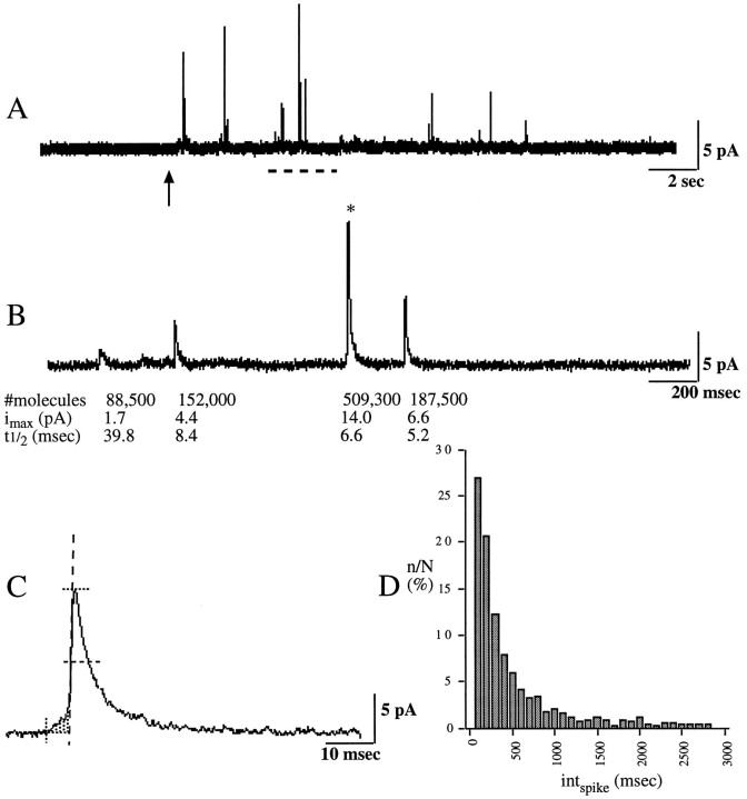
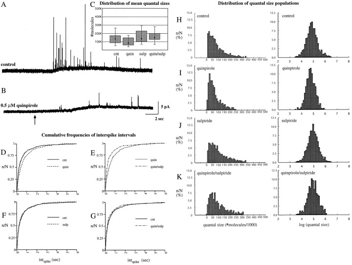
D2-like dopamine autoreceptors regulate dopamine release and are implicated in important actions of antipsychotic drugs and rewarding behaviors. To directly observe the effects of D2 autoreceptors on exocytic neurotransmitter release, we measured quantal release of dopamine from pheochromocytoma PC12 cells that express D2 and D4 autoreceptors. High potassium-evoked secretion in PC12 cells produced a unimodal population of quantal sizes. We found that exposures to the D2-like agonist quinpirole that inhibited tyrosine hydroxylase activity by approximately 50% also reduced quantal size by approximately 50%. The reduced quantal size was blocked by the D2 antagonist sulpiride and reversed by L-DOPA. Quinpirole also decreased the frequency of stimulation-evoked quantal release. Together, these findings indicate effects on quantal neurotransmission by D2-like dopamine autoreceptors previously distinguished as synthesis-modulating autoreceptors that regulate tyrosine hydroxylase activity versus impulse-regulating autoreceptors that modulate membrane potential. The results also provide an initial demonstration of a receptor-mediated mechanism that alters quantal size.
Figures


Similar articles
-
Effects of a partial D2-like receptor agonist on striatal dopamine autoreceptor functioning in preweanling rats.Brain Res. 2006 Feb 16;1073-1074:269-75. doi: 10.1016/j.brainres.2005.12.054. Epub 2006 Jan 19. Brain Res. 2006. PMID: 16427034
-
Native human neocortex release-regulating dopamine D2 type autoreceptors are dopamine D2 subtype.Eur J Neurosci. 1999 Jul;11(7):2351-8. doi: 10.1046/j.1460-9568.1999.00651.x. Eur J Neurosci. 1999. PMID: 10383624
-
Reduced dopamine and glutamate neurotransmission in the nucleus accumbens of quinpirole-sensitized rats hints at inhibitory D2 autoreceptor function.J Neurochem. 2015 Sep;134(6):1081-90. doi: 10.1111/jnc.13209. Epub 2015 Jul 14. J Neurochem. 2015. PMID: 26112331
-
D1 dopamine receptor stimulation enables the postsynaptic, but not autoreceptor, effects of D2 dopamine agonists in nigrostriatal and mesoaccumbens dopamine systems.Synapse. 1989;4(4):327-46. doi: 10.1002/syn.890040409. Synapse. 1989. PMID: 2532422
-
The influence of dopamine autoreceptors on temperament and addiction risk.Neurosci Biobehav Rev. 2023 Dec;155:105456. doi: 10.1016/j.neubiorev.2023.105456. Epub 2023 Nov 3. Neurosci Biobehav Rev. 2023. PMID: 37926241 Free PMC article. Review.
Cited by
-
Effects of atypical antipsychotics and haloperidol on PC12 cells: only aripiprazole phosphorylates AMP-activated protein kinase.J Neural Transm (Vienna). 2010 Oct;117(10):1139-53. doi: 10.1007/s00702-010-0457-9. Epub 2010 Aug 5. J Neural Transm (Vienna). 2010. PMID: 20686905
-
The neuronal monoamine transporter VMAT2 is regulated by the trimeric GTPase Go(2).J Neurosci. 2000 Mar 15;20(6):2131-41. doi: 10.1523/JNEUROSCI.20-06-02131.2000. J Neurosci. 2000. PMID: 10704487 Free PMC article.
-
Phospholipid mediated plasticity in exocytosis observed in PC12 cells.Brain Res. 2007 Jun 2;1151:46-54. doi: 10.1016/j.brainres.2007.03.012. Epub 2007 Mar 12. Brain Res. 2007. PMID: 17408597 Free PMC article.
-
Aberrant extracellular dopamine clearance in the prefrontal cortex exhibits ADHD-like behavior in NCX3 heterozygous mice.FEBS J. 2025 Jan;292(2):426-444. doi: 10.1111/febs.17339. Epub 2024 Dec 3. FEBS J. 2025. PMID: 39624860 Free PMC article.
-
Single-Vesicle Electrochemistry Following Repetitive Stimulation Reveals a Mechanism for Plasticity Changes with Iron Deficiency.Angew Chem Int Ed Engl. 2022 May 9;61(20):e202200716. doi: 10.1002/anie.202200716. Epub 2022 Mar 21. Angew Chem Int Ed Engl. 2022. PMID: 35267233 Free PMC article.
References
-
- Albillos A, Dernick G, Hostmann H, Almers W, Alvarez de Toledo G, Lindau M. The exocytic event in chromaffin cells revealed by patch amperometry. Nature. 1997;389:509–512. - PubMed
-
- Alvarez de Toledo G, Fernandez-Chacon R, Fernandez JM. Release of secretory products during transient vesicle fusion. Nature. 1993;363:554–558. - PubMed
-
- Arbogast LA, Soares MJ, Robertson MC, Voogt JL. A factor(s) from a trophoblast cell line increases tyrosine hydroxylase activity in fetal hypothalamic cell cultures. Endocrinology. 1993;133:111–120. - PubMed
-
- Bauerfeind R, Regnier-Vigouroux A, Flatmark T, Huttner WB. Selective storage of acetylcholine, but not catecholamines, in neuroendocrine synaptic-like microvesicles of early endosomal origin. Neuron. 1993;11:105–121. - PubMed
Publication types
MeSH terms
Substances
Grants and funding
LinkOut - more resources
Full Text Sources