Neurite growth patterns leading to functional synapses in an identified embryonic neuron
- PMID: 9671656
- PMCID: PMC6793058
- DOI: 10.1523/JNEUROSCI.18-15-05652.1998
Neurite growth patterns leading to functional synapses in an identified embryonic neuron
Abstract
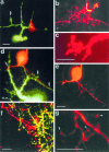
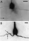
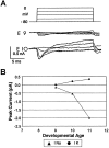
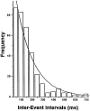
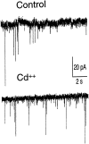
We explored the relationship between neurite outgrowth and the onset of synaptic activity in the central neuropil of the leech embryo in vivo. To follow changes in early morphology and the onset of synaptic activity in the same identified neuron, we obtained whole-cell patch-clamp recordings and fluorescent dye fills from dorsal pressure-sensitive (P) cells, the first neurons that could be reliably identified in the early embryo. We followed the development of the P cell from the first extension of neurites to the elaboration of an adult-like arbor. After the growth of primary neurites, we observed a profuse outgrowth of transient neurites within the neuropil. Retraction of the transient neurites left the primary branches studded with spurs. After a dormant period, stable secondary branches grew apparently from the spurs and became tipped with terminals. At this time, neurites of the Retzius (R) cell, a known presynaptic partner in the adult, were observed to apparently contact the terminals. Although voltage-dependent currents were seen in the P cell at the earliest stage, spontaneous synaptic activity was only observed when terminals had formed. Spontaneous release was observed before evoked release could be detected from the R cell. Our results suggest that transient neurites are formed during an exploratory phase of development, whereas the more precisely timed outgrowth of stable neurites from the spurs signals functional differentiation during synaptogenesis. Because spurs have also been observed in neurons of the mammalian brain, they may constitute a primordial synaptic organizer.
Figures





Similar articles
-
Target cell contact suppresses neurite outgrowth from soma-soma paired Lymnaea neurons.J Neurobiol. 2000 Feb 15;42(3):357-69. doi: 10.1002/(sici)1097-4695(20000215)42:3<357::aid-neu7>3.0.co;2-f. J Neurobiol. 2000. PMID: 10645975
-
Steps in the formation of neurites and synapses studied in cultured leech neurons.Braz J Med Biol Res. 2000 May;33(5):487-97. doi: 10.1590/s0100-879x2000000500002. Braz J Med Biol Res. 2000. PMID: 10775879 Review.
-
Steps in the formation of a bipolar outgrowth pattern by cultured neurons, and their substrate dependence.J Neurobiol. 2002 Feb 5;50(2):106-17. doi: 10.1002/neu.10017. J Neurobiol. 2002. PMID: 11793358
-
Anterograde and retrograde effects of synapse formation on calcium currents and neurite outgrowth in cultured leech neurons.Proc Biol Sci. 1992 Aug 22;249(1325):217-22. doi: 10.1098/rspb.1992.0107. Proc Biol Sci. 1992. PMID: 1360683
-
Serotonin regulation of neurite outgrowth in identified neurons from mature and embryonic Helisoma trivolvis.Perspect Dev Neurobiol. 1998;5(4):373-87. Perspect Dev Neurobiol. 1998. PMID: 10533526 Review.
Cited by
-
On the Basis of Synaptic Integration Constancy during Growth of a Neuronal Circuit.Front Cell Neurosci. 2016 Aug 18;10:198. doi: 10.3389/fncel.2016.00198. eCollection 2016. Front Cell Neurosci. 2016. PMID: 27587998 Free PMC article.
-
Modeling disrupted synapse formation in wolfram syndrome using hESCs-derived neural cells and cerebral organoids identifies Riluzole as a therapeutic molecule.Mol Psychiatry. 2023 Apr;28(4):1557-1570. doi: 10.1038/s41380-023-01987-3. Epub 2023 Feb 7. Mol Psychiatry. 2023. PMID: 36750736 Free PMC article.
-
Exploring the Functional Heterogeneity of Directly Reprogrammed Neural Stem Cell-Derived Neurons via Single-Cell RNA Sequencing.Cells. 2023 Dec 11;12(24):2818. doi: 10.3390/cells12242818. Cells. 2023. PMID: 38132138 Free PMC article.
-
High-throughput kinase inhibitor screening reveals roles for Aurora and Nuak kinases in neurite initiation and dendritic branching.Sci Rep. 2021 Apr 14;11(1):8156. doi: 10.1038/s41598-021-87521-3. Sci Rep. 2021. PMID: 33854138 Free PMC article.
-
Influence of metabolic stress and metformin on synaptic protein profile in SH-SY5Y-derived neurons.Physiol Rep. 2023 Nov;11(22):15852. doi: 10.14814/phy2.15852. Physiol Rep. 2023. PMID: 38010200 Free PMC article.
References
-
- Blagburn JM, Sosa MA, Blanco RE. Specificity of identified central synapses in the embryonic cockroach: appropriate connections form before the onset of spontaneous afferent activity. J Comp Neurol. 1996;373:511–528. - PubMed
-
- Conrad GW, Bee JA, Roche SM, Teillet MA. Fabrication of microscalpels by electrolysis of tungsten wire in a meniscus. J Neurosci Methods. 1993;50:123–127. - PubMed
-
- DeRiemer SA, Macagno ER. Quantitative studies of the growth of neuronal arbors. In: Carew TJ, Kelley DB, editors. Perspectives in neural systems and behavior. Liss; New York: 1989. p. 11.
Publication types
MeSH terms
LinkOut - more resources
Full Text Sources
