. 1998 Jul 21;95(15):8461-6.
doi: 10.1073/pnas.95.15.8461.

The 40-kDa subunit of DNA fragmentation factor induces DNA fragmentation and chromatin condensation during apoptosis

Affiliations

The 40-kDa subunit of DNA fragmentation factor induces DNA fragmentation and chromatin condensation during apoptosis

X Liu et al. Proc Natl Acad Sci U S A. .

Abstract

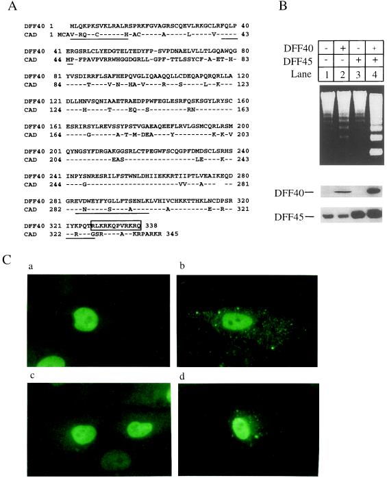
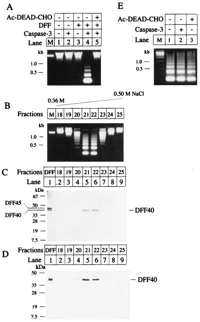
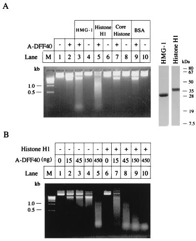
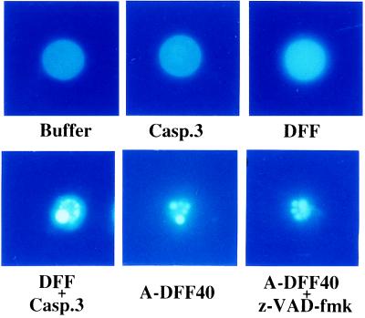
We report here the reconstitution of a pathway that leads to the apoptotic changes in nuclei by using recombinant DNA fragmentation factor (DFF), a heterodimeric protein of 40 and 45 kDa. Coexpression of DFF40 and DFF45 is required to generate recombinant DFF, which becomes activated when DFF45 is cleaved by caspase-3. The cleaved fragments of DFF45 dissociate from the DFF40, the active component of DFF. Purified DFF40 exhibited an intrinsic DNase activity that was markedly stimulated by chromatin-associated proteins histone H1 and high mobility group proteins. DFF40 also triggered chromatin condensation when incubated with nuclei. These data suggest that DFF40 is sufficient to trigger both DNA fragmentation and chromatin condensation during apoptosis.


Figures

Figure 1
Figure 1
Expression of DFF and nuclear localization of DFF40 and DFF45. (A) Amino acid sequences of human DFF40 and mouse CAD (16, 17) are aligned by the Lipman–Pearson method of the dnastar program. The proteolytic peptide sequences from purified DFF40 are underlined. A putative nuclear localization signal at the carboxyl terminus of DFF40 is boxed. The amino acid residues are numbered on the left and right. (B) Human embryonic kidney 293 cells were grown and transfected as described in ref. . The 293 cells were transfected with the vector alone (lanes 1), pDFF40-flag alone (lanes 2), pDFF45 alone (lanes 3), or pDFF40-flag plus pDFF45 (lanes 4). The cells were harvested 36 hr after transfection and the S-100 fraction was prepared as described in ref. . Aliquots (80 μg) of cytosolic proteins were incubated in the presence of caspase-3 with aliquots (7 μl) of hamster liver nuclei for 2 hr at 37°C in 60 μl of buffer A. Genomic DNA was isolated and analyzed as described in ref. . Aliquots (80 μg) of S-100 prepared as described above were also subjected to SDS/10% PAGE and transferred to nitrocellulose filters. The filters were probed either with a rabbit anti-DFF45 antiserum (15) or with M2 anti-FLAG antiserum (1:2000). The antigen–antibody complexes were visualized with an ECL method. (C) CV-1 cells were transfected with pDFF40-flag alone (b), pDFF45 alone (a), or pDFF40-flag plus pDFF45 (c and d). The cells were fixed and subsequently probed with the rabbit anti-DFF45 antiserum or the M2 anti-FLAG antibody. The antigen–antibody complexes were visualized by a fluorescein isothiocyanate method. (a) Cells were transfected with pDFF45 and stained with anti-DFF45 antibody. (b) Cells were transfected with pDFF40-flag and stained with anti-FLAG antibody. (c) Cells were transfected with pDFF45 plus pDFF40-flag and stained with anti-DFF45 antibody. (d) Cells were transfected with pDFF45 plus pDFF40-flag and stained with anti-FLAG antibody.
Figure 2
Figure 2
DFF activation pathway. (A) Recombinant DFF was expressed and purified. An aliquot (7 μl) of hamster liver nuclei was incubated alone (lane 1), with 50 ng of caspase-3 (lane 2), with 500 ng of purified DFF (lane 3), or with 50 ng of caspase-3 plus 500 ng of purified DFF (lane 4) at 37°C for 2 hr in a final volume of 60 μl of buffer A. In lane 5, 50 ng of caspase-3 was preincubated with 1 μM Ac-DEAD-CHO (a tetrapeptide aldehyde inhibitor of caspase-3) at 37°C for 5 min followed by the addition of 500 ng of DFF and 7 μl of nuclei and incubation for 2 hr at 37°C in 60 μl of buffer A. The genomic DNAs were isolated and analyzed as described in ref. . (B) Recombinant DFF was generated and purified as described. An aliquot of 1 mg was incubated with 100 μg of caspase-3 at 30°C for 1 hr. The sample was then purified through a Mono Q 5/5 column (Pharmacia) followed by a Mono S 5/5 column (Pharmacia). The column was eluted with a 30 ml 0.3–0.5 M linear NaCl gradient. Fractions of 1 ml were collected. An aliquot (20 μl) of each fraction was incubated with an aliquot of 7 μl of hamster liver nuclei at 37°C for 2 hr in 60 μl of buffer A. The genomic DNA was isolated, analyzed, and visualized as described in ref. . (C) An aliquot (1 μg) of purified recombinant DFF (lane 1) and aliquots of 30 μl of the indicated Mono S column fractions were subjected to SDS/12% PAGE followed by Coomassie brilliant blue staining. The positions of DFF40 and DFF45 are indicated. (D) An aliquot (20 μl) of the indicated Mono S column fractions was subjected to SDS/12% PAGE and transferred to a nitrocellulose filter. The filter was probed with a rabbit anti-DFF40 antiserum (1:2000) and the antigen–antibody complexes were visualized through an ECL method. (E) An aliquot (30 μl) of the active DFF purified through the Mono S column was incubated with 7 μl of hamster liver nuclei at 37°C for 2 hr in 60 μl of buffer A with buffer alone (lane 1) or in the presence of 50 ng of caspase-3 (lane 2) or 10 μM Ac-DEAD-CHO (lane 3). The genomic DNA was isolated, analyzed, and visualized as described in ref. .
Figure 3
Figure 3
Stimulation of DNase of DFF40 by histone H1 and HMG-1. (A) An aliquot (2 μg) of plasmid DNA (pcDNA3 vector) was incubated with either a 45-ng aliquot of A-DFF40 purified through the Mono S column as described in Fig. 2 in 30 μl of buffer A at 37°C for 2 hr alone (lane 2) or in the presence of 1 μg of bovine HMG-1 (lane 3), 0.5 μg of bovine histone H1 (lane 5), 1 μg of core histone (lane 7), or 1 μg of BSA (lane 9). The plasmid DNA was also incubated with buffer alone (lane 1), 1 μg of bovine HMG-1 (lane 4), 0.5 μg of histone H1 (lane 6), 1 μg of core histone, or 1 μg of BSA (lane 10). The reactions were stopped by adding 5 mM EDTA and the products were directly loading onto a 1.2% agarose gel containing 5 μg/ml ethidium bromide. An aliquot (5 μg) of bovine HMG-1 or histone H1 was also directly subjected to SDS/PAGE followed by Coomassie brilliant blue staining (the right two lanes with molecular mass markers). (B) An aliquot (2 μg) of plasmid DNA was incubated with the indicated amount of A-DFF40 purified through the Mono S column as described in the legend Fig. 2 in the absence (lanes 2–5), or presence (lanes 6–10) of 0.5 μg of histone H1 in a final volume of 30 μl of buffer A at 37°C for 2 hr. The reactions were stopped by adding 5 mM EDTA and the products were directly loaded onto a 1.2% agarose gel containing 5 μg/ml ethidium bromide.
Figure 4
Figure 4
A-DFF40 induces chromatin condensation in nuclei. An aliquot (7 μl) of hamster liver nuclei was incubated at 37°C for 2 hr with buffer alone, with 50 ng of caspase-3, with 500 ng of DFF, with 50 ng of caspase-3 plus 500 ng of DFF, with 300 ng of A-DFF40 purified through the Mono S column as described in the legend of Fig. 2, or with 300 ng of A-DFF40 plus 20 μM z-VAD-fmk as indicated. The nuclei were then stained with 4′,6-diamidino-2-phenylindole (DAPI) and observed under a fluorescence microscope with a DAPI filter.

References

    1. Kerr J F R, Wyllie A H, Currie A R. Br J Cancer. 1972;26:239–257. - PMC - PubMed
    1. Wyllie A H. Nature (London) 1980;284:555–556. - PubMed
    1. Wyllie A H, Kerr J F, Currie A R. Int Rev Cytol. 1980;68:251–305. - PubMed
    1. Wyllie A H, Morris R G, Smith A L, Dunlop D. J Pathol. 1984;142:66–77. - PubMed
    1. Vaux D L. Cell. 1997;90:389–390. - PubMed

Publication types

Associated data