. 1998 Jul 21;95(15):8491-5.
doi: 10.1073/pnas.95.15.8491.

In vivo and in vitro complementation of the N-terminal domain of enzyme I of the Escherichia coli phosphotransferase system by the cloned C-terminal domain

Affiliations

In vivo and in vitro complementation of the N-terminal domain of enzyme I of the Escherichia coli phosphotransferase system by the cloned C-terminal domain

A Fomenkov et al. Proc Natl Acad Sci U S A. .

Abstract

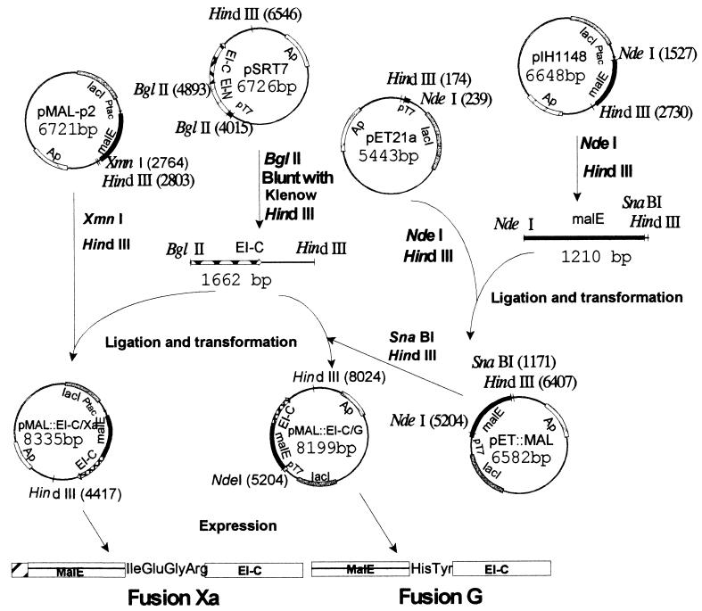
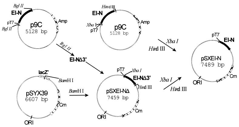
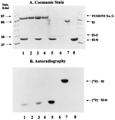
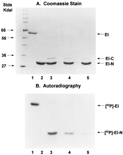
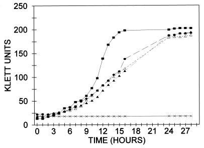
Enzyme I (EI) is the first protein in the phosphoryl transfer sequence from phosphoenolpyruvate (PEP) to sugar in carbohydrate uptake via the bacterial PEP:glycose phosphotransferase system. The EI monomer/dimer transition may regulate the phosphotransferase system because only the EI dimer is autophosphorylated by PEP. We previously showed that the EI monomer comprises two major domains: (i) a compact, protease-resistant N-terminal domain (EI-N), containing the active site His, and (ii) a flexible, protease-sensitive C-terminal domain (EI-C), which is required for EI dimerization. EI-N interacts with the second protein, HPr, and phospho-HPr, but EI-N neither dimerizes nor is phosphorylated by PEP. We report here the molecular cloning and some properties of EI-C. EI-C is rapidly proteolyzed in vivo. Therefore, two different overexpression vectors encoding fusion proteins were constructed. Fusion Xa contains MalE (the maltose-binding protein), the four-amino acid sequence required by protease factor Xa, followed by EI-C. Fusion G contains His-Tyr between MalE and EI-C and is cleaved by the protease genenase. Homogenous EI-C was isolated from fusion G. [32P]PEP phosphorylated EI-N when supplemented with EI-C, fusion Xa, or fusion G. EI-C may act catalytically. Complementation was also demonstrated in vivo. An Escherichia coli ptsI deletion grew on mannitol as the sole source of carbon after it was transformed with two compatible vectors; one vector encoded EI-N and the other encoded fusion Xa or fusion G. The molecular details underlying important properties of EI can now be studied.


Figures

Figure 1
Figure 1
Construction of plasmids for overexpression of fusion proteins. Two vectors (from New England Biolabs) were used for constructing the fusions. plH1148 contains the malE gene terminated by the coding sequences for His-Tyr (the amino acid sequence required for the genenase protease). The malE gene in this vector is deleted for the signal sequence so that the fusion protein is expressed in the cytoplasm. pMAL-p2 contains malE terminated by the coding sequence for Ile-Glu-Gly-Arg (required for the protease factor Xa) and contains the signal sequence so that the fusion protein is secreted into the periplasm. Two overexpression vectors were used for the constructs: pSRT7, which contains ptsI, the gene encoding EI, under control of the inducible T7 polymerase (pT7 promoter), and pET21a, which also contains pT7. pET21a was converted to pET∷MAL by inserting malE, which had been severed from pIH1148 with NdeI and HindIII. A DNA fragment encoding EI-C was cut from pSRT7 with BglII and HindIII, and the desired gene fusions were constructed by inserting the fragment downstream of the malE genes in pMAL-p2 and pET∷MAL, giving the overexpression vectors pMAL∷EI-C/Xa and pMAL∷EI-C/G, respectively.
Figure 2
Figure 2
Construction of plasmid for expression of EI-N for in vivo experiments. The vector pSYX39 (15) contains the pSC101 origin of replication that is compatible with a number of other origins of replication; the vector was cut with BamHI. A DNA fragment was inserted into the vector that encodes a partially truncated EI-N isolated from p9C; the fragment contained the pT7 promoter at the 5′ terminus. The truncation at the 3′ terminus was corrected by cutting p9C with XbaI and HindIII, isolating the fragment, and by inserting it into pSXEI-NΔ, which had also been cut with the two restriction enzymes. The plasmid pSXEI-N contained the gene encoding all EI-N under the control of the pT7 promoter, the Cmr gene, and the desired origin of replication.
Figure 3
Figure 3
Phosphorylation of EI-N and EI. Phosphorylation experiments were conducted as described in Materials and Methods with [32P]PEP as the phosphoryl donor. Protein samples were subjected to SDS/PAGE (16, 17). (A) Coomassie stained gel. (B) Autoradiograph. Standard molecular mass markers are shown at each end of the gel. Incubation mixtures contained the following proteins: Lanes: 1, fusion G (4 μg) and EI-N (4 μg); 2, fusion G (4 μg); 3, fusion Xa (4 μg) and EI-N (4 μg); 4, fusion Xa (4 μg); 5, EI-C (0.4 μg) and EI-N (4 μg); 6, EI-C (0.4 μg); 7, EI (2 μg); 8, EI-N (4 μg).
Figure 4
Figure 4
Effect of concentration of EI-C on phosphorylation of EI-N. The experimental conditions were the same as those used for Fig. 3 (see Materials and Methods) with the following proteins in the incubation mixtures: Lanes: 1, 2 μg EI; 2, 4 μg EI-N; 3, 4 μg EI-N (140 pmol) plus 0.4 μg EI-C (11 pmol); 4, 4 μg EI-N plus 0.2 μg EI-C; 5, 4 μg EI-N plus 0.1 μg EI-C. In the latter, the mol ratio EI-N/EI-C = 51.
Figure 5
Figure 5
Growth of E. coli BL21(DE3) and transformants of BL21(DE3)ΔptsI on mannitol. Cells were grown on a rotary shaker at 37°C in 50-ml Erlenmeyer flasks equipped with side arm tubes for determining turbidity in the Klett colorimeter. The salts (M9) medium (9 ml) was supplemented with mannitol to a 1% concentration. Inocula consisted of 1 ml of cells grown overnight on the same medium. At the end of the growth period, plasmids were isolated from the relevant cells (see text). All cells were double transformants. ■, Control, E. coli BL21(DE3), pSYX39/pET:Mal (expresses intact EI from chromosome and MalE from plasmid). The other transformants were derived from BL21(DE3)ΔptsI: •, pSYX39/pSRT7 (intact EI); ▴, pSXEI-N/ pMal∷EI-C/G (EI-N plus fusion G); ▵, pSXEI-N/pMal∷EI-C/Xa (EI-N plus fusion Xa); ×, pSYX39/pET∷Mal (Mal E).

References

    1. Meadow N D, Fox D K, Roseman S. Annu Rev Biochem. 1990;59:497–542. - PubMed
    1. Postma P W, Lengeler J W, Jacobson G R. Microbiol Rev. 1993;57:543–594. - PMC - PubMed
    1. Saier M H, Reizer J. Mol Microbiol. 1994;13:755–764. - PubMed
    1. Chauvin F, Brand L, Roseman S. Res Microbiol. 1996;147:471–479. - PubMed
    1. Chauvin F, Brand L, Roseman S. J Biol Chem. 1994;269:20270–20274. - PubMed

Publication types

MeSH terms

Substances

LinkOut - more resources