

Identification of viral mutants by mass spectrometry
- PMID: 9671723
- PMCID: PMC21121
- DOI: 10.1073/pnas.95.15.8596
Identification of viral mutants by mass spectrometry
Abstract
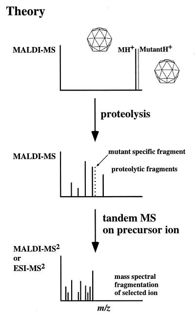
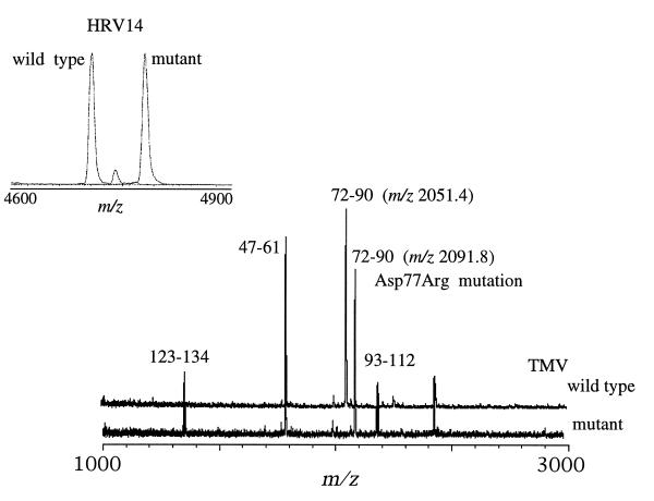
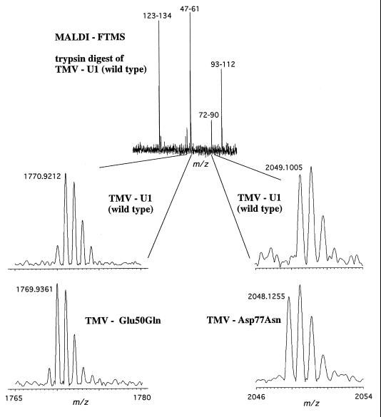
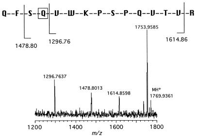
A method to identify mutations of virus proteins by using protein mass mapping is described. Comparative mass mapping was applied to a structural protein of the human rhinovirus Cys1199 --> Tyr mutant and to genetically engineered mutants of tobacco mosaic virus. The information generated from this approach can rapidly identify the peptide or protein containing the mutation and, in cases when nucleic acid sequencing is required, significantly narrows the region of the genome that must be sequenced. High-resolution matrix-assisted laser desorption/ionization (MALDI) mass spectrometry and tandem mass spectrometry were used to identify amino acid substitutions. This method provides valuable information for those analyzing viral variants and, in some cases, offers a rapid and accurate alternative to nucleotide sequencing.
Figures
Similar articles
-
Second-site reversion of a dysfunctional mutation in a conserved region of the tobacco mosaic tobamovirus movement protein.Virology. 1997 May 26;232(1):13-8. doi: 10.1006/viro.1997.8554. Virology. 1997. PMID: 9185584
-
Mapping nucleotides in the 126-kDa protein gene that control the differential symptoms induced by two strains of tobacco mosaic virus.Virology. 1996 Jul 1;221(1):218-25. doi: 10.1006/viro.1996.0368. Virology. 1996. PMID: 8661430
-
Chemically-assisted fragmentation combined with multi-dimensional liquid chromatography and matrix-assisted laser desorption/ionization post source decay, matrix-assisted laser desorption/ionization tandem time-of flight or matrix-assisted laser desorption/ionization tandem mass spectrometry for improved sequencing of tryptic peptides.Eur J Mass Spectrom (Chichester). 2005;11(2):169-79. doi: 10.1255/ejms.734. Eur J Mass Spectrom (Chichester). 2005. PMID: 16046801
-
Elucidation of the genome organization of tobacco mosaic virus.Philos Trans R Soc Lond B Biol Sci. 1999 Mar 29;354(1383):587-91. doi: 10.1098/rstb.1999.0410. Philos Trans R Soc Lond B Biol Sci. 1999. PMID: 10212938 Free PMC article. Review.
-
History of coat protein-mediated protection.Methods Mol Biol. 1998;81:25-38. doi: 10.1385/0-89603-385-6:25. Methods Mol Biol. 1998. PMID: 9760491 Review.
Cited by
-
Mass spectrometry-based identification and whole-genome characterisation of the first pteropine orthoreovirus isolated from monkey faeces in Thailand.BMC Microbiol. 2018 Oct 17;18(1):135. doi: 10.1186/s12866-018-1302-9. BMC Microbiol. 2018. PMID: 30332986 Free PMC article.
-
Getting Hold of the Tobamovirus Particle-Why and How? Purification Routes over Time and a New Customizable Approach.Viruses. 2024 May 30;16(6):884. doi: 10.3390/v16060884. Viruses. 2024. PMID: 38932176 Free PMC article. Review.
-
Mass spectrometry-based proteomics in basic and translational research of SARS-CoV-2 coronavirus and its emerging mutants.Clin Proteomics. 2021 Aug 12;18(1):19. doi: 10.1186/s12014-021-09325-x. Clin Proteomics. 2021. PMID: 34384361 Free PMC article. Review.
-
MASS SPECTROMETRY IN VIROLOGICAL SCIENCES.Mass Spectrom Rev. 2020 Sep;39(5-6):499-522. doi: 10.1002/mas.21617. Epub 2019 Dec 25. Mass Spectrom Rev. 2020. PMID: 31876329 Free PMC article. Review.
-
The unequivocal preponderance of biocomputation in clinical virology.RSC Adv. 2018 May 18;8(31):17334-17345. doi: 10.1039/c8ra00888d. eCollection 2018 May 9. RSC Adv. 2018. PMID: 35539262 Free PMC article. Review.
References
Publication types
MeSH terms
Substances
Grants and funding
LinkOut - more resources
Full Text Sources
Other Literature Sources
