. 1998 Jul 21;95(15):8642-7.
doi: 10.1073/pnas.95.15.8642.

Evidence that phospholipase A2 activity is required for Golgi complex and trans Golgi network membrane tubulation

Affiliations

Evidence that phospholipase A2 activity is required for Golgi complex and trans Golgi network membrane tubulation

P de Figueiredo et al. Proc Natl Acad Sci U S A. .

Abstract

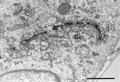
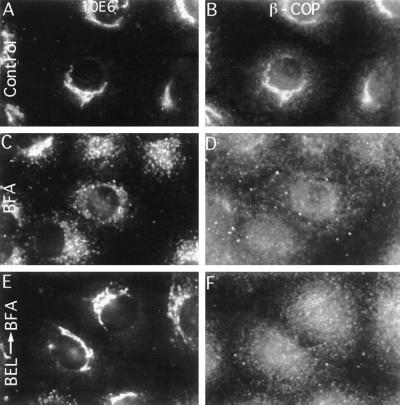
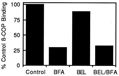
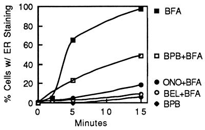
Membrane tubules of uniform diameter (60-80 nm) and various lengths (up to several micrometers) emanate from elements of the Golgi stack and trans Golgi network (TGN). These organelle membrane tubules are thought to be involved in membrane trafficking and maintenance of Golgi/TGN architecture. The number of these tubules, and their frequency of formation, can be greatly enhanced by the fungal metabolite brefeldin A (BFA), an inhibitor of Golgi/TGN-associated coated vesicle formation. We show here that BFA stimulation of Golgi and TGN membrane tubulation, and the resultant retrograde transport of resident Golgi enzymes to the endoplasmic reticulum, was potently inhibited by a number of membrane-permeant antagonists of phospholipase A2 (PLA2; EC 3.1.1.4) activity. In addition, PLA2 inhibitors on their own caused a reversible fragmentation of the Golgi complex into juxtanuclear, stacked cisternal elements. We conclude from these observations that tubulation of Golgi complex and TGN membranes requires a PLA2 activity, and that this activity may participate not only in Golgi tubule-mediated retrograde trafficking to the endoplasmic reticulum, but also in the maintenance of Golgi complex architecture.


Figures

Figure 1
Figure 1
Inhibition of BFA-stimulated Golgi membrane tubulation and reversible fragmentation of intact Golgi complexes by PLA2 inhibitors, as revealed by immunofluorescence of the resident Golgi enzyme αManII. (×720.) (A) Normal control cells. (B) Cells treated with BFA (5 μg/ml for 10 min). (C) Cells pretreated with the PLA2 inhibitor BEL (1 μM for 5 min) before addition of BFA (10 μg/ml) and continued incubation for another 10 min. (D) Cells treated as in C except BEL concentration was 10 μM. (E) Cells treated with the reversible PLA2 inhibitor ONO-RS-082 (ONO) (1 μM), for 30 min. (F) Cells treated with ONO-RS-082 as in E and then washed free of the drug and incubated for another 60 min in normal medium. All incubations were at 37°C.
Figure 2
Figure 2
Effect of the PLA2 inhibitor BEL on αManII localization as revealed by immunoperoxidase staining at the EM level. Cells treated with BEL alone (10 μM for 60 min), although fragmented as shown by immunofluorescence staining, retained typical stacked Golgi cisternae (Gc). (Bar = 1 μm.)
Figure 3
Figure 3
PLA2 inhibitors do not prevent BFA-stimulated redistribution of β-COP from Golgi membranes as shown by double-immunofluorescence labeling with 10E6, a monoclonal antibody against a cis Golgi marker (Left) and polyclonal anti-β-COP antibodies (Right). (A and B) Control cells. (C and D) Cells treated with BFA alone (10 μg/ml for 10 min). (E and F) Cells pretreated with ONO-RS-082 (ONO) (10 μM for 5 min) before addition of BFA (10 μg/ml) and continued incubation for another 10 min. (×540.)
Figure 4
Figure 4
BFA-induced loss of β-COP from isolated Golgi membranes in vitro occurs in the presence of the PLA2 inhibitor BEL. Isolated Golgi complexes were incubated with cytosol alone (Control), cytosol plus BFA (BFA), cytosol plus 25 μM BEL (BEL), or cytosol that was pretreated with BEL (25 μM) before addition of BFA (BEL/BFA). The amount of β-COP on pelleted and washed Golgi membranes was determined by Western blotting.
Figure 5
Figure 5
PLA2 inhibitors prevent tubulation of TGN membranes, but not clathrin redistribution, as revealed by double-immunofluorescence imaging using polyclonal antibodies against the mannose 6-phosphate receptor (Left) and a monoclonal antibody against clathrin heavy chain (Right). (A and B) Control cells. (C and D) Cells treated with BFA alone (10 μg/ml for 10 min). (E and F) Cells pretreated with ONO-RS-082 (ONO) (10 μM for 5 min) before addition of BFA (10 μg/ml for 10 min). (×720.)
Figure 6
Figure 6
Time course of BFA-induced retrograde movement of αManII from the Golgi complex to the ER and its inhibition by various PLA2 inhibitors. Cells were pretreated with various PLA2 inhibitors or solvent controls for 5 min, then BFA (10 μg/ml) was added and cells were incubated for up to 15 min. After treatment, cells were fixed and αManII was localized by immunofluorescence. Cells were then counted to determine the percentage in which αManII exhibited a diffuse, ER-like staining pattern that results from BFA-stimulated, tubule-mediated retrograde transport. BPB, bromophenacyl bromide; ONO, ONO-RS-082.

References

    1. Farquhar M G, Palade G E. Trends Cell Biol. 1998;8:2–10. - PMC - PubMed
    1. Rothman J E, Wieland F T. Science. 1996;272:227–234. - PubMed
    1. Schekman R, Orci L. Science. 1996;271:1526–1533. - PubMed
    1. Mollenhauer H H, Morré D J. J Cell Biol. 1966;29:373–376. - PMC - PubMed
    1. Novikoff P M, Novikoff A B, Quintana N, Hauw J-J. J Cell Biol. 1971;50:859–886. - PMC - PubMed

Publication types