. 1998 Jul 21;95(15):8686-91.
doi: 10.1073/pnas.95.15.8686.

Knockout of the abetalipoproteinemia gene in mice: reduced lipoprotein secretion in heterozygotes and embryonic lethality in homozygotes

Affiliations

Knockout of the abetalipoproteinemia gene in mice: reduced lipoprotein secretion in heterozygotes and embryonic lethality in homozygotes

M Raabe et al. Proc Natl Acad Sci U S A. .

Abstract

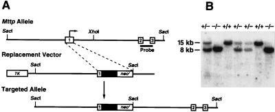
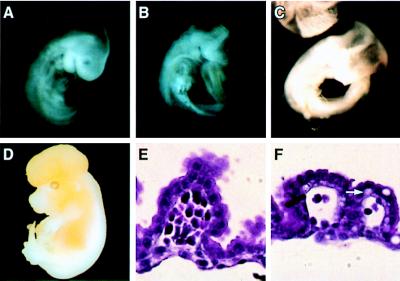
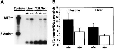
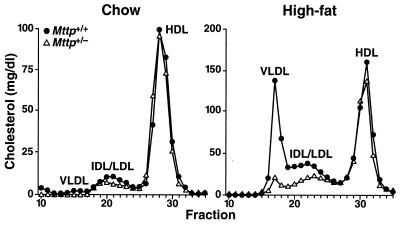
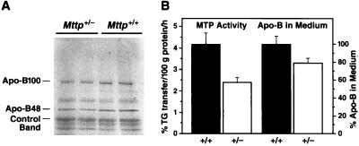
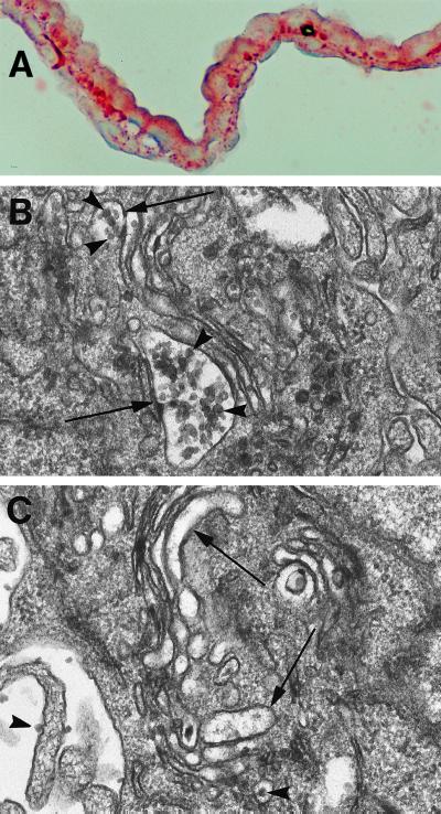
Abetalipoproteinemia, an inherited human disease characterized by a near-complete absence of the apolipoprotein (apo) B-containing lipoproteins in the plasma, is caused by mutations in the gene for microsomal triglyceride transfer protein (MTP). We used gene targeting to knock out the mouse MTP gene (Mttp). In heterozygous knockout mice (Mttp+/- ), the MTP mRNA, protein, and activity levels were reduced by 50%, in both liver and intestine. Compared with control mice (Mttp+/+), chow-fed Mttp+/- mice had reduced plasma levels of low-density lipoprotein cholesterol and had a 28% reduction in plasma apoB100 levels. On a high-fat diet, the Mttp+/- mice exhibited a marked reduction in total plasma cholesterol levels, compared with those in Mttp+/+ mice. Both the livers of adult Mttp+/- mice and the visceral endoderm of the yolk sacs from Mttp+/- embryos manifested an accumulation of cytosolic fat. All homozygous embryos (Mttp-/-) died during embryonic development. In the visceral endoderm of Mttp-/- yolk sacs, lipoprotein synthesis was virtually absent, and there was a marked accumulation of cytosolic fat droplets. In summary, half-normal MTP levels do not support normal levels of lipoprotein synthesis and secretion, and a complete deficiency of MTP causes lethal developmental abnormalities, perhaps because of an impaired capacity of the yolk sac to export lipids to the developing embryo.


Figures

Figure 1
Figure 1
Targeted disruption of the Mttp gene. (A) A map of the Mttp locus, together with the sequence-replacement gene-targeting vector and the targeted Mttp allele. Exons (open boxes), introns (thin line), and start of the MTP ORF (arrow) are indicated. The gene-targeting event replaced exon 1 coding sequences with a cassette containing a neomycin-resistance gene (neo) driven by a phosphoglycerate kinase 1 promoter and a green fluorescent protein cDNA fragment (solid box). Analysis of gene-targeting events and mouse genotyping was performed by Southern blot analysis of SacI-digested genomic DNA, using a 1.3-kb probe (thick bar) extending from exon 2 to exon 3. The probe, which was labeled by random priming, was generated by PCR with oligonucleotides 5′-TGAGCGGCTATACAAGCTCAC-3′ and 5′-CTGGAAGATGCTCTTCTCGC-3′. (B) Southern blot of SacI-digested DNA from E9.5 mouse embryos. The size of the SacI fragment in the targeted allele is 8 kb, versus 15 kb in the wild-type allele.
Figure 2
Figure 2
Phenotype of Mttp−/− embryos. (A) Mttp+/+ embryo at E9.5. (B) An E9.5 Mttp−/− embryo with incomplete anterior closure of the neural tube. (C) An E10.5 Mttp−/− embryo that appeared necrotic, and had incomplete anterior closure of the neural tube. (D) An E14.5 Mttp−/− embryo with exencephalus. (E and F) Hematoxylin and eosin staining of yolk sac membranes from Mttp+/+ (E) and Mttp−/− (F) embryos at E10.5. There were reduced numbers of blood cells in the blood islands of Mttp−/− membranes compared with the Mttp+/+ membranes. Note the vacuolization at the basolateral surfaces of the Mttp−/− visceral endoderm (arrow).
Figure 3
Figure 3
MTP expression levels in Mttp knockout mice. (A) An RNase protection assay for the Mttp mRNA, using liver and yolk sac RNA samples (5 μg of RNA per lane). The first control lane shows the size of the full-length, undigested riboprobes for Mttp and β-actin; the second control lane documents complete digestion of the riboprobes in the presence of RNase but in the absence of cellular RNA. After RNase treatment, the RNA samples were size-fractionated by electrophoresis on 6% denaturing polyacrylamide gels; dried gels were exposed to autoradiographic film and subsequently analyzed with a PhosphorImager. (B) Triglyceride transfer assay with cellular extracts from mouse liver and intestine (n = 10 for each genotype).
Figure 4
Figure 4
Distribution of cholesterol within the plasma lipoproteins of Mttp+/+ and Mttp+/− mice. The plasma lipoproteins were size-fractionated on an FPLC column, and the cholesterol concentration in each fraction was measured enzymatically (15). (Left) Distribution of cholesterol in the plasma of mice fed a chow diet. The study was performed with plasma pooled from three female mice in each group. The FPLC profiles did not suggest that the size of Mttp+/+ and Mttp+/− lipoproteins differed. This conclusion was supported by electron microscopy, which showed the VLDL size distribution to be identical in chow-fed Mttp+/− and Mttp+/+ mice. (Right) Distribution of plasma cholesterol after 5 weeks on a high-fat diet. The study was performed with plasma pooled from four male mice in each group. Once again, electron microscopy revealed no differences in the VLDL size distribution in Mttp+/− and Mttp+/+ mice.
Figure 5
Figure 5
ApoB accumulation in the medium of primary hepatocytes prepared from Mttp+/+ and Mttp+/− mice. (A) Media samples from primary hepatocytes were size-fractionated on SDS-polyacrylamide gels, and the 35S-labeled proteins were visualized by autoradiography. (B) Triglyceride transfer activity and quantification of total apoB secretion in Mttp+/+ and Mttp+/− primary hepatocytes. Triglyceride transfer assays were performed as described (4, 23); the amounts of 35S-labeled apoB48 and apoB100 in the media were quantified with a PhosphorImager and normalized to the amount of a non-apoB protein in the media (“control band”). The amount of total apoB (apoB100 and apoB48) in the medium was 21 ± 5% less in Mttp+/− primary hepatocytes, compared with Mttp+/+ cells (P = 0.05 by t test, n = 4 mice). This experiment was repeated twice, with the same results.
Figure 6
Figure 6
Examination of the yolk sac membranes of mouse embryos at E9.5. (A) Oil red O staining of the yolk sac membrane from an Mttp−/− embryo. (B and C) Electron micrographs of imidazole-stained visceral endoderm cells from the yolk sacs of an Mttp+/− embryo (B) and an Mttp−/− embryo (C). In the Mttp+/− cells, there were invariably clusters of lipid-staining nascent VLDL particles (arrowheads) filling Golgi secretory vesicles (arrows). The lipoproteins were ≈300–800 Å in diameter. The clusters of lipoproteins were invariably absent from Golgi vesicles (arrows) of Mttp−/− yolk sac cells. However, a few lipid-staining particles were identified occasionally in the Golgi compartments and in the adjacent extracellular space (arrowheads) in Mttp−/− yolk sacs. (×34,500.)

References

    1. Kane J P, Havel R J. In: The Metabolic and Molecular Basis of Inherited Disease. Scriver C R, Beaudet A L, Sly W S, Valle D, editors. New York: McGraw–Hill; 1995. pp. 1853–1885.
    1. Illingworth D R, Connor W E, Miller R G. Arch Neurol. 1980;37:659–662. - PubMed
    1. Kayden H J, Traber M G. J Lipid Res. 1993;34:343–358. - PubMed
    1. Wetterau J R, Aggerbeck L P, Bouma M-E, Eisenberg C, Munck A, Hermier M, Schmitz J, Gay G, Rader D J, Gregg R E. Science. 1992;258:999–1001. - PubMed
    1. Shoulders C C, Brett D J, Bayliss J D, Narcisi T M E, Jarmuz A, Grantham T T, Leoni P R D, Bhattacharya S, Pease R J, Cullen P M, et al. Hum Mol Genet. 1993;2:2109–2116. - PubMed

Publication types

LinkOut - more resources