. 1998 Jul 21;95(15):8817-22.
doi: 10.1073/pnas.95.15.8817.

A second major native von Hippel-Lindau gene product, initiated from an internal translation start site, functions as a tumor suppressor

Affiliations

A second major native von Hippel-Lindau gene product, initiated from an internal translation start site, functions as a tumor suppressor

A Schoenfeld et al. Proc Natl Acad Sci U S A. .

Abstract

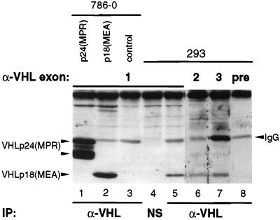
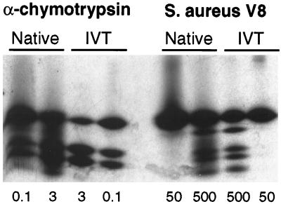
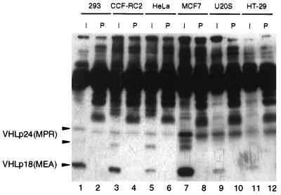
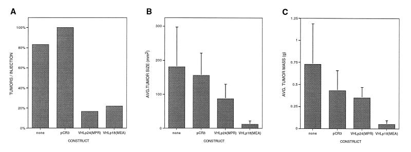
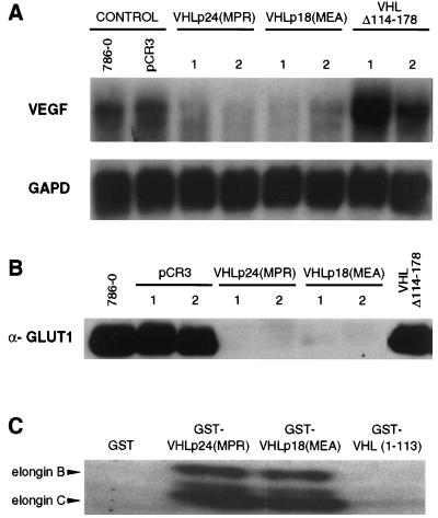
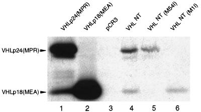
The von Hippel-Lindau (VHL) tumor suppressor gene is inactivated in both sporadic and inherited clear cell renal carcinoma associated with VHL disease. We have identified two distinct native products of the human VHL gene, with apparent molecular masses of 24 and 18 kDa. The 18-kDa VHL protein was more abundant in nearly all cell lines examined. Reintroduction of the 18-kDa VHL gene product into renal carcinoma cells lacking wild-type VHL protein led to down-regulation of vascular endothelial growth factor (VEGF) mRNA and glucose transporter GLUT1 protein and suppressed tumor formation in nude mice. The 18-kDa VHL protein also demonstrated binding to elongins B and C. In an in vitro assay, the second in-frame AUG codon present in VHL mRNA was shown to be necessary and sufficient for production of the 18-kDa VHL protein, consistent with an internal translation mechanism. These data provide evidence for a second major VHL gene product, which contains the functional domains of the VHL gene. Moreover, these results indicate that internal translation initiation is an important mechanism for production of the major VHL protein.


Figures

Figure 1
Figure 1
Identification of native VHL gene products. Total cellular extracts from 786-0 cells stably expressing VHLp24(MPR) (lane 1), VHLp18(MEA) (lane 2), or control 786-0 cells (lane 3) or from 293 cells (lanes 4–8) were immunoprecipitated (IP) with either VHL mAb 12.21 (lanes 1–3, 5–8), or a nonspecific (unrelated) mAb, 12CA5 (NS, lane 4). Immunoprecipitates were separated by electrophoresis on an SDS/12.5% polyacrylamide gel and transferred to a poly(vinylidene difluoride) (PVDF) membrane. The membrane was cut and Western blotting was performed on the pieces by using rabbit polyclonal antisera to peptides in VHL exon 1 (lanes 1–5), exon 2 (lane 6), exon 3 (lane 7), or preimmune rabbit sera (lane 8). VHLp24(MPR) and VHLp18(MEA) are indicated to the left of the blot. Cross-reacting IgG (light chain) is indicated to the right of the blot. NS mAb (lane 4) is subtype IgG2b and did not cross-react with the anti-rabbit IgG secondary antibody.
Figure 2
Figure 2
Peptide mapping of VHLp18(MEA). Native VHL products were immunoprecipitated from metabolically labeled 293 cells, separated on an SDS/13.5% polyacrylamide gel, and visualized by fluorography. Bands comigrating with in vitro translated VHLp18(MEA) were excised from the gel and rehydrated. Partial proteolysis was performed (21) on native and in vitro translated products (IVT) by using amounts of α-chymotrypsin (μg) or S. aureus V8 protease (ng) indicated below the figure. Digestion products were separated on an SDS/15% polyacrylamide gel and visualized by fluorography.
Figure 3
Figure 3
Detection of VHL gene products in cell lines. Immunoprecipitations were performed on indicated cell lines by using either VHL mAb 11E12 (I) or mouse preimmune serum (P), as indicated above lanes. Immune complexes were separated on an SDS/13.5% polyacrylamide gel, transferred to poly(vinylidene difluoride) membrane, and immunoblotted with mAb 12.21. Positions of VHLp24(MPR) and VHLp18(MEA) are indicated to the left of the blot.
Figure 4
Figure 4
Tumor suppression by VHL gene products. Colonies of 786-0 renal carcinoma cells, stably transfected with the indicated plasmids, were assayed for tumor formation in nude mice. Cells (107) of each cell line were trypsinized, washed, resuspended in PBS, and injected into one flank of a nude mouse. Control cells (untransfected 786-0 and pCR3) were injected into the left flank and VHL-expressing cells were injected into the right flank. After 9 weeks, mice were sacrificed and tumors were measured, excised, and weighed. (A) Tumors per injection. Bars represent the number of tumors as a percentage of total injections for each construct [total injections = 18 for pCR3, VHLp24(MPR), and VHLp18(MEA) and 12 for untransfected 786-0 cells]. Three independent clones of VHLp24(MPR) and VHLp18(MEA) and two independent clones of pCR3 were assayed. (B) Average size (mm2) of detected tumors for each construct. (C) Average mass (g) of detected tumors for each construct.
Figure 5
Figure 5
Down-regulation of VEGF and GLUT1 by VHL gene products. 786-0 cells, either untransfected or stably transfected with pCR3, VHLp24(MPR), VHLp18(MEA), or VHL Δ114–178 were assayed for VEGF RNA (A) and GLUT1 protein (B). (A) Northern blot for VEGF. Total RNA was isolated from cell lines (indicated above blot) grown to low confluence (50–70%), and 40 μg of total RNA was loaded per well. Two independent clones of VHLp24(MPR), VHLp18(MEA), and VHL Δ114–178 were assayed. After probing with VEGF, the blot was stripped and rehybridized with a glyceraldehyde-3-phosphate dehydrogenase (GAPD) probe. Probes are indicated to the left of the blot. (B) Anti-GLUT1 Western blot. Each lane was loaded with 25 μg of protein extract from cell lines (indicated above blot). Western blotting was performed with GLUT1 antisera (Alpha Diagnostic International, San Antonio, TX). Two independent clones of pCR3, VHLp24(MPR), and VHLp18(MEA) were assayed. (C) Elongin binding assay. Beads containing 5 μg of GST or GST-VHL fusion proteins (indicated above lanes) were incubated with 20 μl of in vitro translated elongins B and C, washed, separated on an SDS/8–16% polyacrylamide gel, and visualized by fluorography. Positions of the elongin B and C products are indicated to the left.
Figure 6
Figure 6
In vitro translation of VHL gene products. In vitro transcription/translation was performed in the presence of [35S]methionine. Heterologous leader sequence plasmids VHLp24(MPR), VHLp18(MEA), and pCR3 and native 5′ untranslated sequence constructs VHLNT, VHLNT(M54I), and VHLNT(M1I) were used as templates (as indicated above the lanes). Fifteen microliters of the resulting products was immunoprecipitated with mAb 11E12, separated on an SDS/13.5% polyacrylamide gel, and visualized by fluorography. Positions of the VHLp24(MPR) and VHLp18(MEA) products are indicated to the left.

References

    1. Latif F, Tory K, Gnarra J, Yao M, Duh F M, Orcutt M L, Stackhouse T, Kuzmin I, Modi W, Geil L, et al. Science. 1993;260:1317–1320. - PubMed
    1. Linehan W M, Lerman M I, Zbar B. J Am Med Assoc. 1995;273:564–570. - PubMed
    1. Maher E R, Iselius L, Yates J R, Littler M, Benjamin C, Harris R, Sampson J, Williams A, Ferguson S M A, Morton N. J Med Genet. 1991;28:443–447. - PMC - PubMed
    1. Gnarra J R, Tory K, Weng Y, Schmidt L, Wei M H, Li H, Latif F, Liu S, Chen F, Duh F M, et al. Nat Genet. 1994;7:85–90. - PubMed
    1. Whaley J M, Naglich J, Gelbert L, Hsia Y E, Lamiell J M, Green J S, Collins D, Neumann H P, Laidlaw J, Li F P, et al. Am J Hum Genet. 1994;55:1092–1102. - PMC - PubMed

Publication types

LinkOut - more resources