. 1998 Jul 21;95(15):8835-40.
doi: 10.1073/pnas.95.15.8835.

hZAC encodes a zinc finger protein with antiproliferative properties and maps to a chromosomal region frequently lost in cancer

Affiliations

hZAC encodes a zinc finger protein with antiproliferative properties and maps to a chromosomal region frequently lost in cancer

A Varrault et al. Proc Natl Acad Sci U S A. .

Abstract

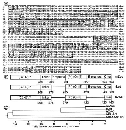
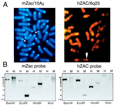
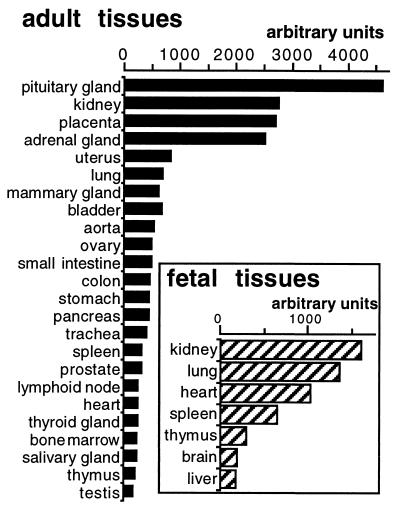
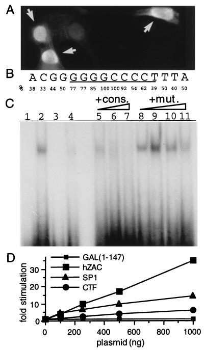
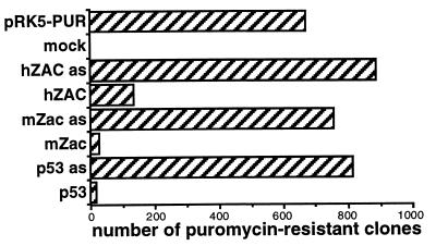
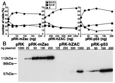
We previously reported the identification of mZac, a novel mouse zinc finger protein that shared with p53 the ability to regulate concomitantly apoptosis and cell cycle progression. We describe here the isolation, chromosomal localization, and functional in vitro characterization of its human homolog. hZAC is a widely expressed zinc finger protein that reveals transactivation and DNA-binding activity. hZAC inhibits tumor cell growth through induction of apoptotic cell death and G1 arrest. Thus hZAC, like its mouse counterpart, displays antiproliferative properties through pathways known to be central to the activity of p53. We mapped hZAC on chromosome 6q24-q25, a region frequently deleted in many solid tumors. Indeed, allelic loss at 6q24-q25 has been shown in breast and ovary cancers, melanomas, astrocytomas, and renal cell carcinomas. Furthermore, Abdollahi et al. [Abdollahi, A., Godwin, A. K., Miller, P. D., Getts, L. A., Schultz, D. C., Tagushi, T., Testa, J. R. & Hamilton, T. C. (1997) Cancer Res. 57, 2029-2034] recently isolated ZAC through its loss of expression in a surface epithelial ovary tumor model and accordingly named it Lot for "lost on transformation." In view of these observations, the functional properties we report here provide further arguments to consider hZAC as a tumor suppressor gene candidate.


Figures

Figure 1
Figure 1
Sequences of the ZAC/LOT family. (A) Sequence alignments of the ZAC/LOT proteins. Human ZAC (hZAC, identical to hLOT1), mouse Zac (mZac), and rat Lot1 (rLot) were aligned according to a Clustal method. Residues that matched hZAC with one distance unit are boxed. (B) Schematic representation of the ZAC/LOT proteins structures. Each domain is indicated as a box: the seven ZF of C2H2 type, the linker region, the Pro repeats present in mZac only, the Pro, Gln and Glu rich region, the Glu clusters absent in hZAC, and the C terminus. The numbering of the amino acid residues is indicated below the boxes. (C) Phylogenetic tree for ZAC/LOT family. Only the ZF domains were taken into account for the analysis.
Figure 2
Figure 2
Localizations of hZAC and mZac genes. (A) Chromosomal localizations. FISH with hZAC probe revealed spots on human chromosome 6 band 6q25; FISH with mZac probe revealed spots on mouse chromosome 10 band 10A2. (B) Southern blots of human and mouse genomic DNA. Human (H) and mouse (M) genomic DNAs were digested with the indicated restriction enzymes, fractionated on a 1% agarose gel, and blotted. The blot was first probed with a radioactive mZac fragment and autoradiographed. Then, the membrane was stripped, and the blot was rehybridized with a hZAC probe and autoradiographed. The same data were obtained with another set of enzymes including NcoI, SacII, SacI, and PstI (not shown).
Figure 3
Figure 3
Human tissue distribution of hZAC. A human RNA blot was hybridized with a hZAC probe and the signal intensity for each dot was measured by using a storage phosphor imaging system (Bio-Rad). In addition, hZAC was weakly expressed in adult peripheric leucocytes, spinal cord, liver, skeletal muscle, and whole brain (less than 150 units). hZAC was expressed in different brain areas: mainly in the occipital lobe, thalamus, and cerebral cortex (100–150 units), in other areas (amygdala, caudate, cerebellum, frontal lobe, hippocampus, medulla oblongata, putamen, substantia nigra, temporal lobe, and subthalamic nuclei) the signals were very weak (<100 units).
Figure 4
Figure 4
hZAC intracellular localization, DNA binding, and transactivation. (A) Nuclear localization of hZAC. Immunocytochemistry of SaOs-2 cells transfected with HA-tagged-hZAC (1 μg plasmid) was performed by using an anti-HA antibody and a fluorescein isothiocyanate-conjugated secondary antibody. As cells were transiently transfected, positive transfected cells (indicated with an arrow) as well as negative nontransfected cells were present on the same sample. (B) Consensus nucleotide sequence for hZAC DNA-binding site. This consensus is derived from the alignment of 13 oligonucleotides selected after screening of a random oligonucleotide library. The frequency of the specific base(s) at each position is indicated. (C) Sequence-specific DNA-binding of GST-hZAC-ZF. An electrophoretic mobility-shift assay was performed. GST-hZAC-ZF (100 ng) was allowed to bind to 40,000 cpm of 32P-labeled double-stranded oligonucleotide containing the consensus binding sequence (GGGGGGCCCC; lane 2) or a mutated sequence where the two most conserved bases at the center of the consensus have been mutated (indicated in bold: GGGGGCGCCC; lane 4). Lanes 1 and 3 represent the control electromobilities of both oligonucleotides in the absence of GST-hZAC-ZF. For competition experiments, GST-hZAC-ZF was preincubated with the indicated competitors: consensus (cons.) or mutated consensus (mut.) oligonucleotide and then allowed to bind to the radio-labeled consensus site. Each competitor was added in molar excess of 1-, 10-,100-, and 1,000-fold in lanes 5 and 8, 6 and 9, 7 and 10, and 11, respectively. (D) Transactivation by hZAC. SaOs-2 cells were transfected with plasmids encoding a fusion protein between the GAL4 DNA-binding domain and either hZAC or the transactivation domains of SP1 or CAAT-box binding transcription factor, together with a luciferase reporter gene under the control of a GAL4 sensitive minimal promoter. Luciferase activity for each condition is indicated as a fold stimulation over basal.
Figure 5
Figure 5
hZAC inhibits colony formation. SaOs-2 cells were transfected with the plasmid encoding the puromycin resistance alone (pRK5-PUR), or together with plasmids encoding hZAC, mZac, and p53 in their sense and antisense (as) orientations. After puromycin selection, the resistant colonies were counted. This experiment is representative of three independent experiments.
Figure 6
Figure 6
hZAC induces a G1 arrest. (A) Cell cycle distribution. SaOs-2 cells were cotransfected with pRK5 encoding CD20 and different amounts of pRK5 encoding either mZac, hZAC, or p53. CD20 positive and propidium iodide-stained cells were analyzed by flow cytometry to measure DNA content. This experiment is representative of three independent experiments. (B) Western blots. SaOs-2 cells were transfected with different amounts of pRK5 encoding mZac, hZAC, or p53, as indicated. Western blot of total cell lysates was performed with anti-HA antibody.
Figure 7
Figure 7
hZAC induces apoptotic cell death. DNA laddering. SaOs-2 cells were transfected with different quantities of vector pRK5 plasmid (1,500 ng, lane 1) or encoding chloramphenicol acetyltransferase (1,500 ng, lane 2), mZac (50, 150, and 500 ng, for lanes 3–5, respectively), hZAC (500, 1,000, and 1,500 ng, for lanes 6–8, respectively), or p53 (100 ng, lane 9). This experiment is representative of three independent experiments.

References

    1. Hartwell L H, Kastan M B. Science. 1994;266:1821–1828. - PubMed
    1. Hussussian C J, Struewing J P, Goldstein A M, Higgins P A, Ally D S, Sheahan M D, Clark W H J, Tucker M A, Dracopoli N C. Nat Genet. 1994;8:15–21. - PubMed
    1. Chandrasekharappa S C, Guru S C, Manickam P, Olufemi S E, Collins F S, Emmert-Buck M R, Debelenko L V, Zhuang Z, Lubensky I A, Liotta L A, et al. Science. 1997;276:404–407. - PubMed
    1. Faienza M F, della Ragione F, Basso G, Coppola B, Miraglia del Giudice E, Schettini F, Iolascon A. Br J Haematol. 1996;93:632–636. - PubMed
    1. Mollenhauer J, Wiemann S, Scheurlen W, Korn B, Hayashi Y, Wilgenbus K K, von Deimling A, Poutska A. Nat Genet. 1997;17:32–39. - PubMed

Publication types

MeSH terms

Associated data