. 1998 Jul 21;95(15):8991-6.
doi: 10.1073/pnas.95.15.8991.

Cloning and characterization of a cAMP-specific cyclic nucleotide phosphodiesterase

Affiliations

Cloning and characterization of a cAMP-specific cyclic nucleotide phosphodiesterase

S H Soderling et al. Proc Natl Acad Sci U S A. .

Abstract

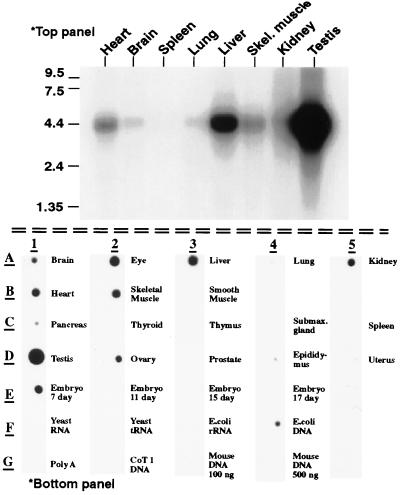
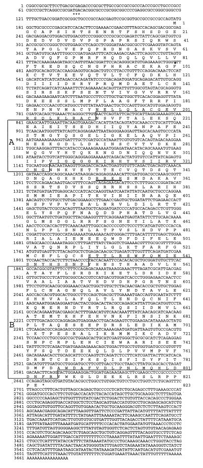
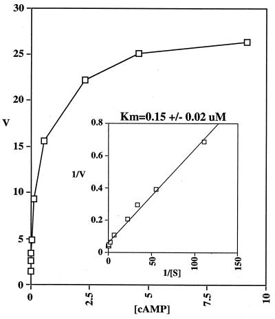
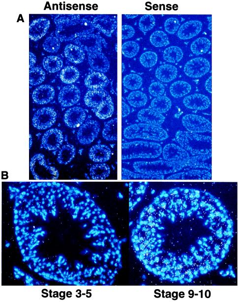
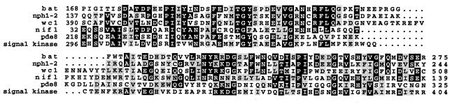
Cyclic nucleotide phosphodiesterases (PDEs) regulate intracellular levels of cAMP and cGMP by hydrolyzing them to their corresponding 5' monophosphates. We report here the cloning and characterization of a novel cAMP-specific PDE from mouse testis. This unique phosphodiesterase contains a catalytic domain that overall shares <40% sequence identity to the catalytic domain of all other known PDEs. Based on this limited homology, this new PDE clearly represents a previously unknown PDE gene family designated as PDE8. The cDNA for PDE8 is 3,678 nucleotides in length and is predicted to encode an 823 amino acid enzyme. The cDNA includes a full ORF as it contains an in-frame stop codon before the start methionine. PDE8 is specific for the hydrolysis of cAMP and has a Km of 0.15 microM. Most common PDE inhibitors are ineffective antagonists of PDE8, including the nonspecific PDE inhibitor 3-isobutyl-1-methylxanthine. Dipyridamole, however, an inhibitor that is generally considered to be relatively specific for the cGMP selective PDEs, does inhibit PDE8 with an IC50 of 4.5 microM. Tissue distribution studies of 22 different mouse tissues indicates that PDE8 has highest expression in testis, followed by eye, liver, skeletal muscle, heart, 7-day embryo, kidney, ovary, and brain in decreasing order. In situ hybridizations in testis, the tissue of highest expression, shows that PDE8 is expressed in the seminiferous epithelium in a stage-specific manner. Highest levels of expression are seen in stages 7-12, with little or no expression in stages 1-6.


Figures

Figure 1
Figure 1
Tissue distribution of PDE8 by (Upper) Northern blot analysis and (Lower) Poly(A) RNA dot blot. Top panel: Northern blot of poly(A) RNA from multiple mouse tissues. A single band corresponding to a message size of 4.4 kb is seen. Bottom panel: Poly(A) RNA dot blot from mouse tissues. PDE8 expression is detected in a variety of tissues. Relative intensity of signal as determined by using the National Institutes of Health image program was testis>eye>skeletal muscle>heart>embryo 7d>kidney>ovary>brain. Note the signal found in the negative control “E. coli DNA” is most likely due to the fact that the probe template was prepared from E. coli hosts.
Figure 2
Figure 2
Nucleotide and amino acid sequence of PDE8. Note the in-frame stop codon above the predicted start methionine is marked by ∗∗∗. Underlined amino acid residues correspond to a nuclear localization consensus sequence. (A) The N-terminal PAS domain is homologous to many signaling proteins (see Fig. 5). (B) The catalytic domain of PDE8. ∗ denotes the stop codon at the end of the ORF.
Figure 3
Figure 3
PDE8 Kinetics. Concentration range of cAMP was 0.01–9.2 μM. Inset is a Lineweaver-Burke plot.
Figure 4
Figure 4
In situ hybridization of mouse testis with PDE8. Approximate stage was determined by using Hoechst 33258 to stain nuclei. (A) Antisense and sense view under fluorescent light to observe the nuclear stain (blue) and bright field to observe probe signal (white dots). Note the low probe signal with the sense probe and the uneven probe signal within the semifierous tubules in the antisense field, indicating a spatial and temporal regulation of PDE8 expression. (B) Representative high magnification fluorescent and bright field of a early stage tubule and late stage tubule. Note the lack of probe signal (white dots) above background at stage 3–5 vs. the intense signal at stage 9–10.
Figure 5
Figure 5
Multiple sequence alignment of PDE PAS/PAC domain with homologous sequences. Abbreviations: Bat; putative bacterio-opsin activator from Halobacterium halobium (accession no. M23247). Nph1–2; nonphototropic hypocotyl 1 from Avena sativa (accession no. AF033097). Wc1; white collar 1 from Neurospora crassa (accession no. X94300). NifL; NifL from Enterobacter agglomerans (accession no. X89104). Signal kinase; putative signal-transducing histidine kinase—from Archaeoglobus fulgidus (accession no. AE000990).

References

    1. Beavo J A. Physiol Rev. 1995;75:725–748. - PubMed
    1. Soderling S H, Bayuga S J, Beavo J A. J Biol Chem. 1998;273:15553–15558. - PubMed
    1. Fisher D A, Smith J F, Pillar J S, St. Denis S H, Cheng J B. J Biol Chem. 1998;273:15559–15564. - PubMed
    1. Kauvar L M. J Neurosci. 1982;2:1347–1358. - PMC - PubMed
    1. Shaulsky G, Escalante R, Loomis W F. Proc Natl Acad Sci USA. 1996;93:15260–15265. - PMC - PubMed

Publication types

Substances

Associated data