

Inducible nitric oxide synthase-deficient mice develop enhanced type 1 cytokine-associated cellular and humoral immune responses after vaccination with attenuated Schistosoma mansoni cercariae but display partially reduced resistance
- PMID: 9673227
- PMCID: PMC108380
- DOI: 10.1128/IAI.66.8.3510-3518.1998
Inducible nitric oxide synthase-deficient mice develop enhanced type 1 cytokine-associated cellular and humoral immune responses after vaccination with attenuated Schistosoma mansoni cercariae but display partially reduced resistance
Abstract
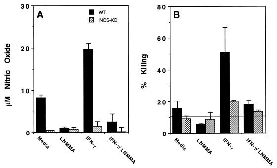
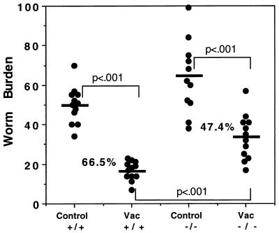
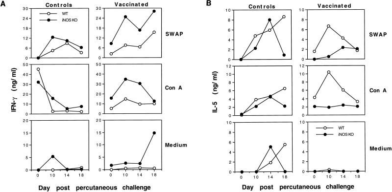
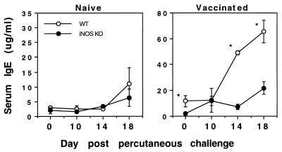
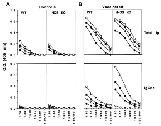
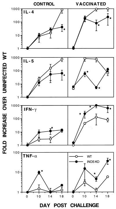
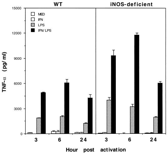
High levels of nitric oxide (NO) are produced by inducible nitric oxide synthase (iNOS) in response to activating signals from Th1-associated cytokines and play an important role in cytotoxicity and cytostasis against many pathogenic microorganisms. In addition to its direct effector function, NO serves as a potent immunoregulatory factor. NO produced by gamma interferon-activated macrophages immobilizes and kills Schistosoma mansoni larvae, and several studies have indicated a role for this pathway in protective immunity against this parasite. The potential regulatory influence of NO in immunity to S. mansoni is less well understood. In this study, we have used iNOS-deficient mice to determine the role of NO in mice vaccinated with irradiated cercariae of S. mansoni. We show by enzyme-linked immunosorbent assay and reverse transcriptase PCR analysis that vaccinated iNOS-deficient mice develop exacerbated type 1 cytokine responses in the lungs, the site where resistance to infection is primarily manifested. In addition, parasite-specific immunoglobulin G2a (IgG2a) and IgG2b antibody responses were significantly increased in vaccinated iNOS-deficient animals and total IgE antibody levels in serum were decreased relative to those in wild-type controls. Surprisingly, since resistance in this vaccine model is largely Th1 dependent and since Th1-related cellular and humoral immune responses were found to be exacerbated in vaccinated iNOS-deficient mice, vaccine-elicited protective immunity against challenge infection was found to be reduced. These findings demonstrate that iNOS plays a paradoxical role in immunity to S. mansoni, both in the effector mechanism of resistance and in the down regulation of the type 1 cytokine response, which is ultimately required for NO production.
Figures

Similar articles
-
Nitric oxide produced in the lungs of mice immunized with the radiation-attenuated schistosome vaccine is not the major agent causing challenge parasite elimination.Immunology. 1998 Jan;93(1):55-63. doi: 10.1046/j.1365-2567.1998.00405.x. Immunology. 1998. PMID: 9536119 Free PMC article.
-
Studies with double cytokine-deficient mice reveal that highly polarized Th1- and Th2-type cytokine and antibody responses contribute equally to vaccine-induced immunity to Schistosoma mansoni.J Immunol. 1999 Jul 15;163(2):927-38. J Immunol. 1999. PMID: 10395689
-
Endothelial cells are activated by cytokine treatment to kill an intravascular parasite, Schistosoma mansoni, through the production of nitric oxide.Proc Natl Acad Sci U S A. 1994 Feb 1;91(3):999-1003. doi: 10.1073/pnas.91.3.999. Proc Natl Acad Sci U S A. 1994. PMID: 7508126 Free PMC article.
-
The debate over the effector function of eosinophils in helminth infection: new evidence from studies on the regulation of vaccine immunity by IL-12.Mem Inst Oswaldo Cruz. 1997;92 Suppl 2:105-8. doi: 10.1590/s0074-02761997000800014. Mem Inst Oswaldo Cruz. 1997. PMID: 9698921 Review.
-
T cell derived cytokines in lung-phase immunity to Schistosoma mansoni.Mem Inst Oswaldo Cruz. 1992;87 Suppl 4:105-9. doi: 10.1590/s0074-02761992000800015. Mem Inst Oswaldo Cruz. 1992. PMID: 1343880 Review.
Cited by
-
Bacille Calmette-Guérin (BCG)-associated inflammation and fibrosis: modulation by recombinant BCG expressing interferon-gamma (IFN-gamma).Clin Exp Immunol. 2000 Jan;119(1):92-8. doi: 10.1046/j.1365-2249.2000.01100.x. Clin Exp Immunol. 2000. PMID: 10606969 Free PMC article.
-
In the absence of CD154, administration of interleukin-12 restores Th1 responses but not protective immunity to Schistosoma mansoni.Infect Immun. 2007 Jul;75(7):3539-47. doi: 10.1128/IAI.00252-07. Epub 2007 May 7. Infect Immun. 2007. PMID: 17485453 Free PMC article.
-
Role of gamma interferon in cellular immune response against murine Encephalitozoon cuniculi infection.Infect Immun. 1999 Apr;67(4):1887-93. doi: 10.1128/IAI.67.4.1887-1893.1999. Infect Immun. 1999. PMID: 10085032 Free PMC article.
-
Nitric oxide-dependent changes in Schistosoma mansoni gene expression.Mol Biochem Parasitol. 2006 Dec;150(2):367-70. doi: 10.1016/j.molbiopara.2006.08.003. Epub 2006 Aug 28. Mol Biochem Parasitol. 2006. PMID: 16962671 Free PMC article. No abstract available.
-
Nitric oxide selectively decreases interferon-gamma expression by activated human T lymphocytes via a cGMP-independent mechanism.Immunology. 1999 Nov;98(3):393-9. doi: 10.1046/j.1365-2567.1999.00895.x. Immunology. 1999. PMID: 10583599 Free PMC article.
References
-
- Albina J E, Abate J A, Henry W L., Jr Nitric oxide production is required for murine resident peritoneal macrophages to suppress mitogen-stimulated T cell proliferation. Role of IFN-gamma in the induction of the nitric oxide-synthesizing pathway. J Immunol. 1991;147:144–148. - PubMed
-
- Albina J E, Cui S, Mateo R B, Reichner J S. Nitric oxide-mediated apoptosis in murine peritoneal macrophages. J Immunol. 1993;150:5080–5085. - PubMed
-
- Bergquist N R. Schistosomiasis vaccine development: approaches and prospects. Mem Inst Oswaldo Cruz Rio J. 1995;90:221–227. - PubMed
MeSH terms
Substances
LinkOut - more resources
Full Text Sources
Molecular Biology Databases
