. 1998 Aug;66(8):3523-6.
doi: 10.1128/IAI.66.8.3523-3526.1998.

CD1 presents antigens from a gram-negative bacterium, Haemophilus influenzae type B

Affiliations

CD1 presents antigens from a gram-negative bacterium, Haemophilus influenzae type B

R M Fairhurst et al. Infect Immun. 1998 Aug.

Abstract

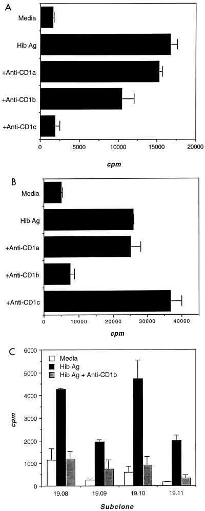
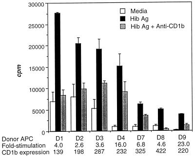
Human CD1 is a family of nonpolymorphic major histocompatibility complex class I-like molecules capable of presenting mycobacterial lipids, including lipoarabinomannan (LAM), to double-negative (DN; CD4(-) CD8(-)) as well as CD8(+) T cells. Structural similarities between LAM and the capsular polysaccharides of gram-negative bacteria led us to consider the latter as candidate CD1 ligands. We derived two CD1-restricted DN T-cell populations which proliferated to Haemophilus influenzae type b (Hib) antigen. One T-cell population also proliferated to proteinase K-treated Hib antigen, suggesting that it recognized a nonpeptide. Our work thus expands the universe of T cell antigens to include nonpeptides distinct from mycobacterial lipids and suggests a potential role for CD1-restricted T cells in immunity to Hib.


Figures

FIG. 1
FIG. 1
Hib Ag-specific, CD1-restricted proliferation of Hib16 (A) and Hib19 (B) T-cell populations and of Hib19 T-cell clones (C). T cells were cocultured with irradiated CD1+ APC and Hib Ag in the presence of blocking antibodies specific for CD1a, CD1b, or CD1c. During the last 4 h of a 72-h incubation, cultures were pulsed with tritiated thymidine, the cellular incorporation of which was determined by scintillation counting and expressed as counts per minute.
FIG. 2
FIG. 2
Hib Ag-specific, CD1b-restricted proliferation of Hib19 T cells. Hib19 T cells were cocultured with irradiated CD1+ APC (isolated from seven unrelated donors [D1 to D4 and D7 to D9]) and Hib Ag in the presence of CD1b blocking antibodies. Proliferation was measured as for Fig. 1. The fold stimulation and mean fluorescence intensity of CD1 expression are indicated for each donor.
FIG. 3
FIG. 3
Validation that proteinase K-treated Hib Ag lacks immunogenic peptides. Hib19 was tested for the ability to respond to Hib Ag and to proteinase K (PK)-treated Hib Ag. Validation that proteinase K-treated Hib Ag lacked immunogenic peptides was achieved by using Hib Ag-specific CD4+ T-cell lines (2, 3, 9, and 11) derived from the same donor. Proliferation was measured as for Fig. 1.

Similar articles

Cited by

References

    1. Anderson P, Smith D H. Isolation of the capsular polysaccharide from culture supernatant of Haemophilus influenzae type b. Infect Immun. 1977;15:472–477. - PMC - PubMed
    1. Arakere G, Lee A L, Frasch C E. Involvement of phospholipid end groups of group C Neisseria meningitidis and Haemophilus influenzae type b polysaccharides in association with isolated outer membranes and in immunoassays. J Bacteriol. 1994;176:691–695. - PMC - PubMed
    1. Beckman E M, Porcelli S A, Morita C T, Behar S M, Furlong S T, Brenner M B. Recognition of a lipid antigen by CD1-restricted alpha beta+ T cells. Nature. 1994;372:691–694. - PubMed
    1. Beckman E M, Brenner M B. MHC class I-like, class II-like and CD1 molecules: distinct roles in immunity. Immunol Today. 1995;16:349–352. - PubMed
    1. Behar S M, Porcelli S A, Beckman E M, Brenner M B. A pathway of costimulation that prevents anergy in CD28− T cells. B7-independent costimulation of CD1-restricted T cells. J Exp Med. 1995;182:2007–2018. - PMC - PubMed

Publication types

MeSH terms