. 1998 Aug;66(8):3591-6.
doi: 10.1128/IAI.66.8.3591-3596.1998.

Receptor-mediated recognition and uptake of iron from human transferrin by Staphylococcus aureus and Staphylococcus epidermidis

Affiliations

Receptor-mediated recognition and uptake of iron from human transferrin by Staphylococcus aureus and Staphylococcus epidermidis

B Modun et al. Infect Immun. 1998 Aug.

Abstract

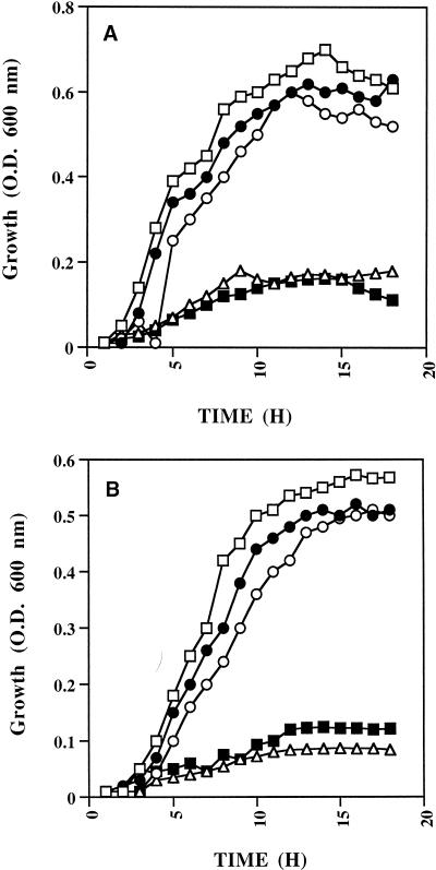
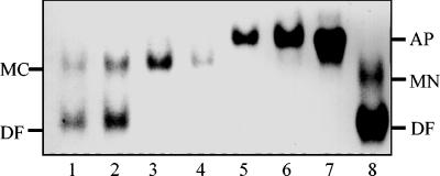
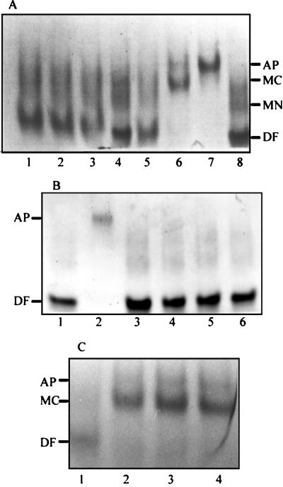
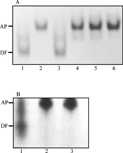
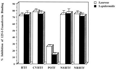
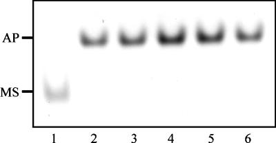
Staphylococcus aureus and Staphylococcus epidermidis both recognize and bind the human iron-transporting glycoprotein, transferrin, via a 42-kDa cell surface protein receptor. In an iron-deficient medium, staphylococcal growth can be promoted by the addition of human diferric transferrin but not human apotransferrin. To determine whether the staphylococcal transferrin receptor is involved in the removal of iron from transferrin, we employed 6 M urea-polyacrylamide gel electrophoresis, which separates human transferrin into four forms (diferric, monoferric N-lobe, and monoferric C-lobe transferrin and apotransferrin). S. aureus and S. epidermidis but not Staphylococcus saprophyticus (which lacks the transferrin receptor) converted diferric human transferrin into its apotransferrin form within 30 min. During conversion, iron was removed sequentially from the N lobe and then from the C lobe. Metabolic poisons such as sodium azide and nigericin inhibited the release of iron from human transferrin, indicating that it is an energy-requiring process. To demonstrate that this process is receptor rather than siderophore mediated, we incubated (i) washed staphylococcal cells and (ii) the staphylococcal siderophore, staphyloferrin A, with porcine transferrin, a transferrin species which does not bind to the staphylococcal receptor. While staphyloferrin A removed iron from both human and porcine transferrins, neither S. aureus nor S. epidermidis cells could promote the release of iron from porcine transferrin. In competition binding assays, both native and recombinant N-lobe fragments of human transferrin as well as a naturally occurring human transferrin variant with a mutation in the C-lobe blocked binding of 125I-labelled transferrin. Furthermore, the staphylococci removed iron efficiently from the iron-loaded N-lobe fragment of human transferrin. These data demonstrate that the staphylococci efficiently remove iron from transferrin via a receptor-mediated process and provide evidence to suggest that there is a primary receptor recognition site on the N-lobe of human transferrin.


Figures

FIG. 1
FIG. 1
Utilization of transferrin-bound iron by S. aureus (A) and S. epidermidis (B). Staphylococci were grown in RPMI (□), iron-depleted RPMI (▵), iron-depleted RPMI supplemented with human diferric transferrin (○), human apotransferrin (■), or ferric chloride (•). Growth was monitored by measuring optical density (O.D.) at 600 nm at 1-h intervals over 18 h.
FIG. 2
FIG. 2
Time course experiment showing (by 6 M urea–polyacrylamide gel electrophoresis) the removal of iron from diferric human transferrin by S. aureus. Staphylococci (108 CFU/ml) were incubated in PBS buffer plus glucose with 250 μg of diferric human transferrin at 37°C for 0 (lane 1), 5 (lane 2), 10 (lane 3), 20 (lane 4), 30 (lane 5), and 60 (lane 6) min. Cells were pelleted by centrifugation, and the supernatant was loaded onto a 6 M urea–polyacrylamide gel. Human apotransferrin (lane 7) and a mixture of human diferric and monoferric N-lobe transferrin (lane 8) are shown as controls. The positions of diferric (DF), monoferric N-lobe (MN), and monoferric C-lobe (MC) transferrins and apotransferrins (AP) are indicated on the right- and left-hand sides of the figure.
FIG. 3
FIG. 3
Gels (6 M urea–polyacrylamide) showing the removal of iron from diferric human transferrin by S. aureus in the presence and absence of glucose (A) and the effect of sodium azide on the receptor-mediated removal of iron from human transferrin by S. aureus (B and C). (A) Staphylococci were incubated in PBS without (lanes 1 to 3) or with (lanes 5 to 7) glucose at 37°C for 0 (lanes 1 and 5), 20 (lanes 2 and 6), and 60 (lanes 3 and 7) min and pelleted by centrifugation, and the supernatant was loaded onto a urea-polyacrylamide gel. As controls, lanes 4 and 8 contain diferric human transferrin. The positions of each of the four forms of transferrin are indicated on the right-hand side. (B) Staphylococci (108 CFU/ml) were incubated in PBS plus glucose at 37°C with diferric human transferrin in the presence of 6 mM sodium azide for 0 (lane 3), 5 (lane 4), 20 (lane 5), and 60 (lane 6) min. Cells were pelleted, and the supernatant was loaded onto the urea-polyacrylamide gel. Diferric human transferrin (lane 1) and human apotransferrin (lane 2) were loaded as controls. (C) Staphylococci (108 CFU/ml) were incubated in PBS plus glucose at 37°C with diferric human transferrin in the presence of 2 mM sodium azide for 0 (lane 1), 5 (lane 2), 20 (lane 3), and 60 (lane 4) min. Cells were pelleted, and the supernatant was loaded onto the urea-polyacrylamide gel. The positions of diferric and monoferric C transferrins and apotransferrins are indicated on the left-hand side. Abbreviations: AP, human apotransferrin; DF, diferric human transferrin; MC, monoferric C-lobe transferrin; MN, monoferric N-lobe transferrin.
FIG. 4
FIG. 4
Gels (6 M urea–polyacrylamide) showing the removal of iron from human (A) and porcine (B) transferrin by the staphylococcal siderophore staphyloferrin A. The siderophore was incubated in PBS plus glucose at 37°C for 60 min. (A) Samples were removed at 0 (lane 3), 5 (lane 4), 20 (lane 5), and 60 (lane 6) min. Lanes 1 and 2 contain diferric transferrin and human apotransferrin as controls. (B) Samples were removed after incubation with porcine transferrin for 0 (lane 1), 20 (lane 2), and 60 (lane 3) min. The positions of diferric transferrin (DF) and apotransferrin (AP) are indicated on the left-hand side.
FIG. 5
FIG. 5
Whole-cell competition binding assay showing the inhibition of binding of human 125I-transferrin to staphylococci by the subtilisin-generated and recombinant N lobe of human transferrin. Staphylococci (108 CFU/ml) were incubated with 125I-transferrin (4 nM) in the presence of 700 nM of one of the following unlabelled transferrins: human (HTf), human C-lobe variant (CVHTf), porcine (POTf), subtilisin generated human N lobe (NSHTf), and recombinant human N lobe (NRHTf). After 30 min at 37°C, bacteria were pelleted and the amount of cell-associated 125I-transferrin was determined. Data presented are the means of three independent experiments + standard deviations (error bars).
FIG. 6
FIG. 6
Gel (6 M urea–polyacrylamide) showing the removal of iron from the N-lobe fragment of human transferrin. S. aureus (108 CFU/ml) were incubated in PBS plus glucose and 250 μg of the recombinant monoferric N lobe for intervals of 0 (lane 1), 5 (lane 2), 10 (lane 3), 20 (lane 4), 30 (lane 5), and 60 (lane 6) min. Bacterial cells were pelleted, and the supernatant was loaded onto the urea gel. The positions of the iron-loaded N lobe (MS) and apoprotein (AP) are indicated on the left-hand side.

Similar articles

Cited by

References

    1. Adhikari P, Berish S A, Nowalk A J, Veraldi K L, Morse S A, Mietzner T A. The fbpABC locus of Neisseria gonorrhoeae functions in the periplasm-to-cytosol transport of iron. J Bacteriol. 1996;178:2145–2149. - PMC - PubMed
    1. Aisen P. Physical biochemistry of the transferrins: update, 1984–1988. Phys Bioinorg Chem Ser. 1989;5:353–371.
    1. Alcantara J, Schryvers A B. Transferrin binding protein 2 interacts with both the N-lobe and C-lobe of ovotransferrin. Microb Pathog. 1996;209:73–85. - PubMed
    1. Archibald F S, DeVoe I W. Iron acquisition by Neisseria meningitidis in vitro. Infect Immun. 1980;27:322–334. - PMC - PubMed
    1. Brock J H, Williams P H, Liceaga J, Woolridge K G. Relative availability of transferrin-bound iron and cell-derived iron to aerobactin-producing and enterochelin-producing strains of Escherichia coli and to other microorganisms. Infect Immun. 1991;59:3185–3190. - PMC - PubMed

Publication types