. 1998 Aug;66(8):3673-81.
doi: 10.1128/IAI.66.8.3673-3681.1998.

Listeria monocytogenes stimulates mucus exocytosis in cultured human polarized mucosecreting intestinal cells through action of listeriolysin O

Affiliations

Listeria monocytogenes stimulates mucus exocytosis in cultured human polarized mucosecreting intestinal cells through action of listeriolysin O

M H Coconnier et al. Infect Immun. 1998 Aug.

Abstract

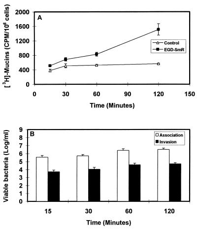
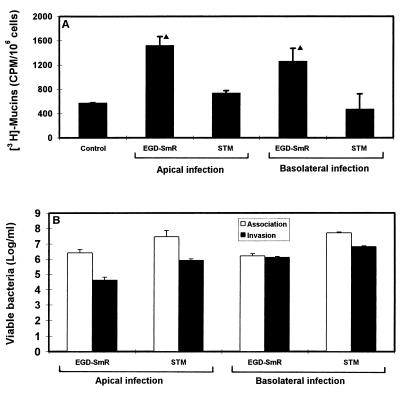
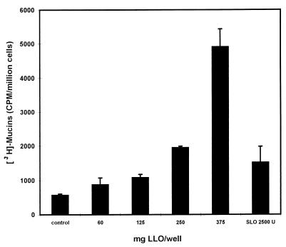
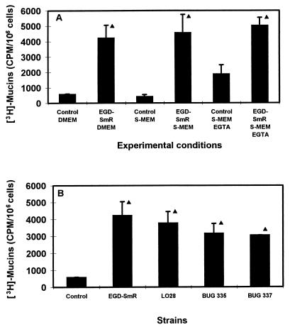
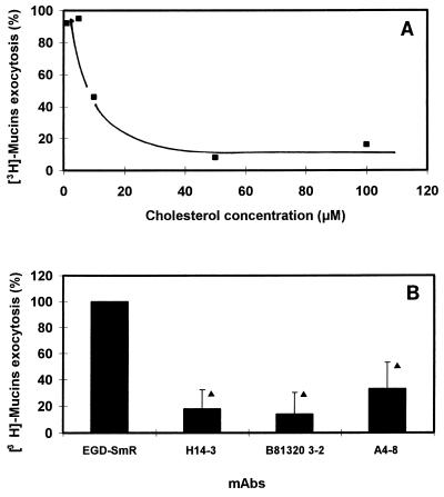
When the intracellular pathogen Listeria monocytogenes infects cultured human mucosecreting polarized HT29-MTX cells apically, it induces the stimulation of mucus exocytosis without cell entry. Using a set of isogenic mutants and purified listeriolysin O (LLO), we identified the L. monocytogenes thiol-activated exotoxin LLO as the agonist of mucus secretion. We demonstrated that the LLO-induced mucus exocytosis did not result from the LLO membrane-damaging activity. We found that LLO-induced mucus exocytosis is an event requiring the binding of LLO to a brush border-associated receptor and membrane oligomerization of the exotoxin. By a pharmacological approach, we demonstrated that no regulatory system or intracellular transducing signal known to be involved in control of mucin exocytosis was activated by LLO. Based on the present data, the stimulatory action of LLO on mucin exocytosis could be accounted for either by an unknown signaling system which remains to be determined or by direct action of LLO with the membrane vesicle components involved in the intracellular vesicular transport of mucins.


Figures

FIG. 1
FIG. 1
Scanning electron micrographs of cultured human mucosecreting HT29-MTX cells infected apically with L. monocytogenes EGD-SmR or treated with L. monocytogenes EGD-SmR-SCS. (A) Infected cells showing secreted mucus at the vicinity of the infecting bacteria. (B) Untreated cells showing a regular brush border. (C and D) Low and high magnifications of cells subjected to L. monocytogenes EGD-SmR-SCS, showing a great increase in mucus secretion. LLO-induced mucin exocytosis stimulation was observed after immunolabeling of secreted mucins at the HT29-MTX cell surface with the anti-mucin M1 MAb (not shown).
FIG. 2
FIG. 2
Mucin exocytosis (A) and bacterial cell association or cell entry (B) in HT29-MTX cells apically infected with 108 CFU of L. monocytogenes EGD-SmR per ml as a function of the time postinfection.
FIG. 3
FIG. 3
Mucin exocytosis (A) and bacterial cell association or cell entry (B) in HT29-MTX cells infected apically or basolaterally with 108 CFU of L. monocytogenes EGD-SmR and S. typhimurium SL1344 per ml. STM, S. typhimurium. Statistical analysis between control and infected cells was performed by Student’s t test: ▴, significant difference (P < 0.01).
FIG. 4
FIG. 4
Dose-dependent induction of mucin exocytosis in HT29-MTX cells subjected apically to purified LLO.
FIG. 5
FIG. 5
Role of the LLO membrane-damaging activity in L. monocytogenes EGD-SmR-SCS-induced stimulation of mucus exocytosis in HT29-MTX cells. (A) Mucus exocytosis promoted by EGD-SmR-SCS applied apically to HT29-MTX cells in the presence of a low-Ca2+ cell culture medium (S-DMEM) and after chelation of the extracellular Ca2+ by EGTA. (B) Mucus exocytosis promoted by the spent culture supernatant of the EGD-SmR, LO28, and mutant strains: BUG 335 (LO28 Trp-491 to Ala) and BUG 337 (LO28 Trp-492 to Ala). ▴, significant difference (P < 0.01) from control.
FIG. 6
FIG. 6
L. monocytogenes EGD-SmR-SCS-induced mucus exocytosis occurs after LLO interaction with an HT29-MTX brush border-associated receptor. (A) Stimulation of mucus exocytosis promoted by EGD-SmR-SCS applied apically to the HT29-MTX cells in the presence of increasing concentrations of cholesterol. (B) Stimulation of mucus exocytosis promoted by EGD-SmR-SCS applied apically to the HT29-MTX cells in the presence of neutralizing MAbs against LLO. MAbs H14-3 (5 μg/ml) and B8B20-3-2 (5 μg/ml) allow LLO binding and prevent cell lysis. A4-8 (10 μg/ml) inhibits both LLO binding and cell lysis. ▴, significant difference (P < 0.01) from EGD-SmR.

Similar articles

Cited by

References

    1. Alouf J E, Geoffroy C. Structure activity relationships in sulphydryl-activated toxins. In: Allouf J E, Fehrenbach F J, Freer J H, Jeljaszewicz J, editors. Bacterial protein toxins. London, United Kingdom: Academic Press, Ltd.; 1984. pp. 165–171.
    1. Augeron C, Voisin T, Maoret J J, Berthon B, Laburthe M, Laboisse C. Neurotensin and neuromedin N stimulate mucin ouput in the human goblet cell clone Cl.16E: a neurotensin receptor-mediated event. Am J Physiol. 1992;262:G470–G476. - PubMed
    1. Bahkdi S, Tranum-Jensen J, Sziegoleit A. Mechanism of membrane damage by streptolysin O. Infect Immun. 1985;47:52–60. - PMC - PubMed
    1. Beubler E, Kollar G, Saria A, Bukhave K, Rask-Madsen J. Involvement of 5-hydroxytryptamine, prostaglandin-E2, and cyclic adenosine monophosphate in cholera toxin-induced fluid secretion in the small intestine of the rat in vivo. Gastroenterology. 1989;96:368–376. - PubMed
    1. Böhm S K, Grady E F, Bunnett N W. Regulatory mechanisms that modulate signalling by G-protein-coupled receptors. Biochem J. 1997;322:1–18. - PMC - PubMed

LinkOut - more resources