. 1998 Aug;66(8):3682-8.
doi: 10.1128/IAI.66.8.3682-3688.1998.

Molecular basis for antigenic variation of a protective strain-specific antigen of Ehrlichia risticii

Affiliations

Molecular basis for antigenic variation of a protective strain-specific antigen of Ehrlichia risticii

B Biswas et al. Infect Immun. 1998 Aug.

Abstract

Ehrlichia risticii, the causative agent of Potomac horse fever, has recently been isolated from many vaccinated horses with typical clinical signs of the disease. The heterogeneity of the E. risticii isolates obtained from the vaccinated horses necessitates the identification of the molecular basis of strain variations to elucidate the vaccine failure and to aid in the development of an efficient vaccine against this disease. As an attempt, two major cross-reacting surface antigen genes of 50- and 85-kDa antigens, present separately in strains 25-D (isolated in 1984) and 90-12 (isolated in 1990 from a vaccinated horse), respectively, were cloned and sequenced. A comparative sequence analysis revealed differences and similarities between these two antigens with strain-specific sizes (SSA). The 2.5- and 1.6-kb genes coding for the 85- and 50-kDa proteins, respectively, contained many different tandem repeats. The identical repeat motifs were more frequent in the middle of both genes, but the numbers and positions of the repeats were altogether different in the genes. Many of these direct repeats of both genes had exact sequence homology and coded for the same amino acids. The homology of the 5'- and 3'-flanking regions of the two genes was greater than that of the regions in the central part of the genes. A comparative analysis of the deduced amino acid sequences of these two antigen genes indicated eight common domains, which were designated identical domains. Although the sequence homologies of these identical domains were the same, the positions of the domains in their respective strains were completely different. This finding might be one of the bases of antigenic variation between the strains. In addition, there were a few unique regions in both antigen genes where no sequence homology existed. These specific regions were designated unique domains. The 50-kDa protein had two such unique domains, and the 85-kDa protein had six such unique domains. The presence of such unique domains contributed to the large size variation of these SSA. The cross-reactivity of recombinant proteins confirmed the presence of conserved epitopes between these two antigens. The SSA have been determined to be apparent protective antigens of E. risticii.


Figures

FIG. 1
FIG. 1
Composite profile of three overlapping clones of the 85-kDa-antigen gene and their positions with respect to the gene. The two overlapping clones, pB85-17 and pB85-11, were identified from the genomic library of strain 90-12. The insert, pCR85-3, was amplified directly from the genomic DNA of strain 90-12 by use of two specific primers, 50-C and 50-D. The sequence information of primer 50-C was obtained from the upstream region of the 50-kDa-antigen gene sequence, whereas that of primer 50-D was obtained from the sequence of clone pB85-17.
FIG. 2
FIG. 2
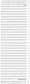
Nucleotide sequence of the 50-kDa-antigen gene and flanking regions and deduced amino acid sequence. Putative −10, −35, and ribosome binding site regions are underlined, and the putative start of transcription is denoted with +1. Regions of dyad symmetry and adjacent thymine-rich regions are underlined.
FIG. 3
FIG. 3
Bar diagrams of direct repeats of 50- and 85-kDa-antigen gene sequences. The repeats are categorized according to their base-pair size, denoted by the numbers (55 to 10) placed under the bars. Each bar was then subdivided according to the sequence homologies of the repeats, as denoted by the block(s) in each bar.
FIG. 4
FIG. 4
Schematic diagram of the diversity and similarity of the 50- and 85-kDa antigens. The numbers at the top show ID. The homology in the amino acid sequences for the corresponding ID regions of the tested antigens varied from more than 94% to less than 79%. The unmarked areas indicate no homology between the two antigens.
FIG. 5
FIG. 5
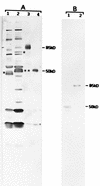
Western blot analysis of recombinant-expressed proteins of the 50- and 85-kDa-antigen genes. Lanes 1 contain the antigens from E. risticii 25-D, and lanes 2 contain the antigens from E. risticii 90-12. Lanes 3 and 4 contain E. coli-expressed recombinant 85- and 50-kDa fusion proteins, respectively. (A) Blot reacted with sera from strain 90-12-infected mice. The asterisk in lane 1 represents the location of the 50-kDa antigen. This antigen is not distinguishable as a separate band, as it overlaps with the 51-kDa antigen band. The single asterisk in lane 2 represents the location of the 85-kDa antigen band, and double asterisks represent the location of the 51-kDa antigen band. The changes in the sizes of the recombinant 85- and 50-kDa proteins (lanes 3 and 4, respectively) are in close agreement with the expected fusion product of the pRSET-ABC expression system, which adds a 3.5-kDa protein mass to its fusion products. (B) Blot reacted with recombinant 85-kDa clone-specific antibody from strain 90-12. The recombinant 85-kDa clone-specific antibody recognized only the 50-kDa antigen in strain 25-D (lane 1) and only the 85-kDa antigen in strain 90-12 (lane 2). Numbers at right are molecular masses in kilodaltons.

Similar articles

Cited by

References

    1. Anderson B E, McDonald G A, Jones D C, Regnery R L. A protective protein antigen of Rickettsia rickettsii has tandemly repeated, near-identical sequences. Infect Immun. 1990;58:2760–2769. - PMC - PubMed
    1. Birnboim H C, Doly J. A rapid alkaline extraction procedure for screening recombinant plasmid DNA. Nucleic Acids Res. 1979;7:1513–1523. - PMC - PubMed
    1. Biswas B, Mukherjee D, Mattingly-Napier B L, Dutta S K. Diagnostic application of polymerase chain reaction for detection of Ehrlichia risticii in equine monocytic ehrlichiosis (Potomac horse fever) J Clin Microbiol. 1991;29:2228–2233. - PMC - PubMed
    1. Dutta S K, Myrup A C, Rice R M, Roble M G, Hammond R C. Experimental reproduction of Potomac horse fever in horses with a newly isolated Ehrlichia organism. J Clin Microbiol. 1985;22:265–269. - PMC - PubMed
    1. Dutta S K, Rice R M, Hughes T D, Savage P K, Myrup A C. Detection of serum antibodies against Ehrlichia risticii in Potomac horse fever by enzyme-linked-immunosorbent assay. Vet Immunol Immunopathol. 1987;14:85–92. - PubMed

Associated data