. 1998 Aug;66(8):3758-66.
doi: 10.1128/IAI.66.8.3758-3766.1998.

Interactions of the invasive pathogens Salmonella typhimurium, Listeria monocytogenes, and Shigella flexneri with M cells and murine Peyer's patches

Affiliations

Interactions of the invasive pathogens Salmonella typhimurium, Listeria monocytogenes, and Shigella flexneri with M cells and murine Peyer's patches

V B Jensen et al. Infect Immun. 1998 Aug.

Abstract

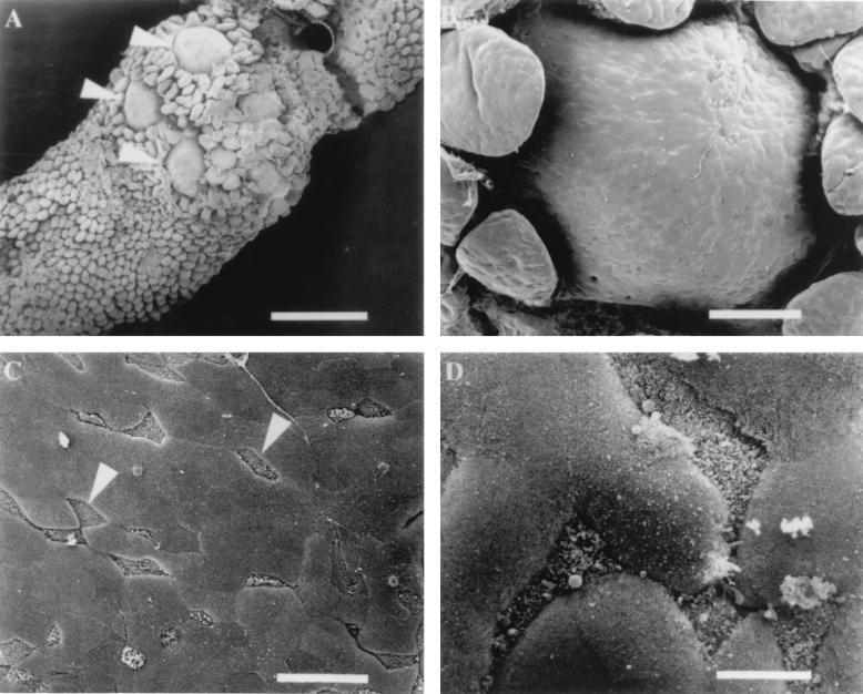
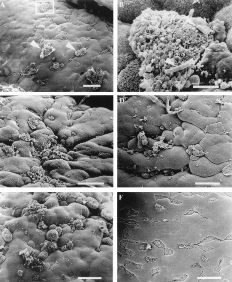
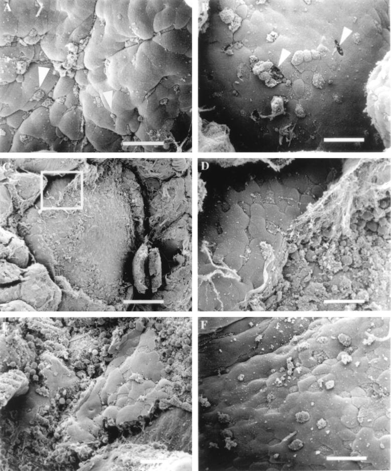
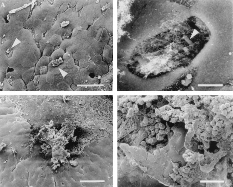
Invasive enteric bacteria must pass through the intestinal epithelium in order to establish infection. It is becoming clear that a common target for intestinal mucosa penetration is the specialized epithelial cell of Peyer's patches, the M cell. In order to gain a better understanding of how bacteria interact with M cells, we have compared the interactions of Salmonella typhimurium, Listeria monocytogenes, and Shigella flexneri with M cells by using a murine ligated-loop model. Our results indicate that S. typhimurium possesses a highly efficient mechanism for M cell entry that targets and destroys these cells, while L. monocytogenes and S. flexneri appear to be internalized into M cells in a less disruptive fashion. Early uptake of Listeria or Shigella into M cells appeared to lead to the death of some cells, as evidenced by the appearance of holes in the intestinal epithelium. At later time points, the follicle-associated epithelium of animals infected with these bacteria displayed extensive destruction. These data indicate that enteric pathogens use different strategies to interact with M cells and initiate infection of a host.


Figures

FIG. 1
FIG. 1
Scanning electron micrographs of murine Peyer’s patch tissue. (A) Luminal side of Peyer’s patch containing several domes (arrowheads) surrounded by villi. Bar, 700 μm. (B) Epithelial surface of a lymphoid follicle dome that contains numerous M cells and enterocytes randomly distributed. Bar, 100 μm. (C) Region of the FAE containing several M cells (arrowheads) that can be distinguished by short microvilli which give an irregular appearance. Bar, 20 μm. (D) Higher magnification of M cells and enterocytes. M cell microvilli create a rough irregular appearance in comparison to adjacent enterocytes which look smooth in texture. Bar, 6 μm.
FIG. 2
FIG. 2
Electron micrographs of mouse FAE infected with invasive S. typhimurium SL1344. (A) Membrane rearrangements are visible on the surfaces of some M cells (arrowheads) 30 min after infection with S. typhimurium SL1344. (B) Higher magnification of the region in the inset from panel A. Rod-shaped organisms (arrowheads) can be observed adhering to M cell debris. Bar, 3 μm. (C) Effects of S. typhimurium 60 min after infection. (D) Tissue infected for 90 min with invasive Salmonella. M cells with a normal apical appearance are difficult to find, and the majority of cells display membrane disruptions and blebbing. (E) Infection of ligated loop with invasive Salmonella for 120 min. A quantitative increase in the number of M cells displaying membrane changes and a qualitative increase in the size of apical membrane disruption are evident. (F) Mouse intestinal tissue inoculated with bacterial growth broth for 30 to 120 min was examined for the appearance of uninfected tissue. The tissue shown has been inoculated with broth for 120 min and appears completely normal. The M cells are clearly visible, and the microvilli have the typical appearance of uninfected tissue. Bar, 20 μm unless otherwise indicated.
FIG. 3
FIG. 3
Effect of L. monocytogenes on murine FAE. (A) Mouse tissue infected for 30 min with L. monocytogenes 10403S. Some M cells display aberrant microvilli that are rounded in appearance (arrowheads). (B) After 60 min of infection, the presence of the bacteria appears to have destroyed the integrity of the FAE by creating holes (arrowheads) in the dome epithelium where M cells once resided. (C) Listeria-induced destruction of the FAE 90 min postinfection. Bar, 100 μm. (D) Higher-magnification view of the inset in Fig. C. At the periphery of the dome, a few M cells and enterocytes are still intact. (E) Tissue infected for 120 min with L. monocytogenes. (F) Tissue infected with the L. monocytogenes listeriolysin O-negative mutant DP-L2161 did not induce epithelial necrosis after 120 min of infection. Bar, 20 μm (unless otherwise indicated).
FIG. 4
FIG. 4
Ligated loops inoculated with invasive S. flexneri 2a. (A) Like L. monocytogenes, S. flexneri induces small changes in the appearance of the M cell surface (arrowheads). In addition, it was possible to occasionally find holes in FAE infected with Shigella. (B) Higher-magnification view of a perforation in the intestinal epithelium with an organism (arrowhead) located within. Bar, 4 μm. (C) Shigella-induced destruction of the FAE. Bar, 40 μm. (D) Higher-magnification view of the damage induced by S. flexneri on the epithelium of the lymphoid follicle. Only a few intact M cells and enterocytes remain intact. Bar, 20 μm unless otherwise indicated.

References

    1. Autenrieth I B, Firsching R. Penetration of M cells and destruction of Peyer’s patches by Yersinia enterocolitica: an ultrastructural and histological study. J Med Microbiol. 1996;44:285–294. - PubMed
    1. Bajaj V, Hwang C, Lee C A. hilA is a novel ompR/toxR family member that activates the expression of Salmonella typhimurium invasion genes. Mol Microbiol. 1995;18:715–727. - PubMed
    1. Bishop D K, Hinrichs D J. Adoptive transfer of immunity to Listeria monocytogenes. The influence of in vitro stimulation on lymphocyte subset requirements. J Immunol. 1987;139:2005–2009. - PubMed
    1. Bockman D E, Cooper M D. Pinocytosis by epithelium associated with lymphoid follicles in the bursa of Fabricius, appendix, and Peyer’s patches. An electron microscopic study. Am J Anat. 1973;136:455–477. - PubMed
    1. Clark M A, Jepson M A, Simmons N L, Booth T A, Hirst B H. Differential expression of lectin-binding sites defines mouse intestinal M cells. J Histochem Cytochem. 1993;41:1679–1687. - PubMed

Publication types

LinkOut - more resources