. 1998 Aug;66(8):3900-8.
doi: 10.1128/IAI.66.8.3900-3908.1998.

Increased levels of intracellular calcium are not required for the formation of attaching and effacing lesions by enteropathogenic and enterohemorrhagic Escherichia coli

Affiliations

Increased levels of intracellular calcium are not required for the formation of attaching and effacing lesions by enteropathogenic and enterohemorrhagic Escherichia coli

C Bain et al. Infect Immun. 1998 Aug.

Abstract

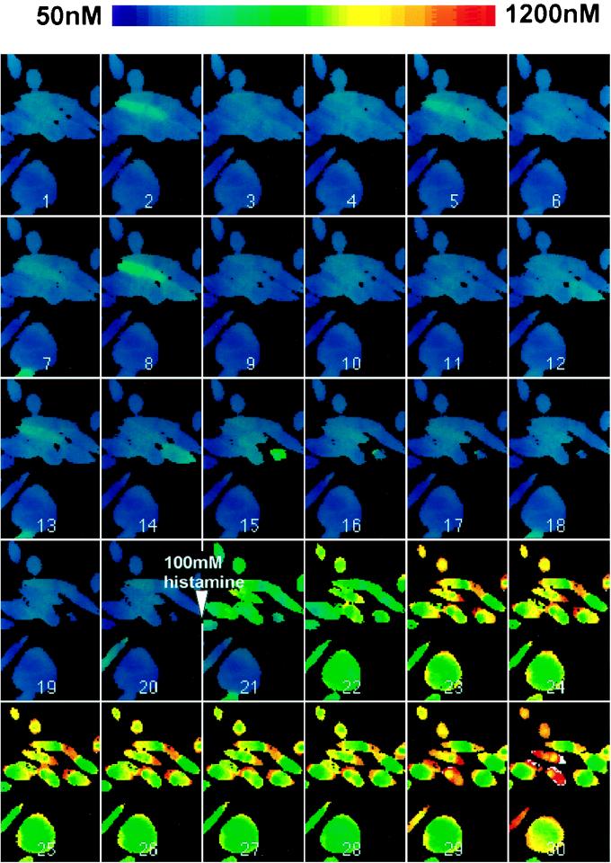
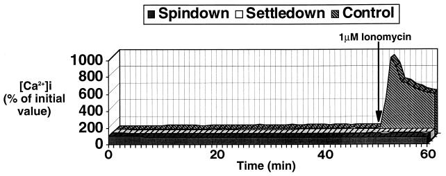
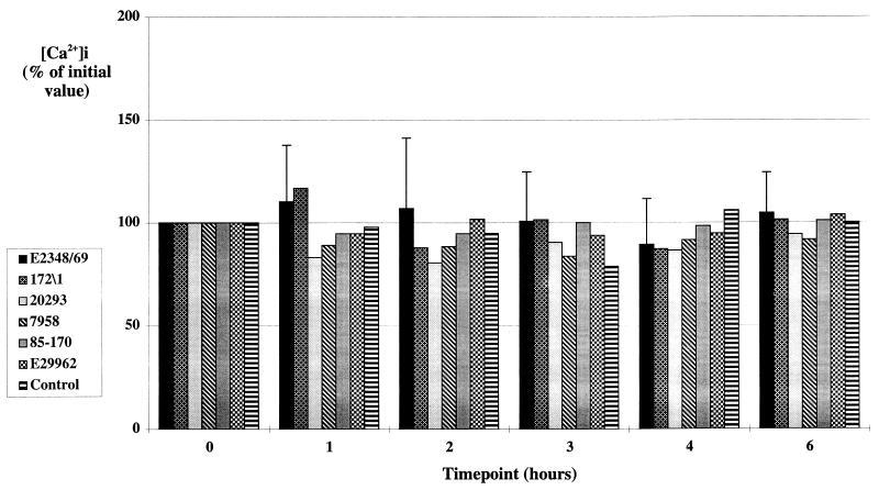
Elevated concentrations of intracellular calcium ([Ca]i) have been implicated as an important signalling event during attaching and effacing (A/E) lesion formation by enteropathogenic Escherichia coli (EPEC). The highly localized nature of the cytoskeletal and cell surface alterations occurring during A/E lesion formation suggests that there should be equally localized EPEC-induced signalling events. To analyze further the calcium responses to infection of HEp-2 cells by EPEC, we employed calcium-imaging fluorescence microscopy, which allows both temporal and spatial measurements of [Ca]i in live cells. Using this imaging technique, not only were we unable to detect any significant elevation in [Ca]i at sites of A/E EPEC adhesion, but, with several different classical EPEC and enterohemorrhagic E. coli (EHEC) strains and three different infection procedures, each of which resulted in extensive A/E bacterial adhesion, we were unable to detect any significant alterations in [Ca]i in infected cells compared to uninfected cells. In addition, chelation of intracellular free calcium with bis-(aminophenoxy)-ethane-N,N,N',N'-tetraacetic acid (BAPTA) did not, as previously reported, prevent A/E lesion formation. We conclude that increased [Ca]i are not required for A/E lesion formation by EPEC and EHEC.


Figures

FIG. 1
FIG. 1
FAS tests performed at the end of typical calcium-imaging experiments. The micrographs show fluorescence (left panels) and corresponding phase-contrast images (right panels) of HEp-2 cell monolayers spindown infected with EPEC strain E2348/69 for 1 h (A), gradually infected with EPEC strain E2348/69 for 6 h (B), gradually infected with EHEC strain E29962 for 6 h (C), and treated with BAPTA and then spindown infected with EPEC strain E2348/69 for 1 h (D). Each infection procedure resulted in good colonization of cells with A/E EPEC (A and B), although adhesion by EHEC strains was significantly less than that by EPEC strains (C); characteristic EPEC-induced actin accretion occurred in BAPTA-treated cells (D). Magnification, ×320.
FIG. 2
FIG. 2
Montage of pseudocolor ratio images taken at 2-min intervals, showing [Ca]i in HEp-2 cells spindown infected with E2348/69 for 1 h. The images show no significant alterations in [Ca]i during the period of A/E lesion formation (images 5 to 10). After 40 min (image 21), 100 mM histamine was added to the cells, resulting in a large increase in [Ca]i.
FIG. 3
FIG. 3
Mean [Ca]i (n = 4) in control HEp-2 cell monolayers and HEp-2 cell monolayers infected for 1 h with EPEC strain E2348/69 by the spindown and settledown procedures. No significant alteration in [Ca]i was seen throughout the 1-h infection period, during which extensive A/E adhesion occurred. An ∼10-fold increase in [Ca]i occurred in control cells following the addition of 1 μM ionomycin, confirming the responsiveness of the cells and the detection system.
FIG. 4
FIG. 4
Mean [Ca]i (n = 2) in control HEp-2 cell monolayers and HEp-2 cell monolayers infected for up to 6 h with four different EPEC strains and two different EHEC strains by using the gradual infection procedure. Values are presented as percentages of the mean [Ca]i in the cells at time zero. No significant change in [Ca]i was seen with any of the six strains over the 6-h time course. Error bars are illustrated for strain E2348/69 and are typical of those for the remainder of the strains.
FIG. 5
FIG. 5
Effect of BAPTA on histamine-induced rises in HEp-2 cell [Ca]i. Histamine (H) (1 mM) was added to both control and BAPTA-treated cells after 7 and 18 min (arrows). Histamine-stimulated rises in [Ca]i were completely abrogated in the BAPTA-treated cells, whereas considerable increases in [Ca]i were seen in the untreated cells.
FIG. 6
FIG. 6
Mean percent cell death (n = 2) in HEp-2 cell monolayers gradually infected with EPEC strain E2348/69 for 6 h. Less than 5% cell death occurred over the 6-h time period of the experiment.

References

    1. Baldwin T J, Lee-Delaunay M B, Knutton S, Williams P H. Calcium-calmodulin dependence of actin accretion and lethality in cultured HEp-2 cells infected with enteropathogenic Escherichia coli. Infect Immun. 1993;61:760–763. - PMC - PubMed
    1. Baldwin T J, Ward W, Aitken A, Knutton S, Williams P H. Elevation of intracellular free calcium levels in HEp-2 cells infected with enteropathogenic Escherichia coli. Infect Immun. 1991;59:1599–1604. - PMC - PubMed
    1. Berridge M J. Elementary and global aspects of calcium signalling. J Exp Med. 1997;200:315–319. - PubMed
    1. Burgess D R, Prum B E. Reevaluation of brush border motility: calcium induces core filament solation and microvillar vesiculation. J Cell Biol. 1982;94:97–107. - PMC - PubMed
    1. Clerc P L, Berthon B, Claret M, Sansonetti P J. Internalization of Shigella flexneri into HeLa cells occurs without an increase in the cytosolic Ca2+ concentration. Infect Immun. 1989;57:2919–2922. - PMC - PubMed

Publication types

LinkOut - more resources