. 1998 Aug;66(8):3925-30.
doi: 10.1128/IAI.66.8.3925-3930.1998.

Passive immunization with antibodies against three distinct epitopes on Plasmodium yoelii merozoite surface protein 1 suppresses parasitemia

Affiliations

Passive immunization with antibodies against three distinct epitopes on Plasmodium yoelii merozoite surface protein 1 suppresses parasitemia

L M Spencer Valero et al. Infect Immun. 1998 Aug.

Abstract

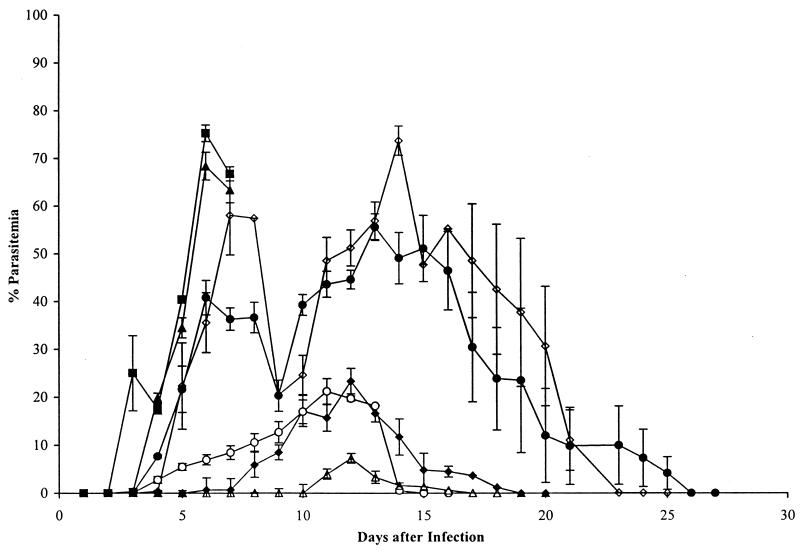
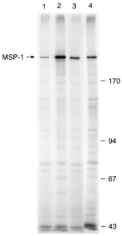
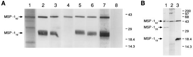
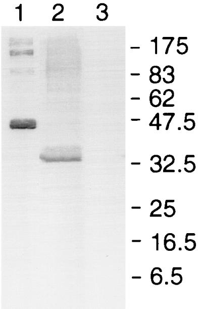
We have produced monoclonal antibodies against Plasmodium yoelii merozoite surface protein 1 (MSP-1) and have assessed their ability to suppress blood stage parasitemia by passive immunization. Six immunoglobulin G antibodies were characterized in detail: three (B6, D3, and F5) were effective in suppressing a lethal blood stage challenge infection, two (B10 and G3) were partially effective, and one (B4) was ineffective. MSP-1 is the precursor to a complex of polypeptides on the merozoite surface; all of the antibodies bound to this precursor and to an approximately 42-kDa fragment (MSP-142) that is derived from the C terminus of MSP-1. MSP-142 is further cleaved to an N-terminal approximately 33-kDa polypeptide (MSP-133) and a C-terminal approximately 19-kDa polypeptide (MSP-119) comprised of two epidermal growth factor (EGF)-like modules. D3 reacted with MSP-142 but not with either of the constituents MSP-133 and MSP-119, B4 recognized an epitope within the N terminus of MSP-133, and B6, B10, F5, and G3 bound to MSP-119. B10 and G3 bound to epitopes that required both C-terminal EGF-like modules for their formation, whereas B6 and F5 bound to epitopes in the first EGF-like module. These results indicate that at least three distinct epitopes on P. yoelii MSP-1 are recognized by antibodies that suppress parasitemia in vivo.


Figures

FIG. 1
FIG. 1
Course of P. yoelii YM infection in groups of 10 BALB/c mice injected intraperitoneally with solutions of MAbs, followed by intravenous challenge with 5 × 10³ parasitized erythrocytes per mouse. Mice in each group received 0.5 mg of IgG in PBS intraperitoneally on days −1, 0, and +1 relative to the day of parasite challenge (day 0). B6 (▵), F5 (⧫), and D3 (○) substantially modified the course of the infection; B10 (•) and G3 (◊) were less effective. The parasitemias in mice treated with B4 (■) and in mice treated with an irrelevant MAb (data not shown) were indistinguishable from that in the group that received PBS (▴). All of the mice cleared the infection, except for those that received B4 and PBS, which died on day 7, and 40% of the mice treated with G3, which died on days 8 and 9. Each point represents the geometric mean parasitemia of mice in each group at the time after parasite challenge, and the vertical bars indicate the standard errors.
FIG. 2
FIG. 2
Immunofluorescence patterns of MAbs reactive with MSP-1 on methanol–acetone-fixed smears of erythrocytes infected with P. yoelii YM. The smears were incubated with MAb B6 (a) or MAb B4 (b), and antibody binding was detected with a second fluorescein isothiocyanate-labelled antibody. (c and d) Corresponding smears stained with DAPI to locate DNA. Note the reaction with schizonts and ring stages in panel a and with schizonts alone in panel b, despite the presence of young parasites detected in panel d by nuclear staining. No specific fluorescence was detected with an irrelevant first antibody or with the second labelled antibody alone (data not shown).
FIG. 3
FIG. 3
The MAbs immunoprecipitate MSP-1 from extracts of 35S-labelled P. yoelii YM-infected erythrocytes solubilized with buffer containing detergent. In this experiment the following MAbs were used: B6 (lane 1), D3 (lane 2), F5 (lane 3), and G3 (lane 4). The immunoprecipitates were analyzed by SDS-PAGE on a 7.5% polyacrylamide gel and detected by fluorography. The position of MSP-1 on the left and the mobilities of standard molecular mass markers (Pharmacia) (in kilodaltons) on the right are indicated. MSP-1 was also precipitated by B4, B10, 25.1, and a polyclonal antiserum to GST–MSP-119 but not by antibodies in normal mouse serum (not shown).
FIG. 4
FIG. 4
The MAbs react with fragments of MSP-1 in an extract of P. yoelii YM merozoites by Western blotting. (A) The samples were subjected to SDS-PAGE (without prior reduction) on a 5 to 15% polyacrylamide gradient gel, blotted onto nitrocellulose and then probed with MAb B4 (lane 1), B6 (lane 2), B10 (lane 3), D3 (lane 4), F5 (lane 5), or G3 (lane 6) or with polyclonal anti-GST–MSP-119 (lane 7) and normal mouse serum (lane 8). The positions of MSP-142 and MSP-119 on the left and the mobilities of standard molecular mass markers (in kilodaltons) on the right are indicated. (B) The merozoites were incubated in vitro to allow processing to continue before analysis. The samples were fractionated on a 12.5% polyacrylamide gel, blotted, and probed with MAbs B4 (lane 1), D3 (lane 2), and F5 (lane 3). The positions of MSP-142, MSP-133, and MSP-119 on the left and the mobilities of standard molecular mass markers (in kilodaltons) on the right are indicated.
FIG. 5
FIG. 5
MAb B4 binds to an epitope in the N-terminal end of MSP-133. Recombinant proteins consisting of MSP-133 sequences fused to GST were fractionated by SDS-PAGE on a 12.5% polyacrylamide gel under reducing conditions, transferred to nitrocellulose, and then probed with the MAb. Lane 1, PyMSP133A (Thr1415 to His1619); lane 2, PyMSP133B (Thr1415 to Lys1513); lane 3, PyMSP133C (Ala1527 to His1619). The mobilities of molecular mass standards (New England Biolabs) (in kilodaltons) are shown. The antibody did not react with GST alone, none of the other MAbs reacted with GST–MSP-133A, and each of the fusion proteins was detected with antibody specific for GST (data not shown).

Similar articles

Cited by

References

    1. Akerstrom B, Brodin T, Reis K, Bjorck L. Protein G: a powerful tool for binding and detection of monoclonal and polyclonal antibodies. J Immunol. 1985;135:2589–2592. - PubMed
    1. Blackman M J, Heidrich H-G, Donachie S, McBride J S, Holder A A. A single fragment of a malaria merozoite surface protein remains on the parasite surface during red cell invasion and is the target of invasion-inhibiting antibodies. J Exp Med. 1990;172:379–382. - PMC - PubMed
    1. Blackman M J, Ling I T, Nicholls S C, Holder A A. Proteolytic processing of the Plasmodium falciparum merozoite surface protein-1 produces a membrane-bound fragment containing two epidermal growth factor-like domains. Mol Biochem Parasitol. 1991;49:29–34. - PubMed
    1. Blackman M J, Scott-Finnigan T J, Shai S, Holder A A. Antibodies inhibit the protease-mediated processing of a malaria merozoite surface protein. J Exp Med. 1994;180:389–393. - PMC - PubMed
    1. Blackman M J, Dennis E D, Hirst E M A, Kocken C H, Scott-Finnigan T J, Thomas A W. Plasmodium knowlesi: secondary processing of the malaria merozoite surface protein-1. Exp Parasitol. 1996;83:229–239. - PubMed

Publication types

MeSH terms