

Manganese inhibits mitochondrial aconitase: a mechanism of manganese neurotoxicity
- PMID: 9675333
- PMCID: PMC4126159
- DOI: 10.1016/s0006-8993(98)00481-8
Manganese inhibits mitochondrial aconitase: a mechanism of manganese neurotoxicity
Abstract
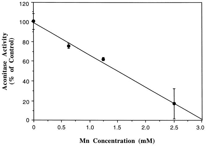
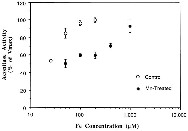
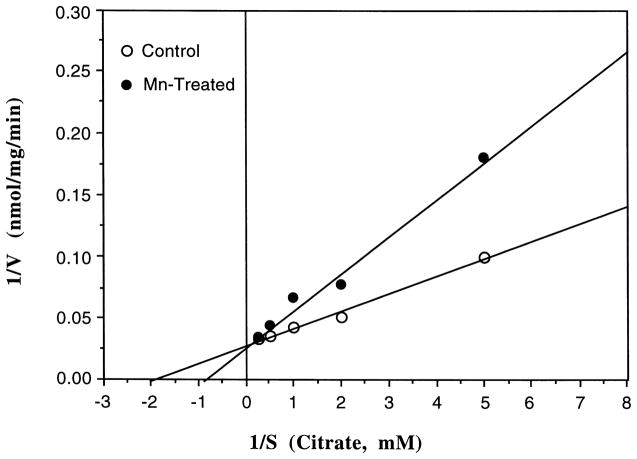
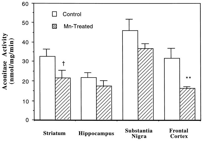
The symptoms of Mn-induced neurotoxicity resemble those of Parkinson's diseases. Since iron (Fe) appears to play a pivotal role in pathophysiology of Parkinson's disease, we set out to test the hypothesis that alterations in Fe-requiring enzymes such as aconitase contribute to Mn-induced neurotoxicity. Mitochondrial fractions prepared from rat brain were preincubated with MnCl2 in vitro, followed by the enzyme assay. Mn treatment significantly inhibited mitochondrial aconitase activity (24% inhibition at 625 microM to 81% at 2.5 mM, p<0.05). The inhibitory effect was reversible and Mn-concentration dependent, and was reversed by the addition of Fe (0.05-1 mM) to the reaction mixture. In an in vivo chronic Mn exposure model, rats received intraperitoneal injection of 6 mg/kg Mn as MnCl2 once daily for 30 consecutive days. Mn exposure led to a region-specific alteration in total aconitase (i.e. , mitochondrial+cytoplasmic): 48.5% reduction of the enzyme activity in frontal cortex (p<0.01), 33.7% in striatum (p<0.0963), and 20.6% in substantia nigra (p<0.139). Chronic Mn exposure increased Mn concentrations in serum, CSF, and brain tissues. The elevation of Mn in all selected brain regions (range between 3.1 and 3.9 fold) was similar in magnitude to that in CSF (3.1 fold) rather than serum (6. 1 fold). The present results suggest that Mn alters brain aconitase activity, which may lead to the disruption of mitochondrial energy production and cellular Fe metabolism in the brain.
Copyright 1998 Elsevier Science B.V. All rights reserved.
Figures
References
-
- Aschner M, Aschner JL. Manganese transport across the blood–brain barrier: relationship to iron homeostasis. Brain Res Bull. 1996;24:857–860. - PubMed
-
- Barbeau A, Inoué N, Cloutier T. Role of manganese in dystonia. Adv Neurol. 1976;14:339–352. - PubMed
-
- Beinert H, Kennedy MC. Aconitase, a two-faced protein: enzyme and iron regulatory factor. FASEB J. 1993;7:1442–1449. - PubMed
-
- Bradford MM. A rapid and sensitive method for the quantification of microgram quantities of protein. Anal Biochem. 1976;72:248–254. - PubMed
-
- Chua AC, Morgan EH. Effects of iron deficiency and iron overload on manganese uptake and deposition in the brain and other organs of the rat. Biol Trace Elem Res. 1996;55:39–54. - PubMed
Publication types
MeSH terms
Substances
Grants and funding
LinkOut - more resources
Full Text Sources
