. 1998 Jul 15;12(14):2131-43.
doi: 10.1101/gad.12.14.2131.

Control of cyclin B1 localization through regulated binding of the nuclear export factor CRM1

Affiliations

Control of cyclin B1 localization through regulated binding of the nuclear export factor CRM1

J Yang et al. Genes Dev. .

Abstract

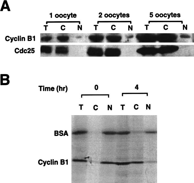
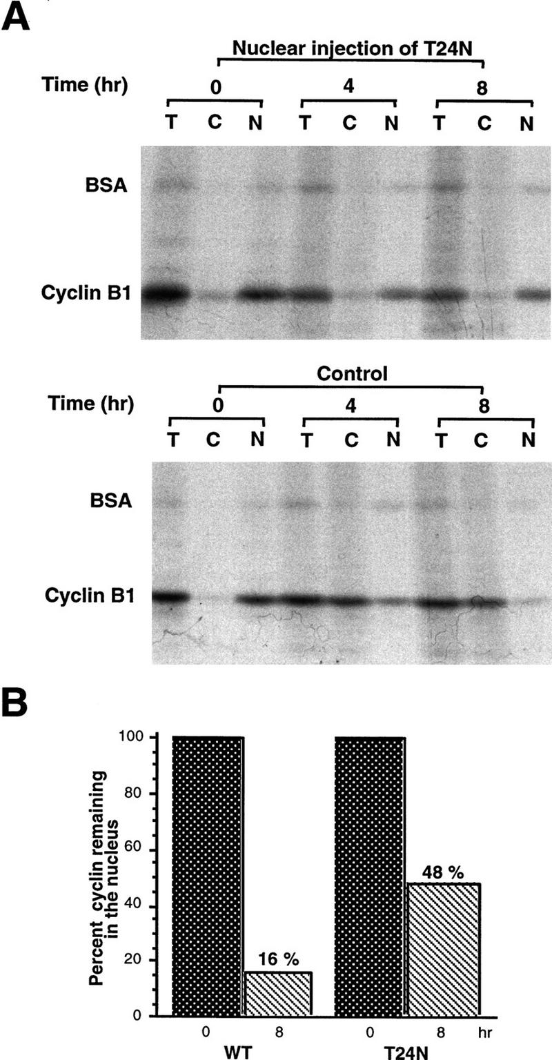
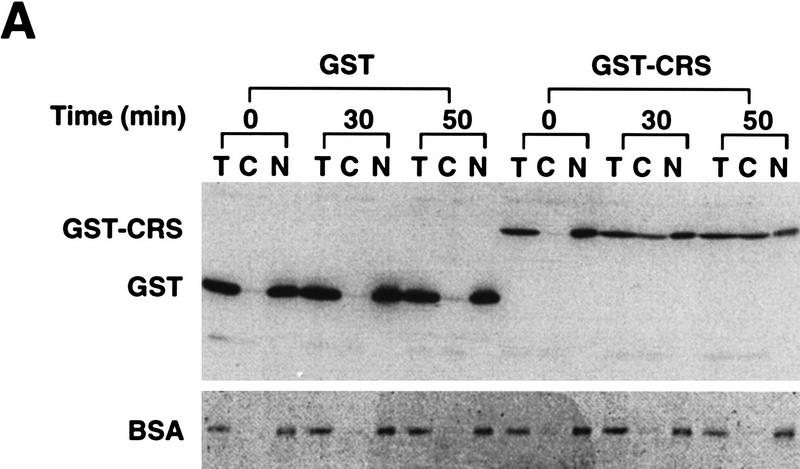
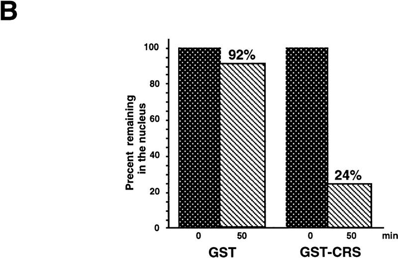
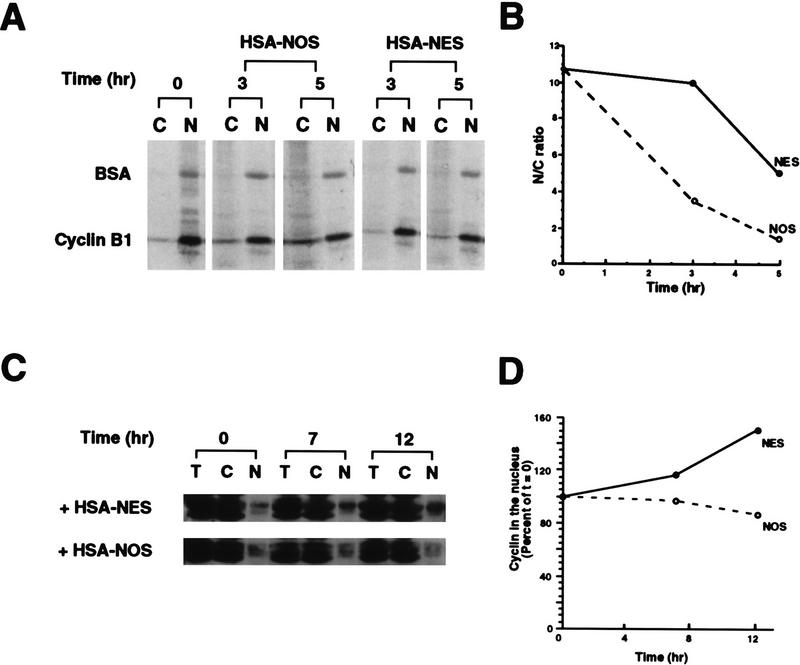
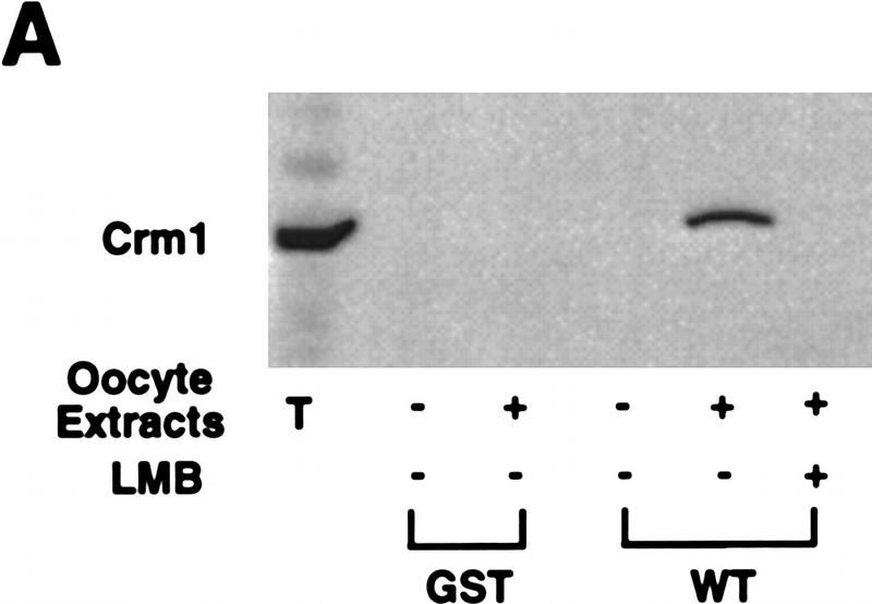
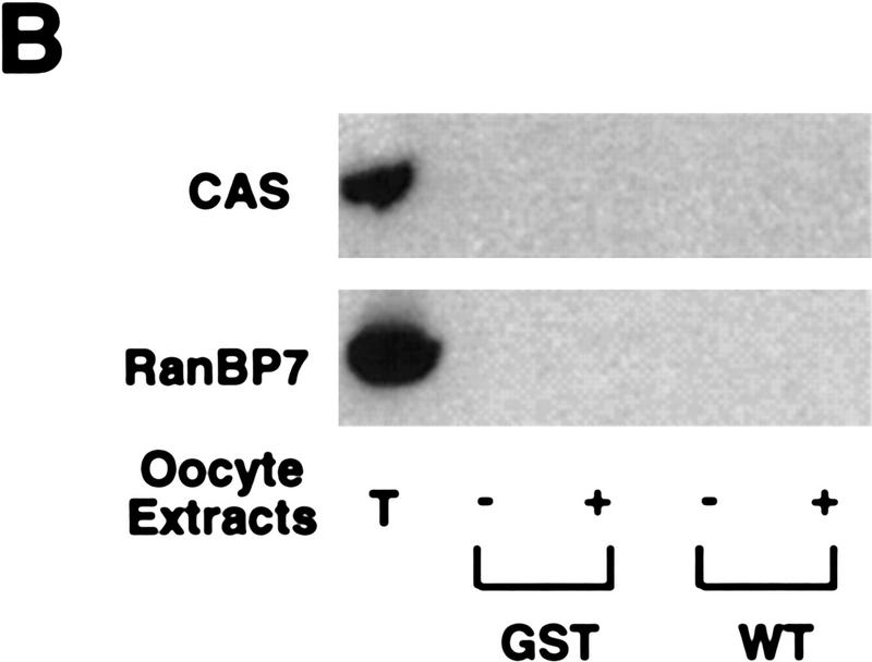
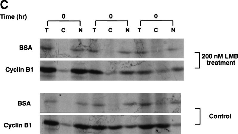
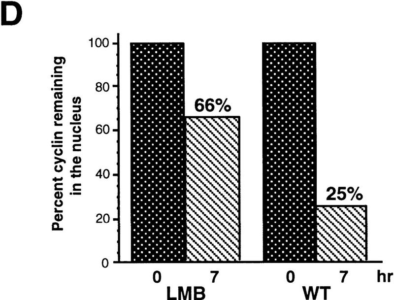
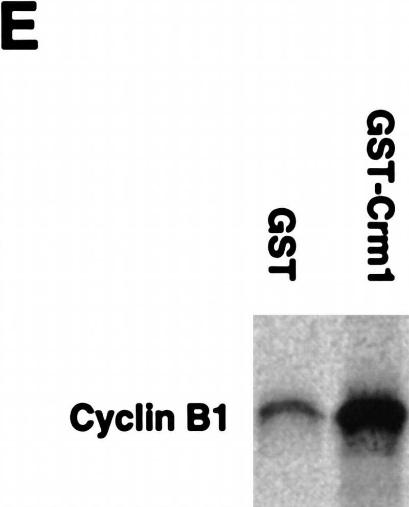
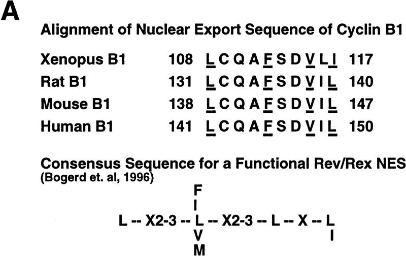
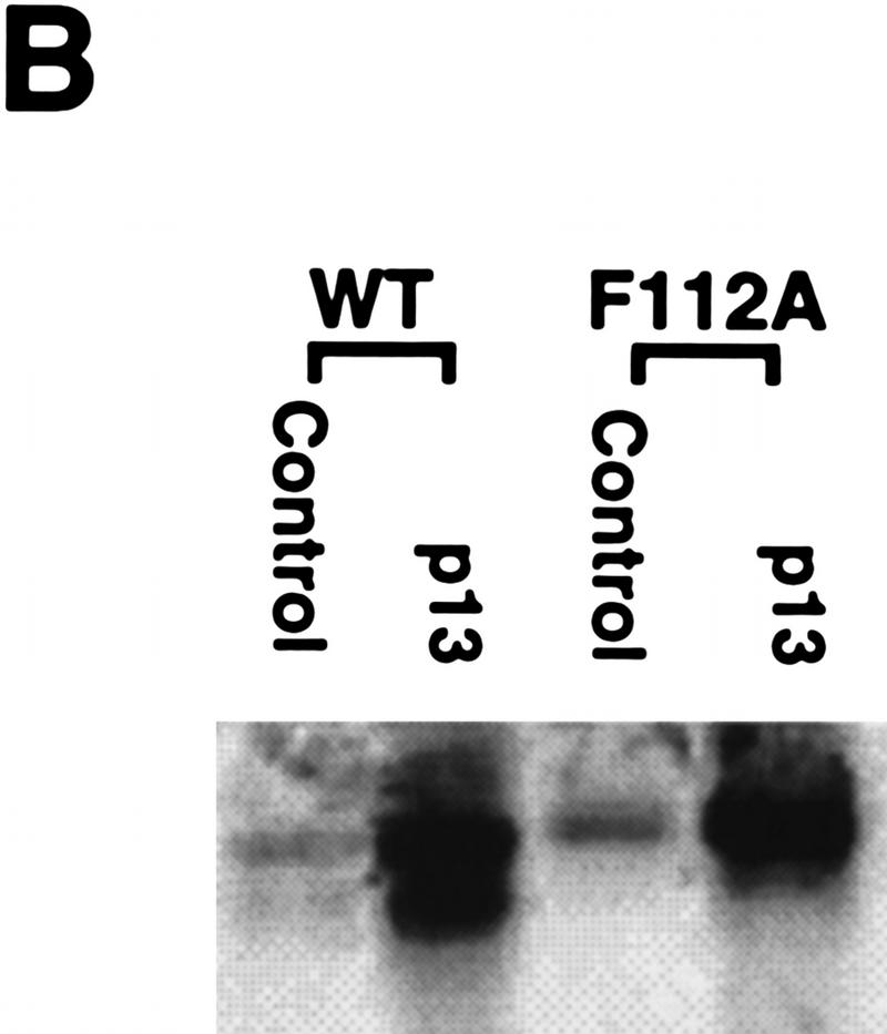
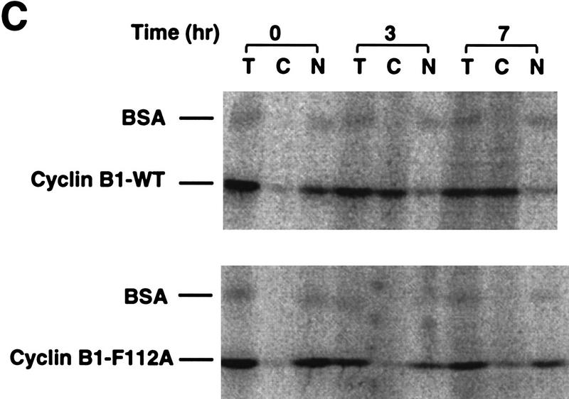
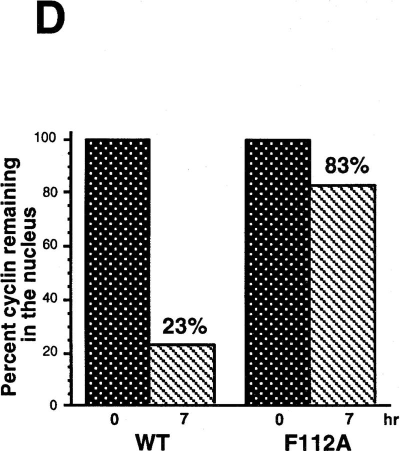
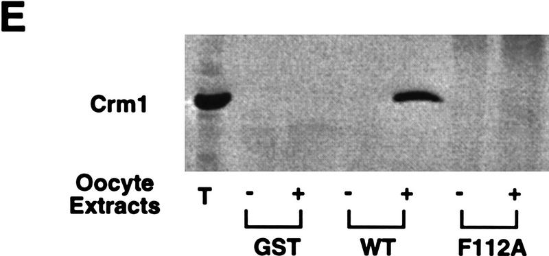
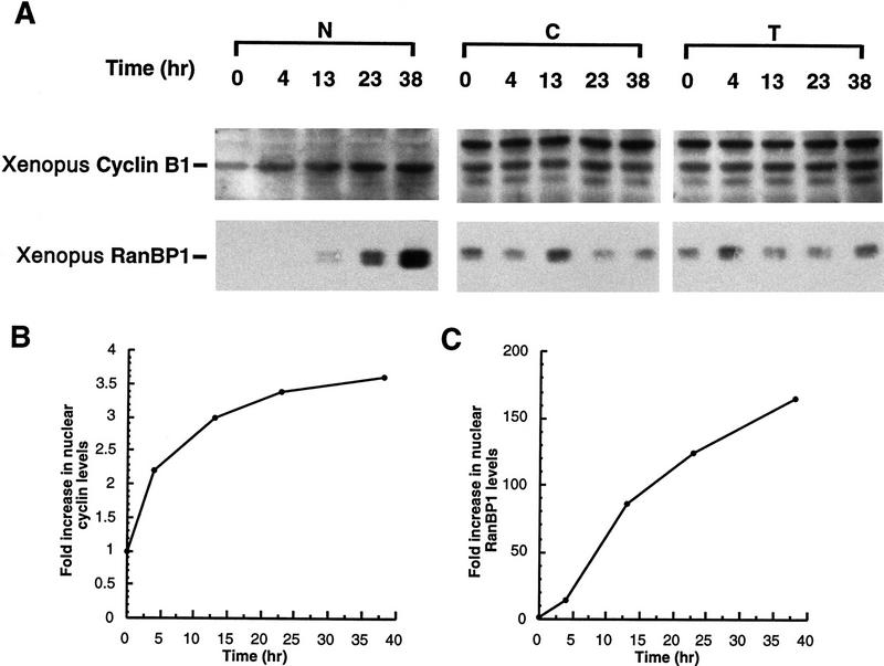
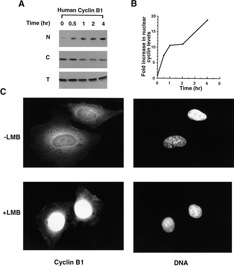
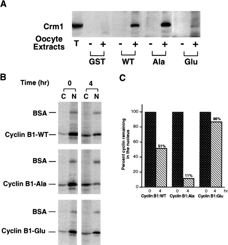
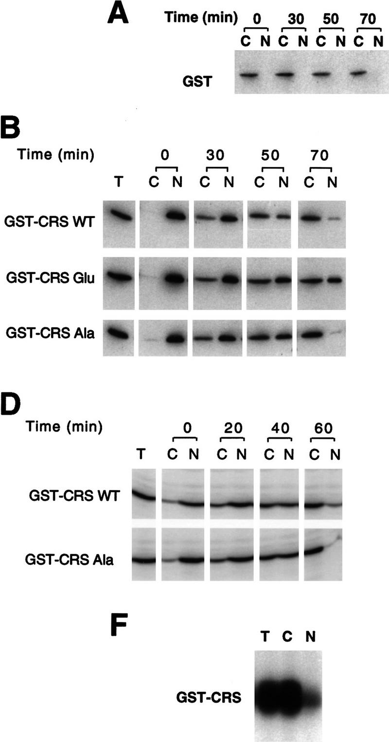
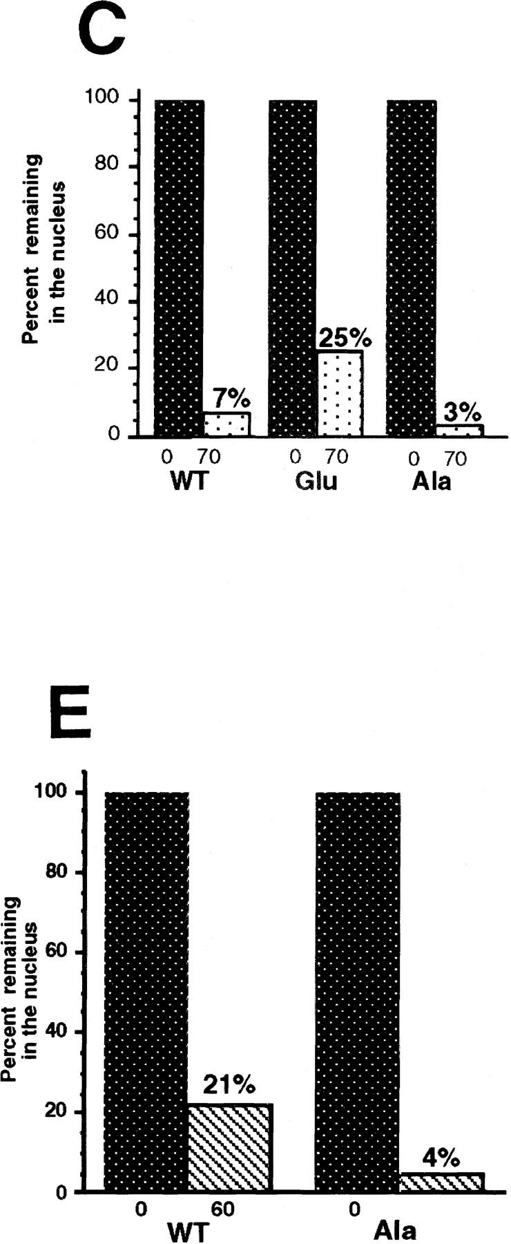
Activation of the Cyclin B/Cdc2 kinase complex triggers entry into mitosis in all eukaryotic cells. Cyclin B1 localization changes dramatically during the cell cycle, precipitously transiting from the cytoplasm to the nucleus at the beginning of mitosis. Presumably, this relocalization promotes the phosphorylation of nuclear targets critical for chromatin condensation and nuclear envelope breakdown. We show here that the previously characterized cytoplasmic retention sequence of Cyclin B1, responsible for its interphase cytoplasmic localization, is actually an autonomous nuclear export sequence, capable of directing nuclear export of a heterologous protein, and able to bind specifically to the recently identified export mediator, CRM1. We propose that the observed cytoplasmic localization of Cyclin B1 during interphase reflects the equilibrium between ongoing nuclear import and rapid CRM1-mediated export. In support of this hypothesis, we found that treatment of cells with leptomycin B, which disrupted Cyclin B1-CRM1 interactions, led to a marked nuclear accumulation of Cyclin B1. In mitosis, Cyclin B1 undergoes phosphorylation at several sites, a subset of which have been proposed to play a role in Cyclin B1 accumulation in the nucleus. Both CRM1 binding and the ability to direct nuclear export were affected by mutation of these phosphorylation sites; thus, we propose that Cyclin B1 phosphorylation at the G2/M transition prevents its interaction with CRM1, thereby reducing nuclear export and facilitating nuclear accumulation.


Figures

Figure 1
Figure 1
Characterization of Cyclin B1 nuclear export in Xenopus oocytes. (A) One, two, or five Xenopus stage VI oocytes were manually dissected into cytoplasmic and nuclear fractions and the distribution of endogenous Cyclin B1 was analyzed by SDS-PAGE followed by Western blotting with anti-Cyclin B1 sera. (T,C,N) Proteins extracted from total oocytes, cytoplasmic or nuclear fractions. Cdc25, which is cytoplasmic in oocytes, was used as a control. (B) Cyclin B1 was translated in reticulocyte lysates in the presence of [35S]methionine and injected into Xenopus oocyte nuclei along with 14C-labeled BSA. At 0 and 4 hr after injection, the oocytes were dissected and successfully injected oocytes were identified by the presence of pink coloring from the reticulocyte lysate. These oocytes were separated into cytoplasmic and nuclear fractions, extracted, and analyzed by SDS-PAGE followed by autoradiography.
Figure 2
Figure 2
A GDP-bound form of Ran inhibits Cyclin B1 nuclear export. (A) A mixture of 35S-labeled Cyclin B1 and 14C-labeled BSA was coinjected into nuclei with recombinant T24N mutant Ran protein (GDP-bound form). Oocytes were dissected 4 hr and 8 hr later and proteins were extracted and analyzed by SDS-PAGE and autoradiography. (B) The bar graph represents a quantitation of the data in A, showing the percentage of Cyclin B1 remaining in nuclei after 0 and 8 hr.
Figure 3
Figure 3
The CRS mediates nuclear export of Cyclin B1. (A) The isolated CRS from Xenopus Cyclin B1 (amino acids 76–125) was linked to the carboxyl terminus of GST. Either GST or GST–CRS protein was injected into oocyte nuclei along with 14C-labeled BSA. Oocytes were dissected at 0, 30, and 50 min after injection. Proteins were analyzed by SDS-PAGE followed by immunoblotting with GST antibody and detection with 125I-labeled protein A (quantitated by PhosphorImager). 14C-Labeled BSA was detected by autoradiography. (B) The graph represents a quantitation of the data in A, showing the percentage of injected proteins remaining in nuclei at 0 and 50 min.
Figure 3
Figure 3
The CRS mediates nuclear export of Cyclin B1. (A) The isolated CRS from Xenopus Cyclin B1 (amino acids 76–125) was linked to the carboxyl terminus of GST. Either GST or GST–CRS protein was injected into oocyte nuclei along with 14C-labeled BSA. Oocytes were dissected at 0, 30, and 50 min after injection. Proteins were analyzed by SDS-PAGE followed by immunoblotting with GST antibody and detection with 125I-labeled protein A (quantitated by PhosphorImager). 14C-Labeled BSA was detected by autoradiography. (B) The graph represents a quantitation of the data in A, showing the percentage of injected proteins remaining in nuclei at 0 and 50 min.
Figure 4
Figure 4
The PKI NES reduces nuclear export and induces nuclear accumulation of Cyclin B1. (A) Human serum albumin was conjugated with the NES peptide from PKI (HSA–NES), or to an inactive scrambled mutant variant of the PKI NES as the unexportable control (HSA–NOS). The 35S-labeled Cyclin B1 and 14C-labeled BSA mixture was injected into oocyte nuclei either with 8 μg/μl HSA–NES or HSA–NOS. Oocytes were dissected at 0, 3, and 5 hr after injection, and proteins were analyzed by SDS-PAGE followed by autoradiography. (B) The data in A are graphed to show the change in ratio of nuclear/cytoplasmic injected Cyclin B1 over a 5-hr time course. (C) HSA–NES or HSA–NOS (16 μg/μl) was injected into oocyte nuclei with 10 μg/μl dextran blue 2000 as an injection marker. Oocytes were dissected at 0, 7, and 12 hr after injection and proteins were analyzed by SDS-PAGE followed by immunoblotting with Cyclin B1 antibody. (D) The amount of cyclin present in the nucleus at time 0 was quantitated; the increase in nuclear cyclin after PKI NES injection is plotted as a percentage of cyclin quantities initially found in the nucleus.
Figure 5
Figure 5
The nuclear export receptor, CRM1, mediates Cyclin B1 nuclear export through binding to CRS. (A) GST or GST–CRS fusion protein was coupled to glutathione–Sepharose beads. Twenty microliters of these resins were incubated in 100 μl of oocyte extract for 1 hr alone or in the presence of 200 nm leptomycin B. The beads were then pelleted and washed three times with oocyte extract (EB) buffer. The bead-bound proteins were analyzed by SDS-PAGE followed by Western blotting with anti-human Crm1 antibody. (B) The same samples as in A (with the exception of the leptomycin B-containing sample) were immunoblotted with antisera directed against Ranbp7 and Cas. (C) Oocytes were incubated with 200 nm leptomycin B in MB buffer for 2 hr before injection. A 35S-labeled Cyclin B1 and 14C-labeled BSA mixture was then injected into oocyte nuclei. Oocytes were dissected at 0, 3, and 7 hr after injection and proteins were analyzed by SDS-PAGE followed by autoradiography. (D) The graph represents a quantitation of the data in C, showing the percentage of injected proteins remaining in nuclei at 0 and 7 hr. (E) Human Crm1 was fused to the carboxyl terminus of GST protein. Either GST alone or GST–Crm1 fusion protein was coupled to glutathione–Sepharose beads. Twenty microliters of each resin was incubated with 2 μl of 35S-labeled Cyclin B1 in 100 μl of buffer with 0.3 μg/μl Ran G19V (a mutant form of Ran that lacks GTPase activity and hence remains constiutively GTP-bound) and 5 μm GTP at room temperature for 1 hr. The beads were then pelleted and washed three times with oocyte extract buffer. The beads-bound proteins were analyzed by SDS-PAGE followed by autoradiography.
Figure 5
Figure 5
The nuclear export receptor, CRM1, mediates Cyclin B1 nuclear export through binding to CRS. (A) GST or GST–CRS fusion protein was coupled to glutathione–Sepharose beads. Twenty microliters of these resins were incubated in 100 μl of oocyte extract for 1 hr alone or in the presence of 200 nm leptomycin B. The beads were then pelleted and washed three times with oocyte extract (EB) buffer. The bead-bound proteins were analyzed by SDS-PAGE followed by Western blotting with anti-human Crm1 antibody. (B) The same samples as in A (with the exception of the leptomycin B-containing sample) were immunoblotted with antisera directed against Ranbp7 and Cas. (C) Oocytes were incubated with 200 nm leptomycin B in MB buffer for 2 hr before injection. A 35S-labeled Cyclin B1 and 14C-labeled BSA mixture was then injected into oocyte nuclei. Oocytes were dissected at 0, 3, and 7 hr after injection and proteins were analyzed by SDS-PAGE followed by autoradiography. (D) The graph represents a quantitation of the data in C, showing the percentage of injected proteins remaining in nuclei at 0 and 7 hr. (E) Human Crm1 was fused to the carboxyl terminus of GST protein. Either GST alone or GST–Crm1 fusion protein was coupled to glutathione–Sepharose beads. Twenty microliters of each resin was incubated with 2 μl of 35S-labeled Cyclin B1 in 100 μl of buffer with 0.3 μg/μl Ran G19V (a mutant form of Ran that lacks GTPase activity and hence remains constiutively GTP-bound) and 5 μm GTP at room temperature for 1 hr. The beads were then pelleted and washed three times with oocyte extract buffer. The beads-bound proteins were analyzed by SDS-PAGE followed by autoradiography.
Figure 6
Figure 6
Cyclin B1 contains a Rev/Rex consensus NES within the CRS region. (A) The NESs of vertebrate Cyclin B1s were aligned and compared with the consensus sequence for a functional Rev/Rex NES. (B) A full-length Cyclin B1 mutant in which the Phe residue at position 112 was changed to Ala was translated in vitro and added to interphase extracts of Xenopus eggs (these contain Cdc2, but lack mitotic cyclins). The 35S-labeled F112A mutant cyclin and wild-type cyclin were similarly retrieved from extracts by p13 Sepharose (which can only bind Cyclin B1 indirectly through interactions with its binding partner, Cdc2), but not by control Sepharose. (C) A full-length Cyclin B1 mutant in which the conserved Phe-112 within CRS was mutated to Ala was translated in reticulocyte lysates in the presence of [35S]methionine and mixed with 14C-labeled BSA, a nonexportable injection control. The mixture was injected into Xenopus oocyte nuclei. After injection (0, 3, and 7 hr), the oocytes were dissected into cytoplasmic and nuclear fractions, proteins were extracted, and samples were then analyzed by SDS-PAGE, followed by PhosphorImager quantitation. (D) The graph represents a quantitation of the data in C, showing the percentage of injected proteins remaining in nuclei at the 0 and 7 hr time points. (E) Phe-112 of Cyclin B1 was mutated to Ala and the isolated CRS containing this mutation was fused to the carboxyl terminus of GST protein. GST alone, the wild-type GST–CRS protein, or F112A mutant GST–CRS protein were coupled to glutathione–Sepharose beads. Twenty microliters of each resin was incubated in oocyte extract for 60 min and the beads were then pelleted and washed three times with oocyte extract buffer. The bead-bound material was analyzed by SDS-PAGE followed by immunoblotting with anti-CRM1 antibody.
Figure 6
Figure 6
Cyclin B1 contains a Rev/Rex consensus NES within the CRS region. (A) The NESs of vertebrate Cyclin B1s were aligned and compared with the consensus sequence for a functional Rev/Rex NES. (B) A full-length Cyclin B1 mutant in which the Phe residue at position 112 was changed to Ala was translated in vitro and added to interphase extracts of Xenopus eggs (these contain Cdc2, but lack mitotic cyclins). The 35S-labeled F112A mutant cyclin and wild-type cyclin were similarly retrieved from extracts by p13 Sepharose (which can only bind Cyclin B1 indirectly through interactions with its binding partner, Cdc2), but not by control Sepharose. (C) A full-length Cyclin B1 mutant in which the conserved Phe-112 within CRS was mutated to Ala was translated in reticulocyte lysates in the presence of [35S]methionine and mixed with 14C-labeled BSA, a nonexportable injection control. The mixture was injected into Xenopus oocyte nuclei. After injection (0, 3, and 7 hr), the oocytes were dissected into cytoplasmic and nuclear fractions, proteins were extracted, and samples were then analyzed by SDS-PAGE, followed by PhosphorImager quantitation. (D) The graph represents a quantitation of the data in C, showing the percentage of injected proteins remaining in nuclei at the 0 and 7 hr time points. (E) Phe-112 of Cyclin B1 was mutated to Ala and the isolated CRS containing this mutation was fused to the carboxyl terminus of GST protein. GST alone, the wild-type GST–CRS protein, or F112A mutant GST–CRS protein were coupled to glutathione–Sepharose beads. Twenty microliters of each resin was incubated in oocyte extract for 60 min and the beads were then pelleted and washed three times with oocyte extract buffer. The bead-bound material was analyzed by SDS-PAGE followed by immunoblotting with anti-CRM1 antibody.
Figure 7
Figure 7
Leptomycin B treatment induces endogenous Cyclin B1 nuclear accumulation in Xenopus oocytes. (A) Oocytes were treated with 200 nm leptomycin B in MB buffer. At the indicated times after treatment, they were dissected into cytoplasmic and nuclear fractions. Proteins were extracted and analyzed by SDS-PAGE followed by immunoblotting with anti-Xenopus Cyclin B1 antibody. Although this antisera cross-reacts with other proteins from the extract, the indicated band is Cyclin B1; this has been confirmed by specific reactivity of the antisera against recombinant Cyclin B1 and not B2, and by observation of the periodic accumulation and destruction of this band in cycling extracts of Xenopus eggs. The samples blotted with anti-cyclin sera were also immunoblotted with anti-Ranbp1 sera, as a positive control protein for nuclear accumulation following leptomycin B treatment. (B) The graph represents a quantitation of the data in A, showing the fold increase of endogenous Cyclin B1 accumulated in nuclei at the indicated times after leptomycin B treatment. For every oocyte nuclear equivalent loaded on the gels, only one-eighth the amount of cytoplasm was loaded to facilitate observation of potential changes in cytoplasmic levels of protein. (C) The graph represents a quantitation of the data in A, showing the fold increase of endogenous RanBP1 in nuclei following leptomycin B treatment.
Figure 8
Figure 8
Leptomycin B treatment induces nuclear accumulation of Cyclin B1 in HeLa cells. (A) HeLa cells were treated with 20 nm leptomycin B. At the indicated times, cells were collected and separated into cytoplasmic and nuclear fractions. Sampled were analyzed by SDS-PAGE followed by Western blot with anti-human Cyclin B1 antibody (Santa Cruz). All of the nuclear or cytoplasmic fraction from 1.5 × 105 HeLa cells were loaded in each lane, as indicated. (B) The graph represents a quantitation of the data in A, showing the fold increase of endogenous Cyclin B1 in HeLa cell nuclei following leptomycin B treatment. Note the much shorter time course shown here than in the Xenopus experiment in 7B (4 hr vs. 40 hr). (C) Either untreated HeLa cells or HeLa cells treated for 4 hr with 20 nm leptomycin B were processed for indirect immunofluorescence with a monoclonal directed against human Cyclin B1, as described in Materials and Methods. (Left) Cyclin B1 immunofluorescence signals in representative interphase cells either treated or untreated with leptomycin B. (Right) The same cells stained with the DNA intercalating dye, Hoechst 33258. Fluorescence was observed and photographed with a 60× Planapochromat objective and CCD camera.
Figure 9
Figure 9
Phosphorylation within the CRS of Cyclin B1 impairs its ability to bind to Crm1 and inhibits Cyclin B1 nuclear export. (A) Mutant variants of the isolated CRS in which Ser-94, Ser-96, Ser-101, and Ser-113 had all been changed to either Ala or Glu were fused to the carboxyl terminus of GST protein. Equal amounts of these fusion proteins or the wild-type GST–CRS protein were coupled to glutathione–Sepharose beads. Twenty microliters of each resin was incubated in oocyte extract for 60 min and the beads were then pelleted and washed three times with oocyte extract buffer. The bead-bound material was analyzed by SDS-PAGE followed by immunoblotting with anti-CRM1 antibody. (B) Wild-type full-length Cyclin B1 or full-length mutant variants in which all four sites of Ser phosphorylation within the CRS had been mutated to either Ala or Glu was translated in the reticulocyte lysate in the presence of [35S]methionine and mixed with 14C-labeled BSA, a nonexportable injection control. The mixture was injected into Xenopus oocyte nuclei. At 0 and 4 hr after injection, the oocytes were dissected into cytoplasmic and nuclear fractions; proteins were extracted and samples were then analyzed by SDS-PAGE and quantitated with a PhosphorImager. (C) The bar graph represents a quantitation of the data in B, showing the percentage of each Cyclin B1 variant remaining in nuclei at 0 and 4 hr. Values were normalized to coinjected 14C-labeled BSA.
Figure 10
Figure 10
Phosphorylation of Cyclin B1 within the CRS directly modulates nuclear export. (A) GST protein was injected into the cytoplasm of oocytes and incubated for 30, 50, or 70 min. Oocytes were dissected into nuclear and cytoplasmic fractions, resolved by SDS-PAGE and immunoblotted with anti-GST sera. Injected GST remains cytoplasmic. (B) Fifty nanograms of GST protein fused to the wild-type, Ala, or Glu mutant CRS sequences was injected into oocyte nuclei along with 14C-labeled BSA. Oocytes were dissected at 0, 30, 50, and 70 min after injection and proteins were analyzed by SDS-PAGE followed by immunoblotting with GST antibody and 125I-labeled protein A or autoradiography to detect the 14C-labeled BSA. (C) Quantitation of the data in B. The 125I-labeled protein A signal was quantitated using a PhosphorImager. The bar graph shows the percent of each injected cyclin variant remaining in the nucleus immediately after and 70 min after injection. (D) GST fused to the wild-type or Ala mutant CRS sequences was injected into oocyte nuclei along with 14C-labeled BSA. Oocytes were dissected at 0, 20, 40, and 60 min after injection and proteins were analyzed by SDS-PAGE followed by Western blotting with GST antibody or autoradiography to detect the 14C-labeled BSA. Approximately 2.5-fold less protein was injected in this experiment than the experiment shown in B. (E) Quantitation of the data in D. The bar graph shows the percent of each injected cyclin variant remaining in the nucleus immediately after and 60 min after injection. (F) Oocytes were dissected into cytoplasmic and nuclear fractions and extracts were prepared from each fraction or from whole oocytes. Each extract was diluted to 1.45 μg/μl. Twenty microliters of GST–CRS wild-type beads was incubated in 45 μl of total (T), cytoplasmic (C), or nuclear (N) oocyte extract, 1 μm cold ATP and 15 μCi of [γ3P]ATP. After 20 min incubation at room temperature, beads were pelleted and washed with extract buffer three times. The bead-bound material was analyzed by SDS-PAGE, developed by PhosphorImager and quantitated.
Figure 10
Figure 10
Phosphorylation of Cyclin B1 within the CRS directly modulates nuclear export. (A) GST protein was injected into the cytoplasm of oocytes and incubated for 30, 50, or 70 min. Oocytes were dissected into nuclear and cytoplasmic fractions, resolved by SDS-PAGE and immunoblotted with anti-GST sera. Injected GST remains cytoplasmic. (B) Fifty nanograms of GST protein fused to the wild-type, Ala, or Glu mutant CRS sequences was injected into oocyte nuclei along with 14C-labeled BSA. Oocytes were dissected at 0, 30, 50, and 70 min after injection and proteins were analyzed by SDS-PAGE followed by immunoblotting with GST antibody and 125I-labeled protein A or autoradiography to detect the 14C-labeled BSA. (C) Quantitation of the data in B. The 125I-labeled protein A signal was quantitated using a PhosphorImager. The bar graph shows the percent of each injected cyclin variant remaining in the nucleus immediately after and 70 min after injection. (D) GST fused to the wild-type or Ala mutant CRS sequences was injected into oocyte nuclei along with 14C-labeled BSA. Oocytes were dissected at 0, 20, 40, and 60 min after injection and proteins were analyzed by SDS-PAGE followed by Western blotting with GST antibody or autoradiography to detect the 14C-labeled BSA. Approximately 2.5-fold less protein was injected in this experiment than the experiment shown in B. (E) Quantitation of the data in D. The bar graph shows the percent of each injected cyclin variant remaining in the nucleus immediately after and 60 min after injection. (F) Oocytes were dissected into cytoplasmic and nuclear fractions and extracts were prepared from each fraction or from whole oocytes. Each extract was diluted to 1.45 μg/μl. Twenty microliters of GST–CRS wild-type beads was incubated in 45 μl of total (T), cytoplasmic (C), or nuclear (N) oocyte extract, 1 μm cold ATP and 15 μCi of [γ3P]ATP. After 20 min incubation at room temperature, beads were pelleted and washed with extract buffer three times. The bead-bound material was analyzed by SDS-PAGE, developed by PhosphorImager and quantitated.

Similar articles

Cited by

References

    1. Atherton-Fessler S, Liu F, Gabrielli B, Lee MS, Peng C-Y, Piwnica-Worms H. Cell cycle regulation of the p34cdc2 Inhibitory kinases. Mol Biol Cell. 1994;5:989–1001. - PMC - PubMed
    1. Bogerd HP, Fridell RA, Benson RE, Hua J, Cullen BR. Protein sequence requirements for function of the human T-cell leukemia virus type 1 Rex nuclear export signal delineated by a novel in vivo randomization-selection assay. Mol Cell Biol. 1996;16:4207–4214. - PMC - PubMed
    1. Corbett AH, Silver PA. Nucleocytoplasmic transport of macromolecules. Microbiol Mol Biol Rev. 1997;61:193–211. - PMC - PubMed
    1. Doye V, Hurt E. From nucleoporins to nuclear pore complexes. Curr Opin Cell Biol. 1997;9:401–411. - PubMed
    1. Dunphy WG, Kumagai A. The cdc25 protein contains an intinsic phosphatase activity. Cell. 1991;67:189–196. - PubMed

Publication types

MeSH terms

LinkOut - more resources