. 1998 Jul 15;12(14):2144-52.
doi: 10.1101/gad.12.14.2144.

Determinants of specificity in TGF-beta signal transduction

Affiliations

Determinants of specificity in TGF-beta signal transduction

Y G Chen et al. Genes Dev. .

Abstract

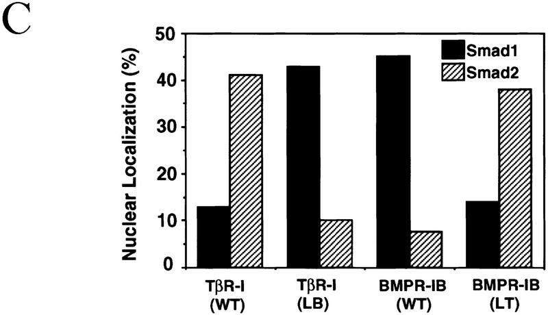
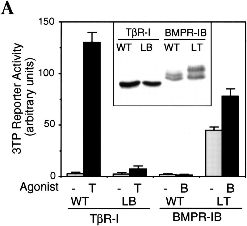
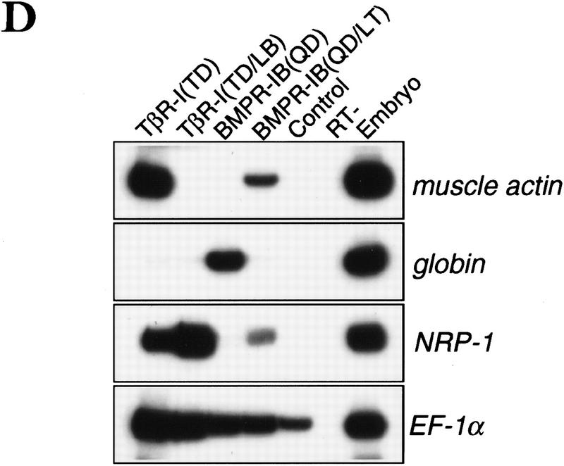
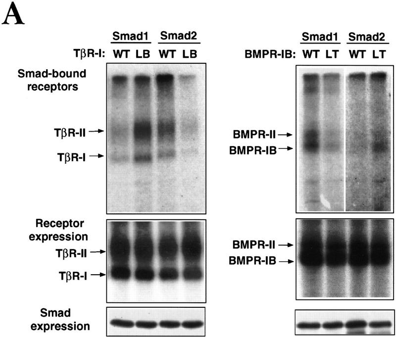
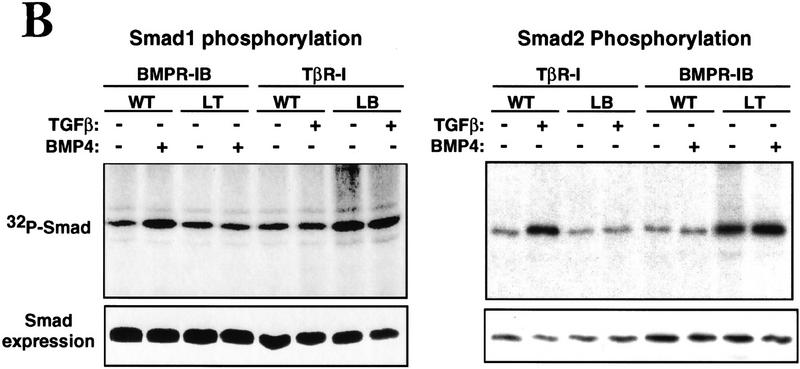
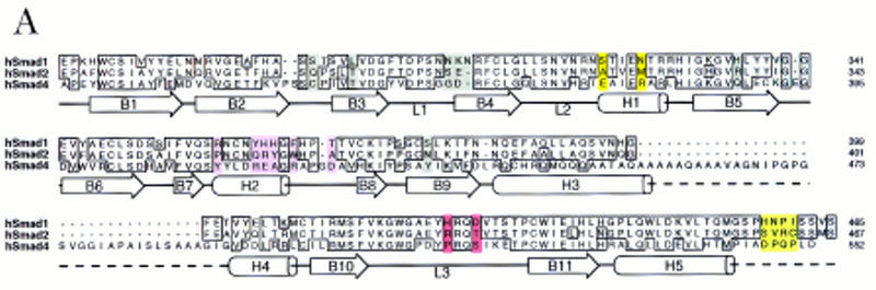
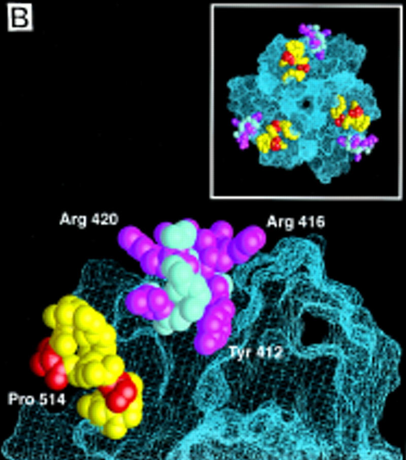
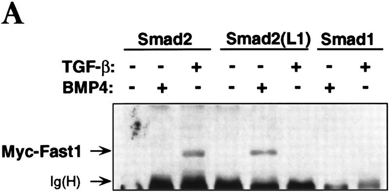
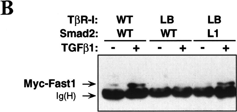
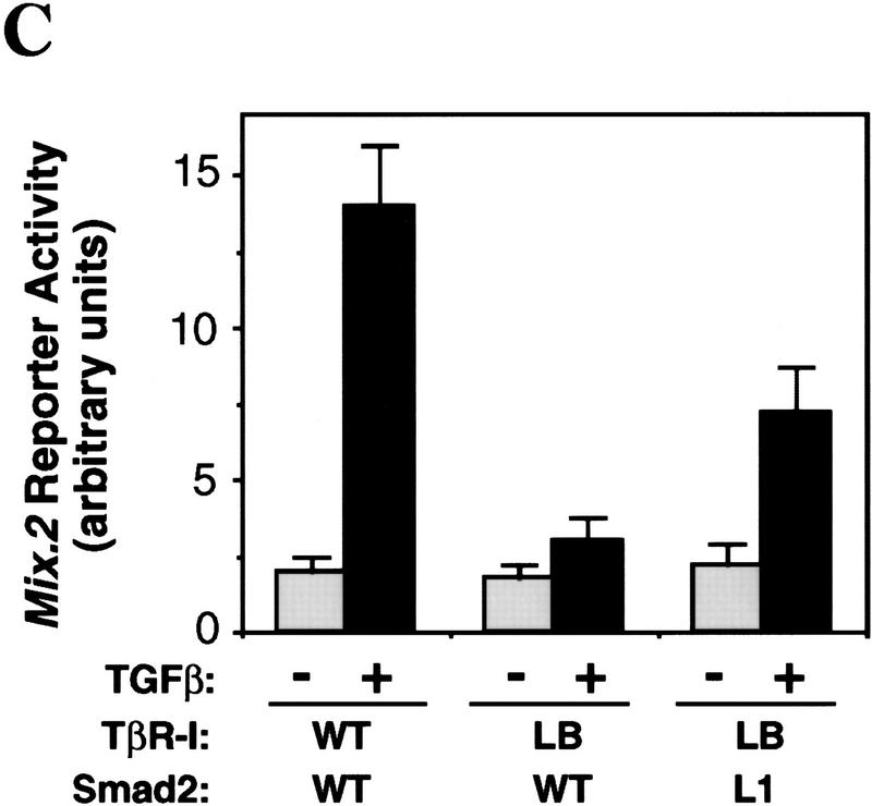
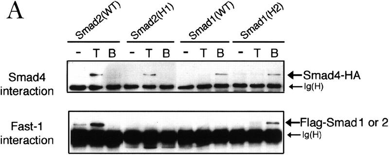
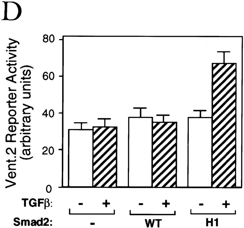
Signal transduction by the TGF-beta family involves sets of receptor serine/threonine kinases, Smad proteins that act as receptor substrates, and Smad-associated transcription factors that target specific genes. We have identified discrete structural elements that dictate the selective interactions between receptors and Smads and between Smads and transcription factors in the TGF-beta and BMP pathways. A cluster of four residues in the L45 loop of the type I receptor kinase domain, and a matching set of two residues in the L3 loop of the Smad carboxy-terminal domain establish the specificity of receptor-Smad interactions. A cluster of residues in the highly exposed alpha-helix 2 of the Smad carboxy-terminal domain specify the interaction with the DNA-binding factor Fast1 and, as a result, the gene responses mediated by the pathway. By establishing specific interactions, these determinants keep the TGF-beta and BMP pathways segregated from each other.


Figures

Figure 1
Figure 1
(A) L45 loop sequences of the TGF-β type I receptor family. Conserved amino acids are boxed. Three groups of functionally related receptors have each a characteristic L45 loop sequence. ALK1 is also known as TSR-1, and ALK2 as ActR-I or Tsk7L. (B) R-Smad association with Smad4. (Scheme) a TGF-β signal transduction pathway with a type II receptor (II), a type I receptor (I), R-Smad phosphorylation (P), Smad4 (4), and a DNA-binding factor (F). COS1 cells were transfected with Flag-tagged Smad1 or Smad2, HA-tagged Smad4, the indicated wild-type (WT) or mutant type I receptors, and the corresponding type II receptors TβR-II or BMPR-II. R-Smad binding to Smad4 was determined after incubation with TGF-β or BMP2. (C) Nuclear translocation of R-Smads induced by wild-type and L45 mutant type I receptors. HepG2 cells were transfected with Flag-tagged Smad1 (solid bars) or Smad2 (hatched bars), the indicated type I receptors, and their corresponding type II receptors. Cells were incubated with TGF-β1 or BMP2 for 1 hr and subjected to anti-Flag immunofluorescence.
Figure 1
Figure 1
(A) L45 loop sequences of the TGF-β type I receptor family. Conserved amino acids are boxed. Three groups of functionally related receptors have each a characteristic L45 loop sequence. ALK1 is also known as TSR-1, and ALK2 as ActR-I or Tsk7L. (B) R-Smad association with Smad4. (Scheme) a TGF-β signal transduction pathway with a type II receptor (II), a type I receptor (I), R-Smad phosphorylation (P), Smad4 (4), and a DNA-binding factor (F). COS1 cells were transfected with Flag-tagged Smad1 or Smad2, HA-tagged Smad4, the indicated wild-type (WT) or mutant type I receptors, and the corresponding type II receptors TβR-II or BMPR-II. R-Smad binding to Smad4 was determined after incubation with TGF-β or BMP2. (C) Nuclear translocation of R-Smads induced by wild-type and L45 mutant type I receptors. HepG2 cells were transfected with Flag-tagged Smad1 (solid bars) or Smad2 (hatched bars), the indicated type I receptors, and their corresponding type II receptors. Cells were incubated with TGF-β1 or BMP2 for 1 hr and subjected to anti-Flag immunofluorescence.
Figure 2
Figure 2
Exchanging the L45 loops switches the signaling specificity of TβR-I and BMPR-IB. (A) Activation of the TGFβ-responsive reporter 3TP-luciferase in TβR-I-defective R1B/L17 cells transfected with wild-type or mutant receptors. Cells were incubated with TGF-β (T) or BMP2 (B), and luciferase activity was determined in triplicate samples. (Inset) HA-tagged receptors immunoprecipitated from metabolically labeled cells as controls. (B) Activation of the A3–CAT reporter containing activin- and TGF-β-responsive Mix.2 elements. R1B/L17 cells were transfected with Fast1 and receptor constructs. TβR-I transfectants were incubated with TGF-β and BMPR-IB transfectants with BMP2, and CAT activity was determined. (C) Activation of the BMP-responsive reporter Vent.2–luciferase in P19 cells transfected with TβR-II and wild-type or mutant TβR-I. Cells were incubated with BMP2 (B) or TGF-β (T), and luciferase activity was determined. (D) Induction of markers of dorsal mesoderm (muscle actin), ventral mesoderm (globin), and neural tissue (NRP-1) in Xenopus embryos. RNAs encoding the indicated constitutively active receptor forms were injected into the animal pole of two-cell embryos. Expression of muscle actin, globin, NRP-1, and EF-1α (as control) in animal caps from these embryos was determined. Animal caps from uninjected embryos (control), whole embryos (embryo), and a sample without reverse transcription (RT−) were included.
Figure 2
Figure 2
Exchanging the L45 loops switches the signaling specificity of TβR-I and BMPR-IB. (A) Activation of the TGFβ-responsive reporter 3TP-luciferase in TβR-I-defective R1B/L17 cells transfected with wild-type or mutant receptors. Cells were incubated with TGF-β (T) or BMP2 (B), and luciferase activity was determined in triplicate samples. (Inset) HA-tagged receptors immunoprecipitated from metabolically labeled cells as controls. (B) Activation of the A3–CAT reporter containing activin- and TGF-β-responsive Mix.2 elements. R1B/L17 cells were transfected with Fast1 and receptor constructs. TβR-I transfectants were incubated with TGF-β and BMPR-IB transfectants with BMP2, and CAT activity was determined. (C) Activation of the BMP-responsive reporter Vent.2–luciferase in P19 cells transfected with TβR-II and wild-type or mutant TβR-I. Cells were incubated with BMP2 (B) or TGF-β (T), and luciferase activity was determined. (D) Induction of markers of dorsal mesoderm (muscle actin), ventral mesoderm (globin), and neural tissue (NRP-1) in Xenopus embryos. RNAs encoding the indicated constitutively active receptor forms were injected into the animal pole of two-cell embryos. Expression of muscle actin, globin, NRP-1, and EF-1α (as control) in animal caps from these embryos was determined. Animal caps from uninjected embryos (control), whole embryos (embryo), and a sample without reverse transcription (RT−) were included.
Figure 3
Figure 3
(A) Receptor–Smad association in COS-1 cells transfected with the indicated type I receptors, the corresponding type II receptors, and Flag-tagged Smad1(1–454) or Smad2(1–456). Receptors were cross-linked to 125I-labeled TGF-β1 (left) or 125I-labeled BMP2 (right). Smad-bound receptors were visualized by anti-Flag immunoprecipitation, SDS-PAGE, and autoradiography (top). Total cell lysates were analyzed to control for receptor expression (middle). Smad expression was controlled by immunoprecipitation from metabolically labeled cells (bottom). (B) Smad phosphorylation was determined in L17 cells transfected with Flag-tagged Smads, the indicated type I receptors, and the corresponding type II receptors. Cells were labeled with [32P]phosphate, incubated with TGF-β1 or BMP2, and immunoprecipitated with anti-Flag.
Figure 3
Figure 3
(A) Receptor–Smad association in COS-1 cells transfected with the indicated type I receptors, the corresponding type II receptors, and Flag-tagged Smad1(1–454) or Smad2(1–456). Receptors were cross-linked to 125I-labeled TGF-β1 (left) or 125I-labeled BMP2 (right). Smad-bound receptors were visualized by anti-Flag immunoprecipitation, SDS-PAGE, and autoradiography (top). Total cell lysates were analyzed to control for receptor expression (middle). Smad expression was controlled by immunoprecipitation from metabolically labeled cells (bottom). (B) Smad phosphorylation was determined in L17 cells transfected with Flag-tagged Smads, the indicated type I receptors, and the corresponding type II receptors. Cells were labeled with [32P]phosphate, incubated with TGF-β1 or BMP2, and immunoprecipitated with anti-Flag.
Figure 4
Figure 4
(A) Sequence alignment of the MH2 domains of Smad1, 2, and 4, with the Smad4 MH2 domain secondary structure elements indicated below. Identical residues are boxed. Subtype-specific residues map to α-helix 1 (yellow), α-helix 2 and its vicinity (purple), the L3 loop (red), and immediately upstream of the carboxy-terminal receptor phosphorylation motif SS(V/M)S (green). The remaining subtype-specific residues (gray) are scattered in the primary sequence but clustered in the crystal structure near the point of connection to the amino-terminal half of the molecule (Shi et al. 1997). (B) A close-up, lateral view of the Smad4 MH2 crystal structure showing the L3 loop (yellow) with subtype specific residues (red) and the α-helix 2 (cyan) with subtype-specific residues (magenta). (Inset) Frontal view of the location of the L3 loop and helix 2 of each MH2 monomer in the crystallographic trimer.
Figure 4
Figure 4
(A) Sequence alignment of the MH2 domains of Smad1, 2, and 4, with the Smad4 MH2 domain secondary structure elements indicated below. Identical residues are boxed. Subtype-specific residues map to α-helix 1 (yellow), α-helix 2 and its vicinity (purple), the L3 loop (red), and immediately upstream of the carboxy-terminal receptor phosphorylation motif SS(V/M)S (green). The remaining subtype-specific residues (gray) are scattered in the primary sequence but clustered in the crystal structure near the point of connection to the amino-terminal half of the molecule (Shi et al. 1997). (B) A close-up, lateral view of the Smad4 MH2 crystal structure showing the L3 loop (yellow) with subtype specific residues (red) and the α-helix 2 (cyan) with subtype-specific residues (magenta). (Inset) Frontal view of the location of the L3 loop and helix 2 of each MH2 monomer in the crystallographic trimer.
Figure 5
Figure 5
Matching receptor L45 loops and R-Smad L3 loops. (A) The L3 loop determines Smad activation by a specific receptor but not Smad interaction with Fast1. COS1 cells were transfected with Flag-tagged Smad constructs, myc-tagged Fast1, and TGF-β receptors or BMP receptors. Cells were incubated with the corresponding receptor ligands, TGF-β1 or BMP4, and Smad association with Fast1 was determined. [Ig(H)] immunoglobulin heavy chain. (B,C) TβR-I(LB) rescues the ability of TGF-β to induce Smad2(L1) association with Fast1 (B) and activation of the A3–luciferase Mix.2 reporter (C). R1B/L17 cells transfected with various constructs, as indicated, were incubated with 0.5 nm TGF-β for 20 hr, and luciferase activity was measured.
Figure 5
Figure 5
Matching receptor L45 loops and R-Smad L3 loops. (A) The L3 loop determines Smad activation by a specific receptor but not Smad interaction with Fast1. COS1 cells were transfected with Flag-tagged Smad constructs, myc-tagged Fast1, and TGF-β receptors or BMP receptors. Cells were incubated with the corresponding receptor ligands, TGF-β1 or BMP4, and Smad association with Fast1 was determined. [Ig(H)] immunoglobulin heavy chain. (B,C) TβR-I(LB) rescues the ability of TGF-β to induce Smad2(L1) association with Fast1 (B) and activation of the A3–luciferase Mix.2 reporter (C). R1B/L17 cells transfected with various constructs, as indicated, were incubated with 0.5 nm TGF-β for 20 hr, and luciferase activity was measured.
Figure 6
Figure 6
The α-helix 2 of Smad2 specifies the interaction with the DNA-binding factor Fast1. (A) Interaction of wild-type R-Smads and helix 2 exchange mutants with Smad4 and Fast1. HA-tagged Smad4 or myc-tagged Fast1 constructs were cotransfected into COS1 cells with the indicated Flag-tagged forms of Smad1 or Smad2. Transfectants were incubated with TGF-β (T) or BMP2 (B) and the associations of R-Smads with Smad4 (top) and with Fast1 (bottom) were determined. The helix 2 exchange mutants bound Smad4 in response to their agonists, but Smad2(H1) lost the ability to associate with Fast1 whereas Smad1(H2) gained the ability to bind Fast1 in response to BMP. (B) Activation of a Mix.2 reporter by wild-type R-Smads and helix 2 exchange mutants. L17 cells were cotransfected with the indicated forms of Smad1 or Smad2, Fast1, the A3-luciferase construct, and TGF-β receptors or BMP receptors. Cells were incubated with the corresponding receptor ligands, and luciferase activity was determined. Smad2(H1) lost the ability to activate the reporter, whereas Smad1(H2) gained the ability to do so in response to BMP. (C) Fast1-dependent activation of a GAL4 reporter by Smad1(H2). L17 cells were cotransfected with the indicated forms of Smad1, a Fast1 fusion with the DNA-binding domain from yeast GAL4, a GAL luciferase reporter, and BMP receptors. Cells were incubated with or without BMP2, and luciferase activity was determined. (D) Activation of the Vent.2–luciferase reporter in P19 cells cotransfected with TβR-I, TβR-II, and the indicated Smad2 constructs. Cells were incubated with or without TGF-β, and luciferase activity was determined in triplicate samples.
Figure 6
Figure 6
The α-helix 2 of Smad2 specifies the interaction with the DNA-binding factor Fast1. (A) Interaction of wild-type R-Smads and helix 2 exchange mutants with Smad4 and Fast1. HA-tagged Smad4 or myc-tagged Fast1 constructs were cotransfected into COS1 cells with the indicated Flag-tagged forms of Smad1 or Smad2. Transfectants were incubated with TGF-β (T) or BMP2 (B) and the associations of R-Smads with Smad4 (top) and with Fast1 (bottom) were determined. The helix 2 exchange mutants bound Smad4 in response to their agonists, but Smad2(H1) lost the ability to associate with Fast1 whereas Smad1(H2) gained the ability to bind Fast1 in response to BMP. (B) Activation of a Mix.2 reporter by wild-type R-Smads and helix 2 exchange mutants. L17 cells were cotransfected with the indicated forms of Smad1 or Smad2, Fast1, the A3-luciferase construct, and TGF-β receptors or BMP receptors. Cells were incubated with the corresponding receptor ligands, and luciferase activity was determined. Smad2(H1) lost the ability to activate the reporter, whereas Smad1(H2) gained the ability to do so in response to BMP. (C) Fast1-dependent activation of a GAL4 reporter by Smad1(H2). L17 cells were cotransfected with the indicated forms of Smad1, a Fast1 fusion with the DNA-binding domain from yeast GAL4, a GAL luciferase reporter, and BMP receptors. Cells were incubated with or without BMP2, and luciferase activity was determined. (D) Activation of the Vent.2–luciferase reporter in P19 cells cotransfected with TβR-I, TβR-II, and the indicated Smad2 constructs. Cells were incubated with or without TGF-β, and luciferase activity was determined in triplicate samples.
Figure 7
Figure 7
Determinants of specificity in TGF-β signal transduction. In the TGF-β or BMP receptor complexes, the type I receptor recognizes and phosphorylates a specific R-Smad, such as Smad2 in the TGF-β pathway or Smad1 in the BMP pathway (Heldin et al. 1997; Massagué 1998). The R-Smad then associates with Smad4 (not shown) and moves into the nucleus. Specific association with the DNA-binding factor Fast1 in the nucleus takes the Smad2–Smad4 complex to specific target genes such as Mix.2, activating their transcription (X. Chen et al. 1996, 1997; Liu et al. 1997). Selection of a R-Smad by a receptor is specified by the type I receptor L45 loop and the R-Smad L3 loop, whereas selection of a DNA-binding factor (such as Fast1 in the case of Smad2) is specified by the α-helix 2 of the R-Smad. Exchanging any of these three elements between the TGF-β and BMP receptors or between Smad1 and Smad2 causes a switch in the signaling specificity of these two pathways. Specific activation of other target genes by Smad1 or Smad2 complexes is presumed to involve different DNA-binding partners.

Similar articles

Cited by

References

    1. Baker J, Harland RM. A novel mesoderm inducer, mMadr-2, functions in the activin signal transduction pathway. Genes & Dev. 1996;10:1880–1889. - PubMed
    1. Candia AF, Watabe T, Hawley SH, Onichtchouck D, Zhang Y, Derynck R, Niehrs C, Cho KW. Cellular interpretation of multiple TGF-β signals: Intracellular antagonism between activin/BVg1 and BMP-2/4 signaling mediated by Smads. Development. 1997;124:4467–4480. - PubMed
    1. Cárcamo J, Weis FMB, Ventura F, Wieser R, Wrana JL, Attisano L, Massagué J. Type I receptors specify growth inhibitory and transcriptional responses to TGF-β and activin. Mol Cell Biol. 1994;14:3810–3821. - PMC - PubMed
    1. Cárcamo J, Zentella A, Massagué J. Disruption of TGF-β signaling by a mutation that prevents transphosphorylation within the receptor complex. Mol Cell Biol. 1995;15:1573–1581. - PMC - PubMed
    1. Cheifetz S, Hernandez H, Laiho M, ten Dijke P, Iwata KK, Massagué J. Distinct transforming growth factor-β receptor subsets as determinants of cellular responsiveness to three TGF-β isoforms. J Biol Chem. 1990;265:20533–20538. - PubMed

Publication types

MeSH terms

Substances

LinkOut - more resources