. 1998 Jul 15;12(14):2153-63.
doi: 10.1101/gad.12.14.2153.

The tumor suppressor Smad4/DPC4 and transcriptional adaptor CBP/p300 are coactivators for smad3 in TGF-beta-induced transcriptional activation

Affiliations

The tumor suppressor Smad4/DPC4 and transcriptional adaptor CBP/p300 are coactivators for smad3 in TGF-beta-induced transcriptional activation

X H Feng et al. Genes Dev. .

Abstract

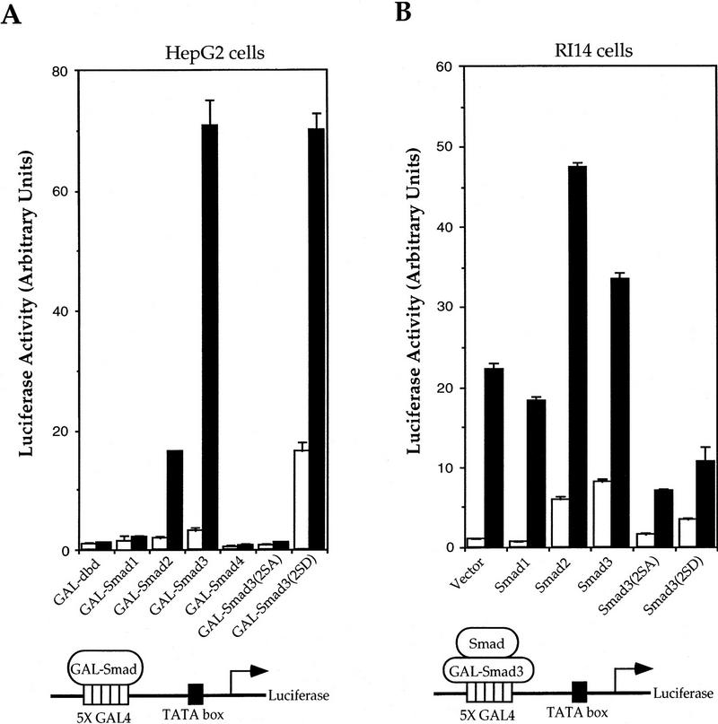
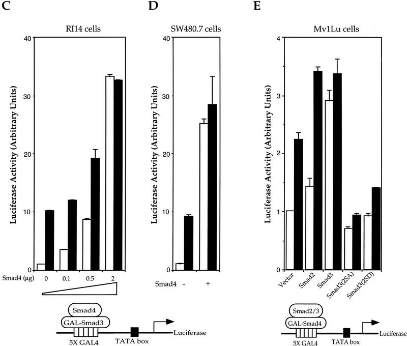
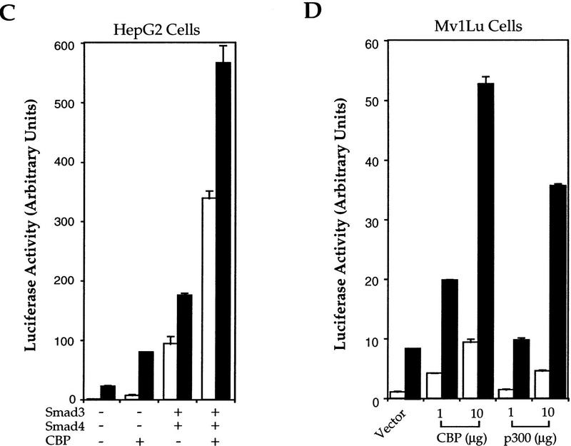
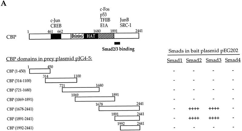
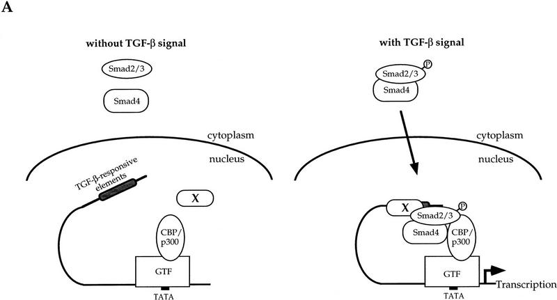
Smads regulate transcription of defined genes in response to TGF-beta receptor activation, although the mechanisms of Smad-mediated transcription are not well understood. We demonstrate that the TGF-beta-inducible Smad3 uses the tumor suppressor Smad4/DPC4 and CBP/p300 as transcriptional coactivators, which associate with Smad3 in response to TGF-beta. The association of CBP with Smad3 was localized to the carboxyl terminus of Smad3, which is required for transcriptional activation, and a defined segment in CBP. Furthermore, CBP/p300 stimulated both TGF-beta- and Smad-induced transcription in a Smad4/DPC4-dependent fashion. Smad3 transactivation and TGF-beta-induced transcription were inhibited by expressing E1A, which interferes with CBP functions. The coactivator functions and physical interactions of Smad4 and CBP/p300 with Smad3 allow a model for the induction of gene expression in response to TGF-beta.


Figures

Figure 1
Figure 1
Transcriptional activity of Smad3 and effect of interactions with Smad2, Smad3, and Smad4 on Smad3-mediated transcription. (A) Smad3 is a TGF-β-inducible transcriptional activator. HepG2 cells were transfected with plasmids encoding the indicated GAL–Smad and the transcriptional activity from the cotransfected GAL4–luciferase reporter plasmid was measured. Assays were performed in the presence or absence of TGF-β. (B) Smad2 and Smad3 potentiate the transactivation activity of GAL–Smad3. RI14 cells were cotransfected with pFR–Luc and the plasmid encoding GAL–Smad3, and expression plasmids for the indicated Smads or mutants. (C) Smad4 is a potent transcriptional coactivator for Smad3. RI14 cells were cotransfected with pFR–Luc, pGAL–Smad3, and indicated amounts of a Smad4 expression plasmid. (D) Smad4 is not essential for transcriptional activity of Smad3. Smad4-deficient SW480.7 cells were cotransfected with pFR–Luc and pGAL–Smad3, without (open bars) or with (solid bars) TβRI (act.). The transcriptional activity was measured in the absence (open bars) or presence (black bars) of a coexpressed constitutively active TβRI. (E) Smad2 and Smad3 stimulate the low transactivation activity of pGAL–Smad4. Mv1Lu cells were cotransfected with pGAL–Smad4 and pFR–Luc, and expression plasmids for the indicated Smads or relevant mutants. (A–C,E) (Open bars) −TGF-β; (solid bars) +TGF-β. Note the lower scale of luciferase activity, when compared to A–D.
Figure 1
Figure 1
Transcriptional activity of Smad3 and effect of interactions with Smad2, Smad3, and Smad4 on Smad3-mediated transcription. (A) Smad3 is a TGF-β-inducible transcriptional activator. HepG2 cells were transfected with plasmids encoding the indicated GAL–Smad and the transcriptional activity from the cotransfected GAL4–luciferase reporter plasmid was measured. Assays were performed in the presence or absence of TGF-β. (B) Smad2 and Smad3 potentiate the transactivation activity of GAL–Smad3. RI14 cells were cotransfected with pFR–Luc and the plasmid encoding GAL–Smad3, and expression plasmids for the indicated Smads or mutants. (C) Smad4 is a potent transcriptional coactivator for Smad3. RI14 cells were cotransfected with pFR–Luc, pGAL–Smad3, and indicated amounts of a Smad4 expression plasmid. (D) Smad4 is not essential for transcriptional activity of Smad3. Smad4-deficient SW480.7 cells were cotransfected with pFR–Luc and pGAL–Smad3, without (open bars) or with (solid bars) TβRI (act.). The transcriptional activity was measured in the absence (open bars) or presence (black bars) of a coexpressed constitutively active TβRI. (E) Smad2 and Smad3 stimulate the low transactivation activity of pGAL–Smad4. Mv1Lu cells were cotransfected with pGAL–Smad4 and pFR–Luc, and expression plasmids for the indicated Smads or relevant mutants. (A–C,E) (Open bars) −TGF-β; (solid bars) +TGF-β. Note the lower scale of luciferase activity, when compared to A–D.
Figure 2
Figure 2
CBP/p300 functions as a transcriptional coactivator for Smad3. (A) CBP and p300 potentiate the transcriptional activity of GAL–Smad3. RI14 cells were cotransfected with pGAL–Smad3 and pFR–Luc, and indicated amounts of expression plasmids for CBP or p300. (B) Smad4 is required for efficient coactivation of Smad3 by CBP. SW480.7 cells were cotransfected with pGAL–Smad3 and pFR–Luc, and indicated amounts of an expression plasmid for Smad4, in the absence (open bar) or presence (hatched bar) of an expression plasmid for CBP. Transfected cells were treated with TGF-β and luciferase values were measured. (C) CBP stimulates Smad3/4-induced transcription from the PAI-1 promoter in the absence or presence of TGF-β. HepG2 cells were cotransfected with the PAI-1 luciferase reporter p800luc, and indicated combinations of expression plasmids for Smad3, Smad4, and CBP. (D) CBP and p300 stimulate TGF-β-induced transcription from the PAI-1 promoter. Mv1Lu cells were cotransfected with the PAI-1 luciferase reporter p800luc, and indicated amounts of expression plasmids for CBP or p300. (E) Smad3 and Smad4 stimulate the transactivation activity of CBP. Mv1Lu and SW480.7 cells were cotransfected with pGAL–CBP(1678–2441), pFR–Luc, and expression plasmids for Smad3 and/or Smad4. (F) The −732 to −635 segment of the PAI-1 promoter mediates TGF-β- and Smad3/4-inducible transcription. Mv1Lu cells were cotransfected with the PAI-1 luciferase reporter pGL5P/97, and indicated combinations of expression plasmids for Smad3 and Smad4. (G) p300, Smad3, and Smad4 participate in a complex assembled at the PAI-1 promoter. Nuclear extracts, prepared from 293 cells, were incubated with the 32P-labeled, 97-bp TGF-β- and Smad3/4-inducible segment of the PAI-1 promoter. Free DNA, DNA–protein (shift), and supershifted (SS) complexes are marked and the nuclear lysates and antibodies are also shown. (A,C–F) (Open bars) −TGF-β; (solid bars) +TGF-β.
Figure 2
Figure 2
CBP/p300 functions as a transcriptional coactivator for Smad3. (A) CBP and p300 potentiate the transcriptional activity of GAL–Smad3. RI14 cells were cotransfected with pGAL–Smad3 and pFR–Luc, and indicated amounts of expression plasmids for CBP or p300. (B) Smad4 is required for efficient coactivation of Smad3 by CBP. SW480.7 cells were cotransfected with pGAL–Smad3 and pFR–Luc, and indicated amounts of an expression plasmid for Smad4, in the absence (open bar) or presence (hatched bar) of an expression plasmid for CBP. Transfected cells were treated with TGF-β and luciferase values were measured. (C) CBP stimulates Smad3/4-induced transcription from the PAI-1 promoter in the absence or presence of TGF-β. HepG2 cells were cotransfected with the PAI-1 luciferase reporter p800luc, and indicated combinations of expression plasmids for Smad3, Smad4, and CBP. (D) CBP and p300 stimulate TGF-β-induced transcription from the PAI-1 promoter. Mv1Lu cells were cotransfected with the PAI-1 luciferase reporter p800luc, and indicated amounts of expression plasmids for CBP or p300. (E) Smad3 and Smad4 stimulate the transactivation activity of CBP. Mv1Lu and SW480.7 cells were cotransfected with pGAL–CBP(1678–2441), pFR–Luc, and expression plasmids for Smad3 and/or Smad4. (F) The −732 to −635 segment of the PAI-1 promoter mediates TGF-β- and Smad3/4-inducible transcription. Mv1Lu cells were cotransfected with the PAI-1 luciferase reporter pGL5P/97, and indicated combinations of expression plasmids for Smad3 and Smad4. (G) p300, Smad3, and Smad4 participate in a complex assembled at the PAI-1 promoter. Nuclear extracts, prepared from 293 cells, were incubated with the 32P-labeled, 97-bp TGF-β- and Smad3/4-inducible segment of the PAI-1 promoter. Free DNA, DNA–protein (shift), and supershifted (SS) complexes are marked and the nuclear lysates and antibodies are also shown. (A,C–F) (Open bars) −TGF-β; (solid bars) +TGF-β.
Figure 3
Figure 3
TGF-β-dependent association of CBP with Smad3 and cooperation of Smad4. (A) TGF-β-dependent association of Smad3 and Smad4 . RI14 cells were transfected with Flag-tagged Smad3 and HA-tagged Smad4, and treated with (+) or without (−) TGF-β. Cell lysates were immunoprecipitated (IP) with anti-Flag antibody, followed by immunoblotting (IB) with anti-HA antibody to detect Smad3-bound Smad4, or with anti-Flag immunoblotting to demonstrate equal expression of Smad3. (B) Smad3 coimmunoprecipitates with CBP after TGF-β-receptor activation. COS-1 cells were transfected with Flag-tagged Smads as shown and HA-tagged CBP, in the absence or presence of a plasmid for an activated TβRI. Immunoprecipitation with anti-Flag antibody was followed by anti-HA immunostaining to detect Smad-bound CBP. The control panel shows the expression levels of Smad3 and Smad4. (C) TGF-β-dependent CBP–Smad3 interaction requires the carboxy-terminal SSXS site of Smad3. RI14 cells were transfected with the indicated combination of plasmids expressing GAL–CBP(1678–2441) and Flag–Smad3 or Flag–Smad3(2SA). Immunoprecipitation with anti-Flag antibody was followed by immunostaining with anti-GAL4 (BabCO) to detect Smad-bound CBP. The control panel shows the Smad3 expression levels. (D) TGF-β-dependent interaction of Smad3 and Smad4 with CBP in mammalian two-hybrid assays. Plasmids for GAL-fused CBP segments, in combination with VP16–Smad plasmids, as indicated, were transfected into RI14 cells, together with the luciferase reporter plasmid pFR–Luc. The interactions were measured by luciferase expression in the absence (−, open bars) or presence (+, solid bars) of TGF-β. GAL–DNA-binding domain is the control containing only the GAL4 DNA-binding domain, not fused to a CBP segment. (E) Smad4 enhances the association of Smad3 with CBP in mammalian two-hybrid assays. Smad4-deficient SW480.7 cells were cotransfected with plasmids for GAL-fused CBP segments and VP16–Smd3, in the absence (open bars) or presence (solid bars) of an activated TβRI plasmid, and the CBP–Smad3 association was scored in the absence or presence of coexpressed Smad4.
Figure 3
Figure 3
TGF-β-dependent association of CBP with Smad3 and cooperation of Smad4. (A) TGF-β-dependent association of Smad3 and Smad4 . RI14 cells were transfected with Flag-tagged Smad3 and HA-tagged Smad4, and treated with (+) or without (−) TGF-β. Cell lysates were immunoprecipitated (IP) with anti-Flag antibody, followed by immunoblotting (IB) with anti-HA antibody to detect Smad3-bound Smad4, or with anti-Flag immunoblotting to demonstrate equal expression of Smad3. (B) Smad3 coimmunoprecipitates with CBP after TGF-β-receptor activation. COS-1 cells were transfected with Flag-tagged Smads as shown and HA-tagged CBP, in the absence or presence of a plasmid for an activated TβRI. Immunoprecipitation with anti-Flag antibody was followed by anti-HA immunostaining to detect Smad-bound CBP. The control panel shows the expression levels of Smad3 and Smad4. (C) TGF-β-dependent CBP–Smad3 interaction requires the carboxy-terminal SSXS site of Smad3. RI14 cells were transfected with the indicated combination of plasmids expressing GAL–CBP(1678–2441) and Flag–Smad3 or Flag–Smad3(2SA). Immunoprecipitation with anti-Flag antibody was followed by immunostaining with anti-GAL4 (BabCO) to detect Smad-bound CBP. The control panel shows the Smad3 expression levels. (D) TGF-β-dependent interaction of Smad3 and Smad4 with CBP in mammalian two-hybrid assays. Plasmids for GAL-fused CBP segments, in combination with VP16–Smad plasmids, as indicated, were transfected into RI14 cells, together with the luciferase reporter plasmid pFR–Luc. The interactions were measured by luciferase expression in the absence (−, open bars) or presence (+, solid bars) of TGF-β. GAL–DNA-binding domain is the control containing only the GAL4 DNA-binding domain, not fused to a CBP segment. (E) Smad4 enhances the association of Smad3 with CBP in mammalian two-hybrid assays. Smad4-deficient SW480.7 cells were cotransfected with plasmids for GAL-fused CBP segments and VP16–Smd3, in the absence (open bars) or presence (solid bars) of an activated TβRI plasmid, and the CBP–Smad3 association was scored in the absence or presence of coexpressed Smad4.
Figure 4
Figure 4
The association of CBP and Smad3 is mediated by carboxy-terminal domains of both proteins. (A) Yeast two-hybrid assays demonstrate the interaction of Smad2 and Smad3, but not Smad1 and Smad4, with carboxy-terminal sequences (aa 1891–2441) of CBP. Smad–CBP interactions were detected by measuring β-galactosidase activity. (−) Lack of detectable interaction; (++++) very strong interaction. The structural organization of CBP is shown with the previously characterized location of sequences required for interaction with the proteins shown. Our results now allow the localization of sequences required for association with Smad2 and Smad3. (B) Localization of the CBP-interacting sequences in Smad3 using yeast two-hybrid. Interactions were scored by measuring β-galactosidase activity from negative (−) to strongly positive (+++) and not determined (n.d.). The correlation with in vitro binding to GST-fused Smad3 or its fragments is also shown. Besides some previously defined functions shown on the schematic diagram of Smad3, the sequences that mediate interaction with CBP have now been localized to the carboxyl domain and require the carboxy-terminal sequence. (C) Direct interaction of 35S-labeled CBP(1678–2441) with GST–Smad3 and GST–Smad3C, but not GST–Smad4. Results are summarized in B.
Figure 4
Figure 4
The association of CBP and Smad3 is mediated by carboxy-terminal domains of both proteins. (A) Yeast two-hybrid assays demonstrate the interaction of Smad2 and Smad3, but not Smad1 and Smad4, with carboxy-terminal sequences (aa 1891–2441) of CBP. Smad–CBP interactions were detected by measuring β-galactosidase activity. (−) Lack of detectable interaction; (++++) very strong interaction. The structural organization of CBP is shown with the previously characterized location of sequences required for interaction with the proteins shown. Our results now allow the localization of sequences required for association with Smad2 and Smad3. (B) Localization of the CBP-interacting sequences in Smad3 using yeast two-hybrid. Interactions were scored by measuring β-galactosidase activity from negative (−) to strongly positive (+++) and not determined (n.d.). The correlation with in vitro binding to GST-fused Smad3 or its fragments is also shown. Besides some previously defined functions shown on the schematic diagram of Smad3, the sequences that mediate interaction with CBP have now been localized to the carboxyl domain and require the carboxy-terminal sequence. (C) Direct interaction of 35S-labeled CBP(1678–2441) with GST–Smad3 and GST–Smad3C, but not GST–Smad4. Results are summarized in B.
Figure 5
Figure 5
Model of Smad3/Smad4/CBP interactions at the promoter and inhibition of TGF-β- and Smad3/4-induced transcription by E1A. (A) A working model for TGF-β-inducible cooperation between Smad2/3, Smad4, and CBP/p300. Nuclear factor X is a hypothetical DNA-binding protein bound to the TGF-β-responsive elements, although Smad2/3 or Smad4 may directly bind to DNA. GTF represents a group of transcription factors for general transcription associated with RNA polymerase II. (B) E1A inhibits transcription from the PAI-1 promoter, induced by Smad3 and Smad4. Smad4-deficient SW480.7 cells were cotransfected with p800luc, and indicated combinations of expression plasmids for Smad3, Smad4, and E1A. (C) E1A, but not E1AΔ2–36, which is incapable of CBP association, inhibits TGF-β-induced transcription from the PAI-1 promoter. E1A928 has a point mutation at aa 928 and does not interact with pRB but still binds to CBP. Mv1Lu cells were cotransfected with p800luc, and wild-type or mutant E1A. (D) E1A, but not E1AΔ2–36, blocks TGF-β-induced transcription activity of GAL–Smad3 and the transactivation of GAL–Smad3 by CBP. Mv1Lu cells were cotransfected with pFR-Luc, and wild-type or mutant E1A, and CBP. (B–D) (Open bars) −TGF-β; (solid bars) +TGF-β.
Figure 5
Figure 5
Model of Smad3/Smad4/CBP interactions at the promoter and inhibition of TGF-β- and Smad3/4-induced transcription by E1A. (A) A working model for TGF-β-inducible cooperation between Smad2/3, Smad4, and CBP/p300. Nuclear factor X is a hypothetical DNA-binding protein bound to the TGF-β-responsive elements, although Smad2/3 or Smad4 may directly bind to DNA. GTF represents a group of transcription factors for general transcription associated with RNA polymerase II. (B) E1A inhibits transcription from the PAI-1 promoter, induced by Smad3 and Smad4. Smad4-deficient SW480.7 cells were cotransfected with p800luc, and indicated combinations of expression plasmids for Smad3, Smad4, and E1A. (C) E1A, but not E1AΔ2–36, which is incapable of CBP association, inhibits TGF-β-induced transcription from the PAI-1 promoter. E1A928 has a point mutation at aa 928 and does not interact with pRB but still binds to CBP. Mv1Lu cells were cotransfected with p800luc, and wild-type or mutant E1A. (D) E1A, but not E1AΔ2–36, blocks TGF-β-induced transcription activity of GAL–Smad3 and the transactivation of GAL–Smad3 by CBP. Mv1Lu cells were cotransfected with pFR-Luc, and wild-type or mutant E1A, and CBP. (B–D) (Open bars) −TGF-β; (solid bars) +TGF-β.

References

    1. Abdollah S, Macías-Silva M, Tsukazaki T, Hayashi H, Attisano L, Wrana JL. TβRI phosphorylation of Smad2 on Ser465 and Ser467 is required for Smad2-Smad4 complex formation and signaling. J Biol Chem. 1997;272:27678–27685. - PubMed
    1. Avantaggiati ML, Ogryzko V, Gardner K, Giordano A, Levine A, Kelly K. Recruitment of p300/CBP in p53-dependent signal pathways. Cell. 1997;89:1175–1184. - PubMed
    1. Candia A, Watabe T, Hawley S, Onichtchouk D, Zhang Y, Derynck R, Niehrs C, Cho KWY. Cellular interpretation of multiple TGF-β signals: Intracellular antagonism between activin/BVg1 and BMP-2/4 signaling mediated by Smads. Development. 1997;124:4467–4480. - PubMed
    1. Chen F, Weinberg RA. Biochemical evidence for the autophosphorylation and transphosphorylation of transforming growth factor β receptor kinases. Proc Natl Acad Sci. 1995;92:1565–1569. - PMC - PubMed
    1. Chen X, Weisberg E, Fridmacher V, Watanabe M, Naco G, Whitman M. Smad4 and FAST-1 in the assembly of activin-responsive factor. Nature. 1997;389:85–89. - PubMed

Publication types

MeSH terms