. 1998 Jul 15;12(14):2188-99.
doi: 10.1101/gad.12.14.2188.

DNA end-independent activation of DNA-PK mediated via association with the DNA-binding protein C1D

Affiliations

DNA end-independent activation of DNA-PK mediated via association with the DNA-binding protein C1D

U Yavuzer et al. Genes Dev. .

Abstract

DNA-dependent protein kinase (DNA-PK), which is involved in DNA double-strand break repair and V(D)J recombination, is comprised of a DNA-targeting component termed Ku and an approximately 465-kD catalytic subunit, DNA-PKcs. Although DNA-PK phosphorylates proteins in the presence of DSBs or other discontinuities in the DNA double helix in vitro, the possibility exists that it is also activated in other circumstances via its association with additional proteins. Here, through use of the yeast two-hybrid screen, we discover that the recently identified high affinity DNA binding protein C1D interacts with the putative leucine zipper region of DNA-PKcs. Furthermore, we show that C1D can interact with DNA-PK in mammalian cells and that C1D is a very effective DNA-PK substrate in vitro. Finally, we establish that C1D directs the activation of DNA-PK in a manner that does not require DNA termini. Therefore, these studies provide a function for C1D and suggest novel mechanisms for DNA-PK activation in vivo.


Figures

Figure 1
Figure 1
C1D interacts with the LZ region of DNA-PKcs. (A) Schematic diagram of DNA-PKcs. The kinase domain (hatched box) and the LZ region used as a bait in the yeast two-hybrid system are indicated. Mutations introduced into the LZ motif to produce constructs, Mut1LZcs and Mut2LZcs, are indicated by asterisks, and the leucine residues of the motif are underlined. (B) C1D fused to VP16 activation domain (Prey) was transformed into L40 strain together with LexA–LZcs, LexA, or other LexA fusion proteins (Lamin and Daughterless). Interactions were measured by β-galactosidase activity. (C) C1D fused to the carboxy-terminal DNA-binding region of LexA (Bait), and the LZcs was fused to the VP16 activation domain. (D) Mutations that disrupt the LZ motif abolish the DNA-PKcs—C1D two-hybrid interaction.
Figure 1
Figure 1
C1D interacts with the LZ region of DNA-PKcs. (A) Schematic diagram of DNA-PKcs. The kinase domain (hatched box) and the LZ region used as a bait in the yeast two-hybrid system are indicated. Mutations introduced into the LZ motif to produce constructs, Mut1LZcs and Mut2LZcs, are indicated by asterisks, and the leucine residues of the motif are underlined. (B) C1D fused to VP16 activation domain (Prey) was transformed into L40 strain together with LexA–LZcs, LexA, or other LexA fusion proteins (Lamin and Daughterless). Interactions were measured by β-galactosidase activity. (C) C1D fused to the carboxy-terminal DNA-binding region of LexA (Bait), and the LZcs was fused to the VP16 activation domain. (D) Mutations that disrupt the LZ motif abolish the DNA-PKcs—C1D two-hybrid interaction.
Figure 2
Figure 2
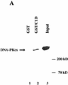
C1D and DNA-PKcs interact in vitro. (A) GST or GST–C1D (0.5 μg in each case) was incubated with 5 μg of purified DNA-PK and subjected to GST pull-down assays, and Western blots were performed with a DNA-PKcs antibody. Quantitation reveals that ∼10% of DNA-PKcs binds to GST–C1D in this experiment, suggestive of a 1:1 binding ratio. (B) GST pull-downs were performed as in A (for lane 3, the pull-down reaction included ethidium bromide), then proteins were immunoblotted with antibodies against Ku or DNA-PKcs.
Figure 2
Figure 2
C1D and DNA-PKcs interact in vitro. (A) GST or GST–C1D (0.5 μg in each case) was incubated with 5 μg of purified DNA-PK and subjected to GST pull-down assays, and Western blots were performed with a DNA-PKcs antibody. Quantitation reveals that ∼10% of DNA-PKcs binds to GST–C1D in this experiment, suggestive of a 1:1 binding ratio. (B) GST pull-downs were performed as in A (for lane 3, the pull-down reaction included ethidium bromide), then proteins were immunoblotted with antibodies against Ku or DNA-PKcs.
Figure 3
Figure 3
C1D is a strong substrate for DNA-PK in vitro. (A) C1D refolded on a linear plasmid (C1D-L) or p53 was incubated with purified DNA-PK, and in vitro kinase assays were performed as described in Materials and Methods. Linear DNA was added to the reaction for lane 4. (B) C1D refolded on supercoiled plasmid (C1D-Sc) was incubated with purified DNA-PK as described above. Where indicated, ethidium bromide or LY294002 were incubated with proteins for 30 min before starting the kinase reaction. (C) As indicated, highly purified DNA-PKcs and/or Ku was incubated with C1D-Sc and kinase assays were performed.
Figure 3
Figure 3
C1D is a strong substrate for DNA-PK in vitro. (A) C1D refolded on a linear plasmid (C1D-L) or p53 was incubated with purified DNA-PK, and in vitro kinase assays were performed as described in Materials and Methods. Linear DNA was added to the reaction for lane 4. (B) C1D refolded on supercoiled plasmid (C1D-Sc) was incubated with purified DNA-PK as described above. Where indicated, ethidium bromide or LY294002 were incubated with proteins for 30 min before starting the kinase reaction. (C) As indicated, highly purified DNA-PKcs and/or Ku was incubated with C1D-Sc and kinase assays were performed.
Figure 4
Figure 4
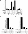
C1D activates DNA-PK when bound to a supercoiled plasmid. (A) DNA-PK activity was tested by standard DNA-PK peptide assays (see Materials and Methods) in the presence of the indicated components. In reactions 8 and 9, PARP or BSA were used instead of C1D-Sc. A total of 800 ng of C1D-Sc was subjected to sequential phenol/chloroform extraction (φ/CHCl3) and the DNA was precipitated, resuspended in a small amount of water, and added into reaction 7 (this amount of DNA is in excess of the amount in 100 ng of C1D-Sc; lanes 5,6,10). Where indicated, 100 ng of excess supercoiled (Sc) or linear (L) DNA was added. (B) Proteins were incubated with the indicated antibodies for 30 min on ice and the kinase reaction was started by adding [γ32P]ATP. The anti-C1D antiserum used in this experiment was raised against the GST–C1D fusion and, therefore, is a distinct derivative from the recombinant His-tagged version of C1D used in the kinase assay. (C) DNA-PK peptide phosphorylation assays were conducted with the indicated components. To generate the Rxn DNA, a standard kinase assay was conducted containing C1D-Sc, DNA-PKcs, and Ku, then the DNA was retrieved by φ/CHCl3 extraction and precipitation.
Figure 5
Figure 5
C1D can activate DNA-PK to phosphorylate full-length p53, and this activation is C1D-dependent. (A) DNA-PK, C1D-Sc, and p53 were incubated as described in Materials and Methods. Where indicated, 100 ng of linear (L) or supercoiled (Sc) plasmid was included. (B) Assays were conducted as in A, except that proteins were incubated with the indicated antibodies before reactions were initiated by the addition of [γ-32P]ATP.
Figure 5
Figure 5
C1D can activate DNA-PK to phosphorylate full-length p53, and this activation is C1D-dependent. (A) DNA-PK, C1D-Sc, and p53 were incubated as described in Materials and Methods. Where indicated, 100 ng of linear (L) or supercoiled (Sc) plasmid was included. (B) Assays were conducted as in A, except that proteins were incubated with the indicated antibodies before reactions were initiated by the addition of [γ-32P]ATP.
Figure 6
Figure 6
C1D and DNA-PKcs interact in mammalian cells. (A) Lysates from untransfected (lane 6) or HA–C1D-transfected COS cells (lanes 15) were immunoprecipitated with preimmune (PI), anti-DNA-PKcs, or anti-HA antisera. For lanes 3 and 4, the cells were treated with 15 Gy of ionizing irradiation before lysate preparation. Western blotting was performed by an anti-HA antibody. The higher band is the IgG light chain (25 kD). (B) Lysates from transfected and untransfected cells were immunoprecipitated with the antisera described in A, and the Western blot was developed by anti-DNA-PKcs antibody.
Figure 6
Figure 6
C1D and DNA-PKcs interact in mammalian cells. (A) Lysates from untransfected (lane 6) or HA–C1D-transfected COS cells (lanes 15) were immunoprecipitated with preimmune (PI), anti-DNA-PKcs, or anti-HA antisera. For lanes 3 and 4, the cells were treated with 15 Gy of ionizing irradiation before lysate preparation. Western blotting was performed by an anti-HA antibody. The higher band is the IgG light chain (25 kD). (B) Lysates from transfected and untransfected cells were immunoprecipitated with the antisera described in A, and the Western blot was developed by anti-DNA-PKcs antibody.
Figure 7
Figure 7
C1D is induced in response to IR. (A) Total cell lysate from cells with or without prior exposure to 15 Gy of IR was resolved by SDS-PAGE and transferred onto nitrocellulose. Western blotting was performed by antibodies raised against a His-tagged C1D (α-C1D 1) or GST–C1D (α-C1D 2) fusion proteins. (B) C1D transcript expression in nonirradiated and irradiated cells. RT–PCR was performed on RNA extracted from irradiated or nonirradiated cells, and cDNA amplified by primers described in Materials and Methods. GAPDH was used as an internal control.
Figure 7
Figure 7
C1D is induced in response to IR. (A) Total cell lysate from cells with or without prior exposure to 15 Gy of IR was resolved by SDS-PAGE and transferred onto nitrocellulose. Western blotting was performed by antibodies raised against a His-tagged C1D (α-C1D 1) or GST–C1D (α-C1D 2) fusion proteins. (B) C1D transcript expression in nonirradiated and irradiated cells. RT–PCR was performed on RNA extracted from irradiated or nonirradiated cells, and cDNA amplified by primers described in Materials and Methods. GAPDH was used as an internal control.

Similar articles

Cited by

References

    1. Alt FW, Oltz EM, Young F, Gorman J, Taccioli G, Chen J. VDJ recombination. Immunol Today. 1992;13:306–314. - PubMed
    1. Anderson CW, Lees-Miller SP. The nuclear serine/threonine protein kinase DNA-PK. Crit Rev Eukaryot Gene Expr. 1992;2:283–314. - PubMed
    1. Avramova Z, Tsanev R. Stable DNA protein complexes in eukaryotic chromatin. J Mol Biol. 1987;196:437–440. - PubMed
    1. Bannister AJ, Kouzarides T. CBP-induced stimulation of c-Fos activity is abrogated by E1A. EMBO J. 1995;14:4758–4762. - PMC - PubMed
    1. Berezney R. Organization and function of the nuclear matrix. Chrom Non-Histone Proteins. 1984;4:119–179.

Publication types

MeSH terms

LinkOut - more resources