. 1998 Jul 27;142(2):377-89.
doi: 10.1083/jcb.142.2.377.

Mistargeting of the lectin ERGIC-53 to the endoplasmic reticulum of HeLa cells impairs the secretion of a lysosomal enzyme

Affiliations

Mistargeting of the lectin ERGIC-53 to the endoplasmic reticulum of HeLa cells impairs the secretion of a lysosomal enzyme

F Vollenweider et al. J Cell Biol. .

Abstract

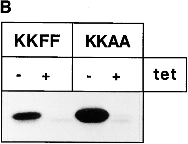
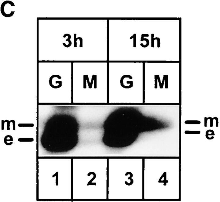
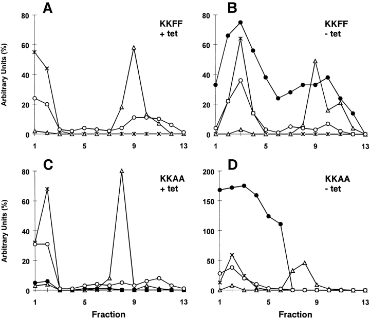
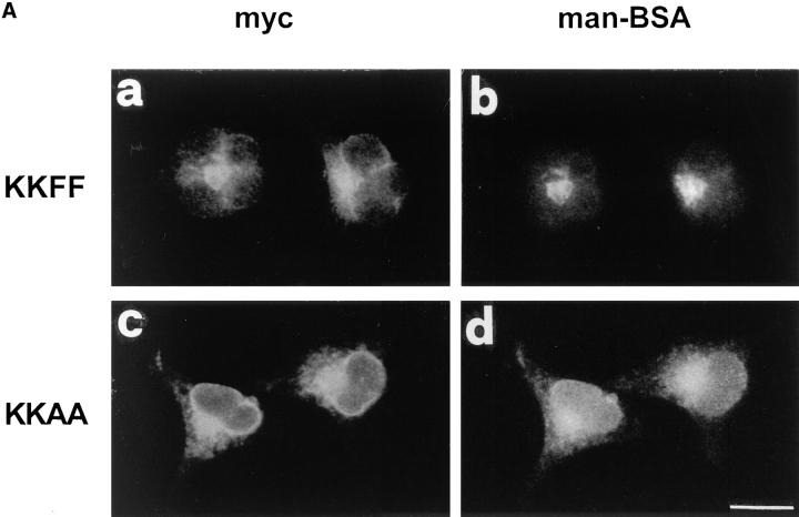
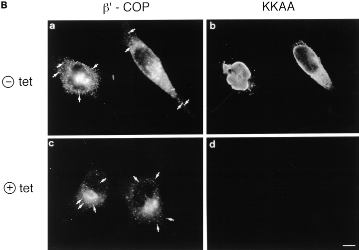
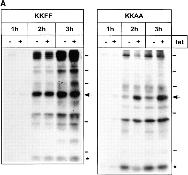
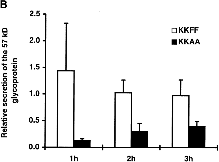
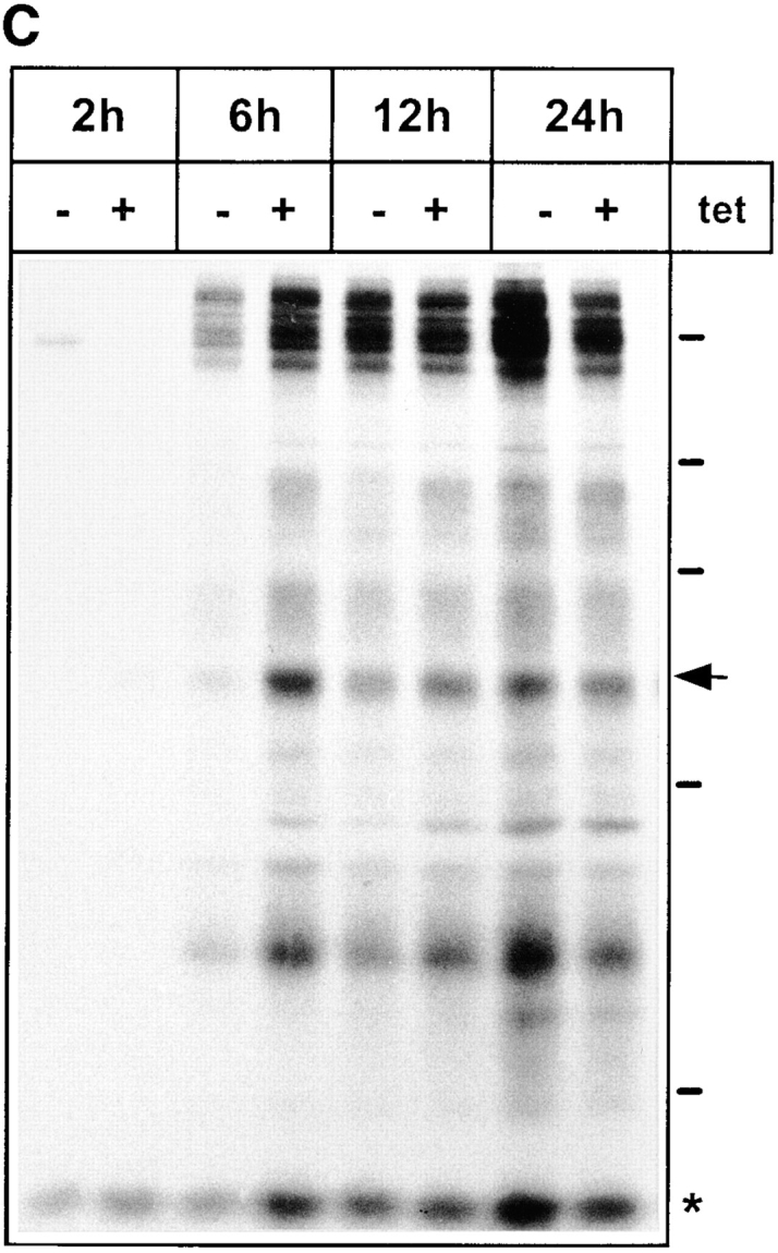
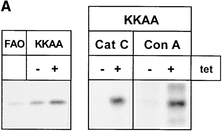
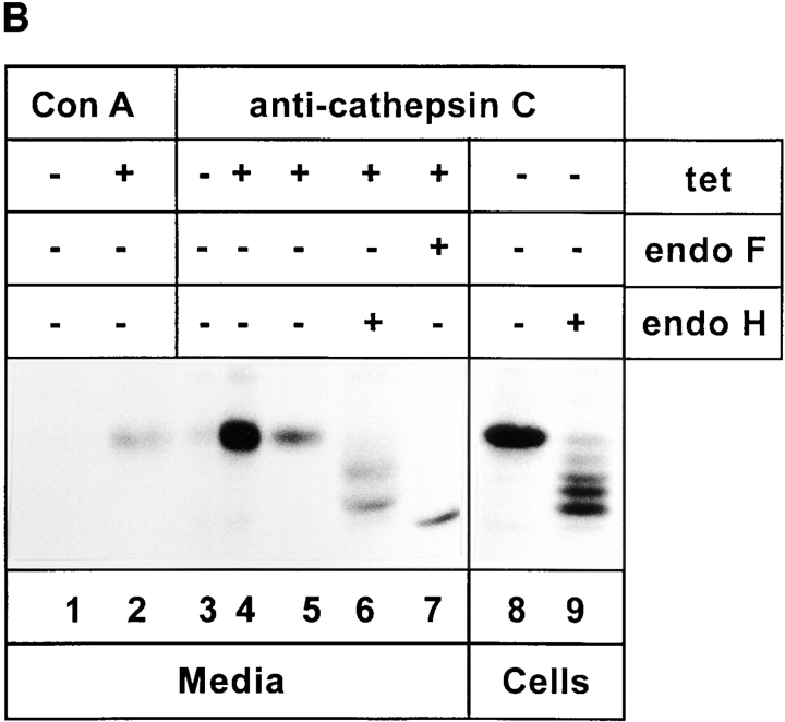
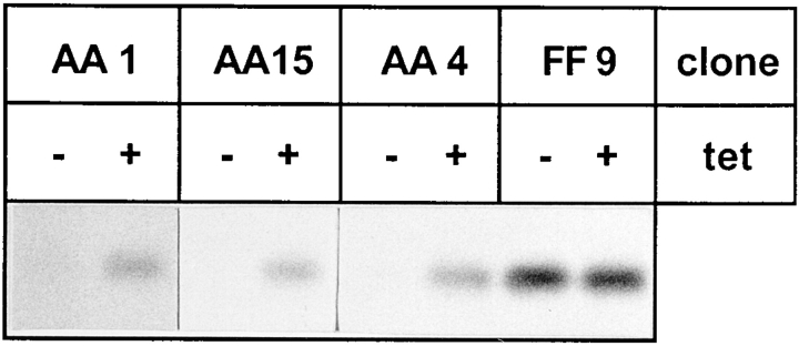
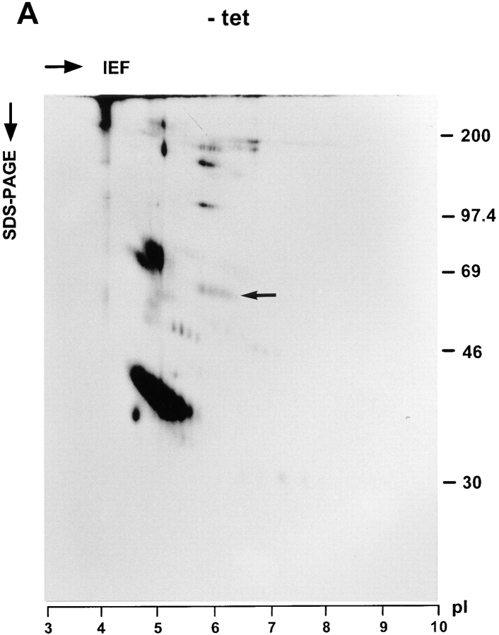
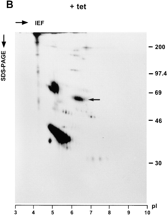
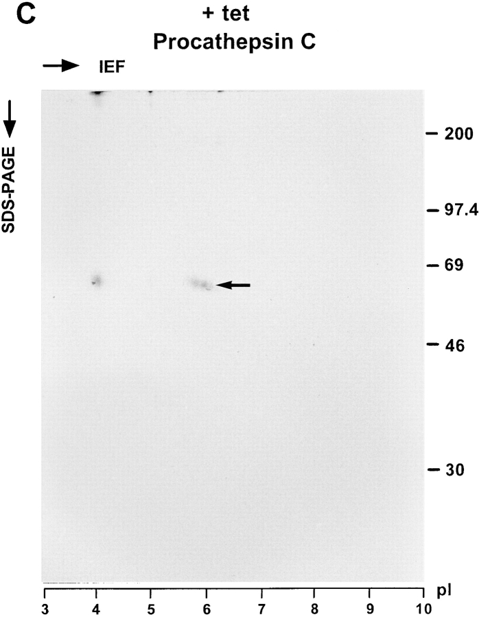
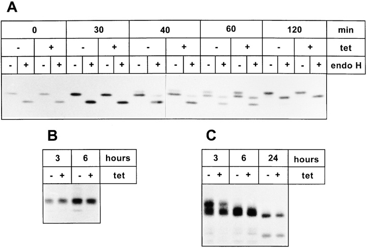
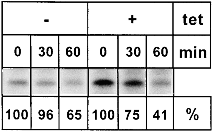
ERGIC-53, a homo-oligomeric recycling protein associated with the ER-Golgi intermediate compartment (ERGIC), has properties of a mannose-selective lectin in vitro, suggesting that it may function as a transport receptor for glycoproteins in the early secretory pathway. To investigate if ERGIC-53 is involved in glycoprotein secretion, a mutant form of this protein was generated that is incapable of leaving the ER. If expressed in HeLa cells in a tetracycline-inducible manner, this mutant accumulated in the ER and retained the endogenous ERGIC-53 in this compartment, thus preventing its recycling. Mistargeting of ERGIC-53 to the ER did not alter the gross morphology of the early secretory pathway, including the distribution of beta'-COP. However, it impaired the secretion of one major glycoprotein, identified as the precursor of the lysosomal enzyme cathepsin C, while overexpression of wild-type ERGIC-53 had no effect on glycoprotein secretion. Transport of two other lysosomal enzymes and three post-Golgi membrane glycoproteins was unaffected by inactivating the recycling of ERGIC-53. The results suggest that the recycling of ERGIC-53 is required for efficient intracellular transport of a small subset of glycoproteins, but it does not appear to be essential for the majority of glycoproteins.


Figures

Figure 1
Figure 1
Expression of KKFF and KKAA in stably transformed HtTA-1 HeLa cells. (A) Schematic representation of the two ERGIC-53 constructs used in this study. Both carry a c-myc epitope after the signal sequence cleavage site (Itin et al., 1995b ). KKFF is the wild-type ERGIC-53. KKAA has the luminal and transmembrane domains of ERGIC-53 and a mutated cytoplasmic domain. The amino acid residues of the cytosolic tails are indicated in the single letter code. (B) Tet-dependent inducibility of the KKFF and KKAA constructs. Cells were incubated for 48 h ± tet, pulsed for 30 min with [35S]methionine, and immunoprecipitated with an mAb against ERGIC-53, and the proteins were separated by 4–10% SDS-PAGE followed by fluorography. (C) Coimmunoprecipitation of the KKAA construct with endogenous ERGIC-53. KKAA cells were cultured in the absence of tet in medium containing [35S]methionine for the indicated times. Immunoprecipitation with an antibody against the myc epitope (M, present only in the KKAA construct) coprecipitated endogenous ERGIC-53 (e) with the KKAA construct (m) (lanes 2 and 4). For reference, immunoprecipitation was also done with G1/93 (G), which recognizes an epitope present on both the KKAA construct and endogenous ERGIC-53 (lanes 1 and 3). The unequal signals are due to the known low immunoprecipitation efficiency of the anti-myc antibody. Proteins were visualized by fluorography after SDS-PAGE.
Figure 1
Figure 1
Expression of KKFF and KKAA in stably transformed HtTA-1 HeLa cells. (A) Schematic representation of the two ERGIC-53 constructs used in this study. Both carry a c-myc epitope after the signal sequence cleavage site (Itin et al., 1995b ). KKFF is the wild-type ERGIC-53. KKAA has the luminal and transmembrane domains of ERGIC-53 and a mutated cytoplasmic domain. The amino acid residues of the cytosolic tails are indicated in the single letter code. (B) Tet-dependent inducibility of the KKFF and KKAA constructs. Cells were incubated for 48 h ± tet, pulsed for 30 min with [35S]methionine, and immunoprecipitated with an mAb against ERGIC-53, and the proteins were separated by 4–10% SDS-PAGE followed by fluorography. (C) Coimmunoprecipitation of the KKAA construct with endogenous ERGIC-53. KKAA cells were cultured in the absence of tet in medium containing [35S]methionine for the indicated times. Immunoprecipitation with an antibody against the myc epitope (M, present only in the KKAA construct) coprecipitated endogenous ERGIC-53 (e) with the KKAA construct (m) (lanes 2 and 4). For reference, immunoprecipitation was also done with G1/93 (G), which recognizes an epitope present on both the KKAA construct and endogenous ERGIC-53 (lanes 1 and 3). The unequal signals are due to the known low immunoprecipitation efficiency of the anti-myc antibody. Proteins were visualized by fluorography after SDS-PAGE.
Figure 2
Figure 2
Subcellular fractionation on Nycodenz gradients of KKFF (A and B) and KKAA (C and D) HeLa cells cultured in the presence (+tet) or absence (−tet, 48 h) of tetracycline. Fractions were collected from bottom (fraction 1) to top (fraction 13). The amount of marker protein in each fraction was assessed by immunoblotting. The blots were quantified by phosphorimaging, and total counts in the gradient were set to 100% with the exception of the KKFF and the KKAA constructs in B and D, which were drawn in relation to the endogenously expressed ERGIC-53. open circles, endogenous ERGIC-53; filled circles, transfected ERGIC-53 construct; triangles, cis/medial-Golgi marker GPP130; X, ER marker p63.
Figure 3
Figure 3
(A) Binding of fluorescein-labeled mannosylated bovine serum albumin (man-BSA). Transformed HeLa cell lines expressing KKFF and KKAA were incubated 48 h in the absence of tet, fixed/ permeabilized with paraformaldehyde/Triton X-100, and tested for binding of man-BSA (Itin et al., 1996). KKFF (a) and KKAA (c) were visualized with anti-myc followed by a rhodamine-labeled secondary antibody. Man-BSA binding was photographed in the fluorescein channel (b and d). (B) Localization of β′-COP and the KKAA construct (± tet) by double immunofluorescence microscopy. KKAA cells were fixed with paraformaldehyde, permeabilized with saponine, and subjected to immunolabeling. β′-COP (a and c) was detected with a specific rabbit antibody followed by a fluorescein-conjugated secondary antibody. The KKAA construct (b and d) was visualized with an mAb against the c-myc epitope followed by a rhodamine-conjugated secondary antibody. Note that the distribution of β′-COP is identical regardless of whether the KKAA construct is expressed (a and b) or not (c and d). Arrows indicate ERGIC clusters. Bars: (A) 15 μm; (B) 7 μm.
Figure 3
Figure 3
(A) Binding of fluorescein-labeled mannosylated bovine serum albumin (man-BSA). Transformed HeLa cell lines expressing KKFF and KKAA were incubated 48 h in the absence of tet, fixed/ permeabilized with paraformaldehyde/Triton X-100, and tested for binding of man-BSA (Itin et al., 1996). KKFF (a) and KKAA (c) were visualized with anti-myc followed by a rhodamine-labeled secondary antibody. Man-BSA binding was photographed in the fluorescein channel (b and d). (B) Localization of β′-COP and the KKAA construct (± tet) by double immunofluorescence microscopy. KKAA cells were fixed with paraformaldehyde, permeabilized with saponine, and subjected to immunolabeling. β′-COP (a and c) was detected with a specific rabbit antibody followed by a fluorescein-conjugated secondary antibody. The KKAA construct (b and d) was visualized with an mAb against the c-myc epitope followed by a rhodamine-conjugated secondary antibody. Note that the distribution of β′-COP is identical regardless of whether the KKAA construct is expressed (a and b) or not (c and d). Arrows indicate ERGIC clusters. Bars: (A) 15 μm; (B) 7 μm.
Figure 4
Figure 4
Secretion of glycoproteins in KKFF and KKAA HeLa cells. (A) Cells were incubated for 48 h ± tet, labeled for 30 min with [35S]methionine and chased for 1, 2, and 3 h. Glycoproteins were isolated with Con A beads and separated by 10% SDS-PAGE. 14C-labeled molecular mass markers are indicated at the right margin (200, 97.4, 66, 46, and 30 kD). The arrows indicate the position of the 57-kD protein. *, band used as a reference for quantification in B. The apparent absence of the 66-kD protein in the KKFF panel (3 h, −tet) is due to an artifactual inhomogeneity of the gel. (B) Quantification of the delay in secretion of the 57-kD glycoprotein. The amount of the 57-kD glycoprotein was determined by densitometry of fluorograms and normalized. Shown is the relative secretion as determined by dividing the value obtained for −tet by that for +tet. Values are means ± SEM of at least three independent experiments. white bars, KKFF; black bars, KKAA. (C) Tet- dependent difference in the secretion of the 57-kD protein disappears with increased chase times. KKAA cells were treated ± tet for 48 h, pulsed for 30 min with [35S]methionine, and chased for 2, 6, 12, and 24 h. Glycoproteins were isolated from the culture medium and analyzed by SDS-PAGE as in A.
Figure 4
Figure 4
Secretion of glycoproteins in KKFF and KKAA HeLa cells. (A) Cells were incubated for 48 h ± tet, labeled for 30 min with [35S]methionine and chased for 1, 2, and 3 h. Glycoproteins were isolated with Con A beads and separated by 10% SDS-PAGE. 14C-labeled molecular mass markers are indicated at the right margin (200, 97.4, 66, 46, and 30 kD). The arrows indicate the position of the 57-kD protein. *, band used as a reference for quantification in B. The apparent absence of the 66-kD protein in the KKFF panel (3 h, −tet) is due to an artifactual inhomogeneity of the gel. (B) Quantification of the delay in secretion of the 57-kD glycoprotein. The amount of the 57-kD glycoprotein was determined by densitometry of fluorograms and normalized. Shown is the relative secretion as determined by dividing the value obtained for −tet by that for +tet. Values are means ± SEM of at least three independent experiments. white bars, KKFF; black bars, KKAA. (C) Tet- dependent difference in the secretion of the 57-kD protein disappears with increased chase times. KKAA cells were treated ± tet for 48 h, pulsed for 30 min with [35S]methionine, and chased for 2, 6, 12, and 24 h. Glycoproteins were isolated from the culture medium and analyzed by SDS-PAGE as in A.
Figure 5
Figure 5
The sequence of three peptides isolated from the 57-kD protein can be accommodated within the amino acid sequence of the precursor of the human lysosomal peptidase procathepsin C (Paris et al., 1995). Shown is part of the deduced partial amino acid sequence of human procathepsin C and the sequence of three tryptic peptides determined by automatic sequencing (bold and underlined).
Figure 6
Figure 6
The inefficiently secreted 57-kD protein is procathepsin C. (A, left) Procathepsin C expression in FAO and HeLa cells. FAO and KKAA (± tet, 48 h) cells were labeled for 30 min with [35S]methionine and immunoprecipitated with an antibody against rat cathepsin C. Immunoprecipitated proteins were separated by 10% SDS-PAGE. (Right) Glycoproteins isolated by Con A beads and immunoprecipitation of procathepsin C. KKAA ± tet (48 h) cells were pulsed for 30 min and chased for 2 h. Secreted glycoproteins released from Con A beads (Con A) were lyophilized, immunoprecipitated with the cathepsin C antibody, and separated by 10% SDS-PAGE (Cat C). (B) Direct immunoprecipitation of procathepsin C from the culture medium and digestion with glycosidases. KKAA in the presence or absence of tet (48 h) cells were pulsed for 30 min and chased for 2 h. The medium (lanes 1–7) was subjected to methanol precipitation, and the precipitate was resuspended and immunoprecipitated with the cathepsin C antibody. Immunoprecipitates were digested with endo H under nonlimiting (lane 6) and limiting (lane 9, sample was from detergent-solubilized cells) conditions or with endo F as indicated and separated by SDS-PAGE.
Figure 6
Figure 6
The inefficiently secreted 57-kD protein is procathepsin C. (A, left) Procathepsin C expression in FAO and HeLa cells. FAO and KKAA (± tet, 48 h) cells were labeled for 30 min with [35S]methionine and immunoprecipitated with an antibody against rat cathepsin C. Immunoprecipitated proteins were separated by 10% SDS-PAGE. (Right) Glycoproteins isolated by Con A beads and immunoprecipitation of procathepsin C. KKAA ± tet (48 h) cells were pulsed for 30 min and chased for 2 h. Secreted glycoproteins released from Con A beads (Con A) were lyophilized, immunoprecipitated with the cathepsin C antibody, and separated by 10% SDS-PAGE (Cat C). (B) Direct immunoprecipitation of procathepsin C from the culture medium and digestion with glycosidases. KKAA in the presence or absence of tet (48 h) cells were pulsed for 30 min and chased for 2 h. The medium (lanes 1–7) was subjected to methanol precipitation, and the precipitate was resuspended and immunoprecipitated with the cathepsin C antibody. Immunoprecipitates were digested with endo H under nonlimiting (lane 6) and limiting (lane 9, sample was from detergent-solubilized cells) conditions or with endo F as indicated and separated by SDS-PAGE.
Figure 7
Figure 7
Inefficient secretion of procathepsin C is not due to clonal variation. Cells of clones KKAA 1, KKAA 15, KKAA 4 (used in this study), and KKFF 9 cells were cultured in the presence or absence of tet for 48 h followed by labeling for 30 min with [35S]methionine and a chase of 2 h. Proteins released into the culture medium were methanol precipitated. The precipitates were resuspended and immunoprecipitated with anti–cathepsin C antibody. The immunoprecipitates were separated by SDS-PAGE and radiolabeled proteins were visualized by fluorography.
Figure 8
Figure 8
2D gel analysis of secreted glycoproteins. KKAA cells were cultured in the absence (A) or presence (B) of tet for 48 h. Thereafter, the cells were labeled for 30 min with [35S]methionine and chased for 3 h. Secreted glycoproteins were isolated with Con A beads, methanol precipitated, and subjected to isoelectric focusing (15 h) in the first dimension using a 3–10 linear immobiline pH gradient. 10% SDS-PAGE was used for the second dimension. As a reference, cathepsin C was immunoprecipitated from the medium of KKAA cells (+tet) that had been labeled in parallel and subjected to the same 2D gel separation procedure (C). Proteins were visualized by fluorography.
Figure 8
Figure 8
2D gel analysis of secreted glycoproteins. KKAA cells were cultured in the absence (A) or presence (B) of tet for 48 h. Thereafter, the cells were labeled for 30 min with [35S]methionine and chased for 3 h. Secreted glycoproteins were isolated with Con A beads, methanol precipitated, and subjected to isoelectric focusing (15 h) in the first dimension using a 3–10 linear immobiline pH gradient. 10% SDS-PAGE was used for the second dimension. As a reference, cathepsin C was immunoprecipitated from the medium of KKAA cells (+tet) that had been labeled in parallel and subjected to the same 2D gel separation procedure (C). Proteins were visualized by fluorography.
Figure 9
Figure 9
Intracellular transport of identified glycoproteins in KKAA cells (treated in the presence or absence of tet for 48 h). (A) Transferrin receptor: Cells were pulsed for 10 min with [35S]methionine and chased as indicated. The receptor was immunoprecipitated from the Triton X-100–solubilized cells and digested (or not) with endo H. (B) Cathepsin D: Cells were metabolically labeled for 1 h followed by a chase for 3 or 6 h. The culture medium was precipitated with methanol. Cathepsin D was immunoprecipitated from the resuspended methanol precipitate. (C) α-Glucosidase: Cells were metabolically labeled for 1 h and chased as indicated, and the enzyme was immunoprecipitated after cell solubilization with Triton X-100. The immunoprecipitates were separated by SDS-PAGE and visualized by fluorography.
Figure 10
Figure 10
Overexpression of the KKAA construct delays ER exit of procathepsin C. KKAA cells were cultured in presence or absence of tet for 48 h and pulse-labeled with [35S]methionine and chased for the indicated times. The cells were homogenized, and membranes were separated by Nycodenz gradient centrifugation (see Materials and Methods). Fractions 1–5 containing the ER marker p63 were pooled and Triton X-100 solubilized, and procathepsin C was immunoprecipitated. The immunoprecipitates were separated by SDS-PAGE, and procathepsin C was quantified from the fluorogram. The start signal after the pulse was set to 100%. All the lanes of this figure originate from the same fluorogram and were identically exposed. They had to be rearranged for reproduction purposes.

References

    1. Arar C, Carpentier V, Le Cear J-P, Monsigny M, Legrand A, Roche AC. ERGIC-53, a membrane protein of the endoplasmic reticulum-Golgi intermediate compartment, is identical to MR60, an intracellular mannose-specific lectin of myelomonocytic cells. J Biol Chem. 1995;270:3551–3553. - PubMed
    1. Aridor M, Bannykh SI, Rowe T, Balch WE. Sequential coupling between COPII and COPI vesicle coats in endoplasmic reticulum to Golgi transport. J Cell Biol. 1995;131:875–893. - PMC - PubMed
    1. Balch WE, McCaffery JM, Plutner H, Farquhar MG. Vesicular stomatitis virus glycoprotein is sorted and concentrated during export from the endoplasmic reticulum. Cell. 1994;76:841–852. - PubMed
    1. Banfiled DK, Louis C, Rabouille C, Warren G, Pelham HR. Localization of Sed5, a putative vesicle targeting molecule, to the cis-Golgi network involves both its transmembrane and cytoplasmic domains. J Cell Biol. 1994;127:357–371. - PMC - PubMed
    1. Bannykh SI, Balch WE. Membrane dynamics at the endoplasmic reticulum–Golgi interface. J Cell Biol. 1997;138:1–4. - PMC - PubMed

Publication types

MeSH terms