. 1998 Jul 27;142(2):443-55.
doi: 10.1083/jcb.142.2.443.

Iqg1p, a yeast homologue of the mammalian IQGAPs, mediates cdc42p effects on the actin cytoskeleton

Affiliations

Iqg1p, a yeast homologue of the mammalian IQGAPs, mediates cdc42p effects on the actin cytoskeleton

M A Osman et al. J Cell Biol. .

Abstract

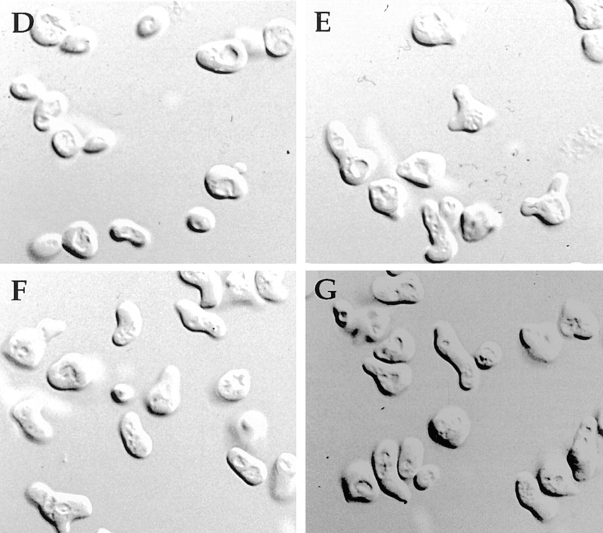
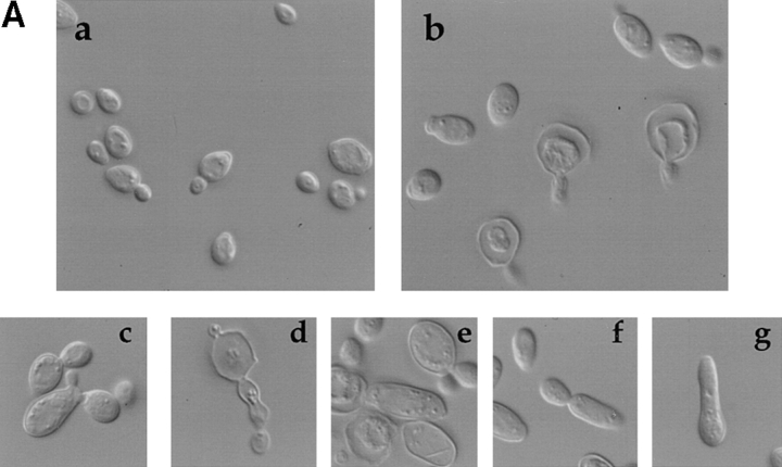
The Rho-type GTPase Cdc42p has been implicated in diverse cellular functions including cell shape, cell motility, and cytokinesis, all of which involve the reorganization of the actin cytoskeleton. Targets of Cdc42p that interface the actin cytoskeleton are likely candidates for mediating cellular activities. In this report, we identify and characterize a yeast homologue for the mammalian IQGAP, a cytoskeletal target for Cdc42p. The yeast IQGAP homologue, designated Iqg1p, displays a two-hybrid interaction with activated Cdc42p and coimmunoprecipitates with actin filaments. Deletion of IQG1 results in a temperature-sensitive lethality and causes aberrant morphologies including elongated and round multinucleated cells. This together with its localization at the mother-bud neck, suggest that Iqg1p promotes budding and cytokinesis. At restrictive temperatures, the vacuoles of the mutant cells enlarge and vesicles accumulate in the bud. Interestingly, Iqg1p shows two-hybrid interactions with the ankyrin repeat-containing protein, Akr1p (Kao, L.-R., J. Peterson, J. Ruiru, L. Bender, and A. Bender. 1996. Mol. Cell. Biol. 16:168-178), which inhibits pheromone signaling and appears to promote cytokinesis and/or trafficking. We also show two-hybrid interactions between Iqg1p and Afr1p, a septin-binding protein involved in projection formation (Konopka, J.B., C. DeMattei, and C. Davis. 1995. Mol. Cell. Biol. 15:723-730). We propose that Iqg1p acts as a scaffold to recruit and localize a protein complex involved in actin-based cellular functions and thus mediates the regulatory effects of Cdc42p on the actin cytoskeleton.


Figures

Figure 1
Figure 1
Homologous regions of IQGAP proteins. (A) Schematic representation of domain composition. (B) The CHD. IR, IQGAP repeats; IQ, four repeats of the calmodulin-binding motif. The WW domain known to bind proline-rich regions in signaling molecules is less conserved in Iqg1p. (C) The Ras-GAP related domain (GRD); the highly conserved subdomains (blocks 1–3; Weissbach et al., 1994) are underlined.
Figure 2
Figure 2
Iqg1p concentrates at sites of cell growth and at the septum. Indirect immunofluorescence localization of Iqg1p in cells transformed with plasmid pA1 encoding the full-length IQG1 gene that complements the phenotypes of iqg1Δ cells. Log phase cells grown in selective media at 30°C were prepared and stained with anti-HA antibodies as described in Materials and Methods.
Figure 3
Figure 3
Growth and polarity defects in wild-type and iqg1Δ cells. (A) Phenotypes of iqg1Δ cells at 23°C. Wild-type (a) and iqg1Δ cells (b–g) were grown in YPD to log phase, briefly sonicated and visualized by Nomarski optics at the same magnification. Phenotype of wild-type (B) and iqg1Δ cells (C) at restrictive temperature. Cells were grown at 23°C and shifted to 37°C for 2 h, and then visualized by Nomarski optics at the same magnification. Haploid wild-type (D) and iqg1Δ (E–G) cells were grown at 23°C and treated with α factor as described in Materials and Methods, and visualized by Nomarski optics. (H) Halo assay; sensitivity to pheromone-induced growth arrest of iqg1Δ cells. Filter disks containing 15 μl of 40 μg/ml α factor were spotted onto lawns of wild-type (left) and iqg1Δ (right) cells on YPD plates, incubated for 3 d at 23°C, and then photographed.
Figure 3
Figure 3
Growth and polarity defects in wild-type and iqg1Δ cells. (A) Phenotypes of iqg1Δ cells at 23°C. Wild-type (a) and iqg1Δ cells (b–g) were grown in YPD to log phase, briefly sonicated and visualized by Nomarski optics at the same magnification. Phenotype of wild-type (B) and iqg1Δ cells (C) at restrictive temperature. Cells were grown at 23°C and shifted to 37°C for 2 h, and then visualized by Nomarski optics at the same magnification. Haploid wild-type (D) and iqg1Δ (E–G) cells were grown at 23°C and treated with α factor as described in Materials and Methods, and visualized by Nomarski optics. (H) Halo assay; sensitivity to pheromone-induced growth arrest of iqg1Δ cells. Filter disks containing 15 μl of 40 μg/ml α factor were spotted onto lawns of wild-type (left) and iqg1Δ (right) cells on YPD plates, incubated for 3 d at 23°C, and then photographed.
Figure 4
Figure 4
Immunofluorescence localization of Cdc42p in α factor–arrested and budding iqg1Δ cells. (A) MATa wild-type cells treated with α factor. (B) MATa iqg1Δ cells treated with α factor. (C) Wild-type (MO1) cells. (D) iqg1Δ (MO4) cells. (E) Nomarski image of an elongated bud attached to the mother cell in iqg1Δ (MO4). (F) Cdc42p localization at presumptive septa (arrows).
Figure 5
Figure 5
Actin filament organization defects caused by iqg1Δ mutation. The images show rhodamine-phalloidin staining of MO1 (wild-type) and MO4 (iqg1Δ) budding cells grown at 30°C. (A) Budding wild-type cells. (B and C) Budding iqg1Δ cells. (D) Large iqg1Δ cell with an elongated bud; arrows pointing at ridges of the presumptive septa locations.
Figure 8
Figure 8
Chitin deposition in wild-type and iqg1Δ cells. Diploid cells grown at 30°C were stained with calcofluor to visualize chitin deposition of MO1 (wild-type) and MO4 (iqg1Δ) cells. (A) Wild-type cells. (B and C) iqg1Δ cells. All cells were photographed at the same magnification.
Figure 9
Figure 9
The iqg1Δ cells accumulate nuclei. (A and B) Wild-type (MO1) cells. (C–F) iqg1Δ cells. Nomarski optics reveal wild-type and iqg1Δ cell morphology at 30°C (A and C). DAPI staining of DNA in iqg1Δ cells reveals the number and location of the nuclei (D–F). Wild-type (G) and iqg1Δ cells (H) stained with Yol1/34 anti-tubulin antibodies.
Figure 7
Figure 7
Immunofluorescence localization of calmodulin in wild-type and iqg1Δ cells treated with α factor. Cells grown at 30°C were treated with α factor and prepared for indirect immunofluorescence as described in Materials and Methods. (A) MATa wild-type cells. (B) MATa iqg1Δ (MO2) cells.
Figure 6
Figure 6
Iqg1p coimmunoprecipitates with actin filaments. Total cell lysate from a CB001 strain harboring the pA1 plasmid encoding the HA-Iqg1p was prepared as described in Materials and Methods. Immunoprecipitation was performed overnight at 4°C in the absence (−) or presence (+) of 25 μM phalloidin. Precipitated proteins were resolved on a 10% SDS-PAGE and immunoblotted with antibodies recognizing actin (top panel) and HA-tagged Iqg1p (bottom panel). Vector with HA-tag and lacking the IQG1 gene did not precipitate actin. Similarly, a vector carrying IQG1 gene and lacking HA tag did not precipitate actin in the presence of phalloidin.
Figure 10
Figure 10
Flow cytometry reveals DNA content of 2C or more at all temperatures tested in iqg1Δ cells. Wild-type and iqg1Δ cells (MO2) were grown at 23°C until early log phase, and then incubated for 4 h at 23°, 30°, or 37°C. DNA content of individual cells was measured by flow cytometry as described in Materials and Methods.
Figure 11
Figure 11
Electron micrographs showing iqg1Δ cells accumulate vesicles in the bud. Cells were grown to early log phase at 23°C and shifted to 37°C for 3 h. Thin-sectioning electron microscopy was done as described in Materials and Methods. (A) iqg1Δ cell. (B) Wild-type cell. (C) The bud in A at higher magnification.
Figure 12
Figure 12
Summary of Iqg1p interactions and proposed functional outcome. Interactions between Iqg1p Cdc42p, actin, Akr1p, and Afr1p (this study); Afr1p and Cdc12p (Konopka et al., 1995); Akr1p, Bem1p, and Gcs1p (Kao et al., 1996).

Similar articles

Cited by

References

    1. Adachi H, Takahashi Y, Hasebe T, Shirouzu M, Yokoyama S, Sutoh K. DictyosteliumIQGAP-related protein specifically involved in the completion of cytokinesis. J Cell Biol. 1997;137:891–898. - PMC - PubMed
    1. Adams, A.E.M., and J.R. Pringle. 1991. Staining of actin with fluorochrome-conjugated phalloidin. Methods in Enzymology. 194:729–731. - PubMed
    1. Adams AEM, Johnson DI, Longnecker RM, Sloat BF, Pringle JR. CDC42 and CDC43, two additional genes involved in budding and the establishment of cell polarity in the yeast Sacharomyces cerevisiae. . J Cell Biol. 1990;111:131–142. - PMC - PubMed
    1. Ausubel, F.M., R. Brent, R.E. Kingston, D.D. Moore, J.G. Seidman, J.A. Smith, and K. Struhl. 1992. Current Protocols in Molecular Biology. John Wiley and Sons Ltd., New York.
    1. Bagrodia S, Derijard B, Davis RJ, Cerione RA. Cdc42 and PAK-mediated signaling leads to jun kinase and p38 mitogen-activated protein kinase activation. J Biol Chem. 1995;270:27995–27998. - PubMed

Publication types

MeSH terms