. 1998 Jul 27;142(2):473-84.
doi: 10.1083/jcb.142.2.473.

Cyclic nucleotide-gated channels on the flagellum control Ca2+ entry into sperm

Affiliations

Cyclic nucleotide-gated channels on the flagellum control Ca2+ entry into sperm

B Wiesner et al. J Cell Biol. .

Abstract

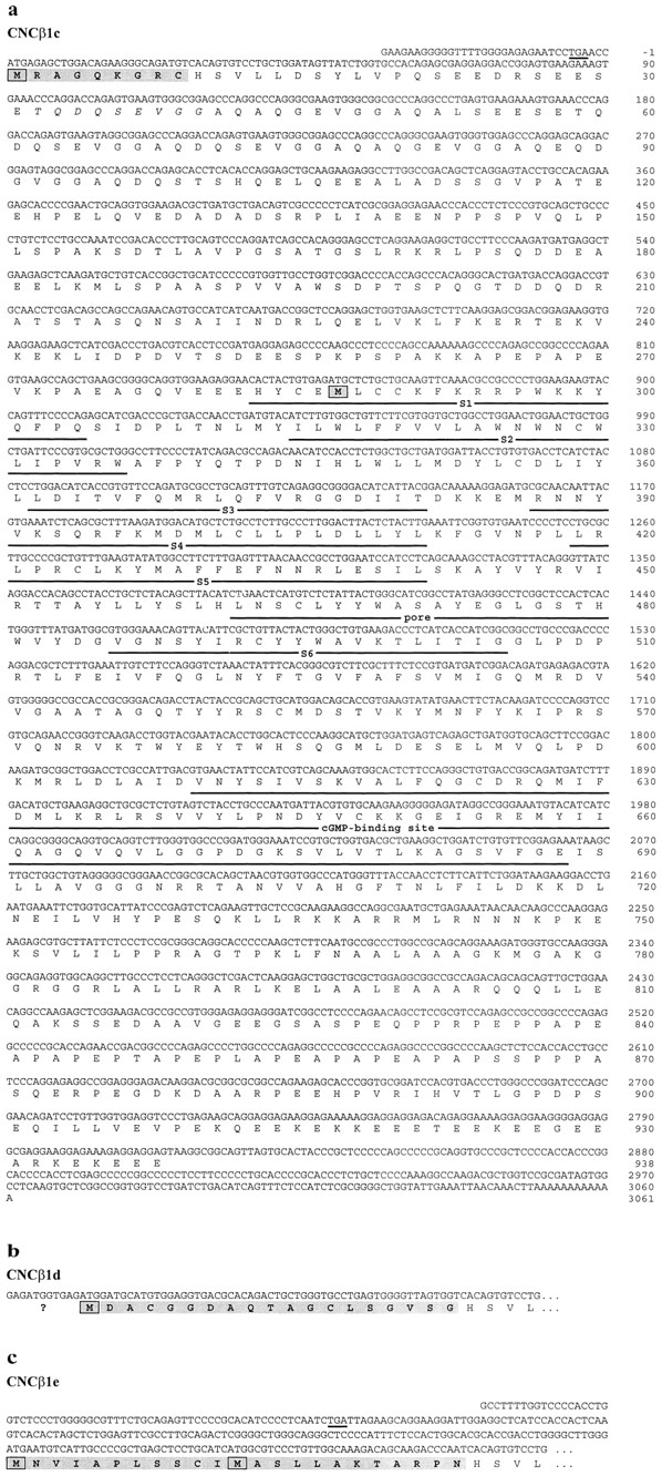
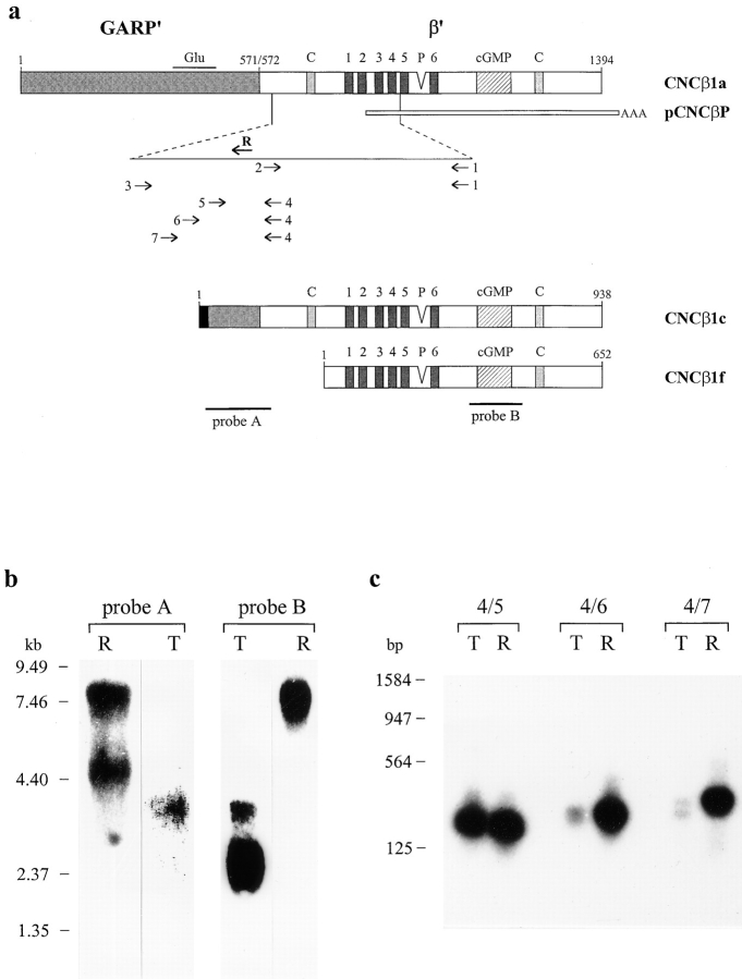
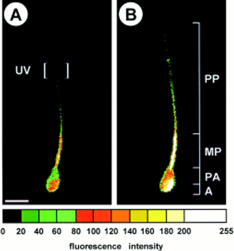
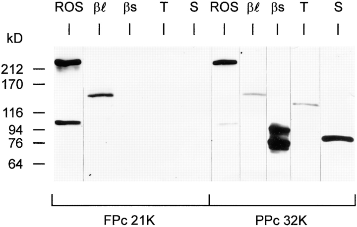
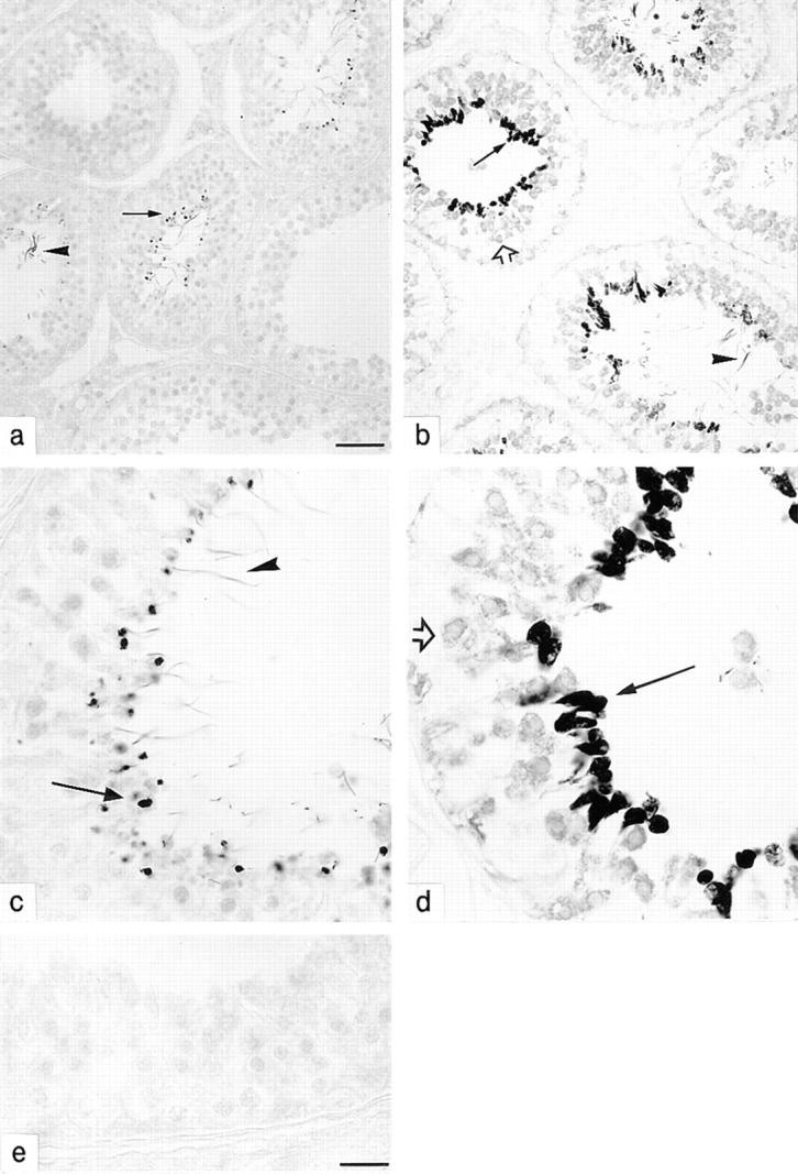
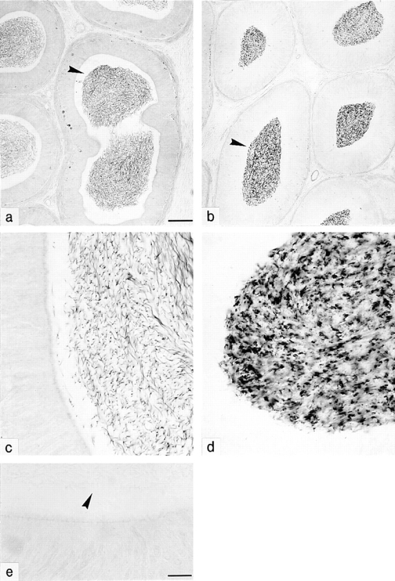
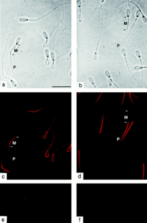
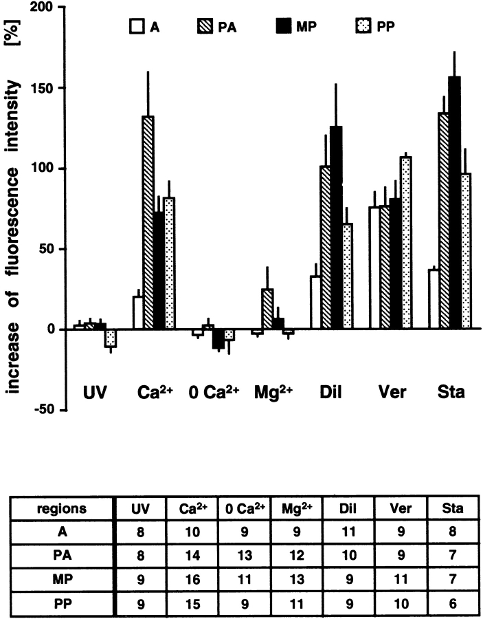
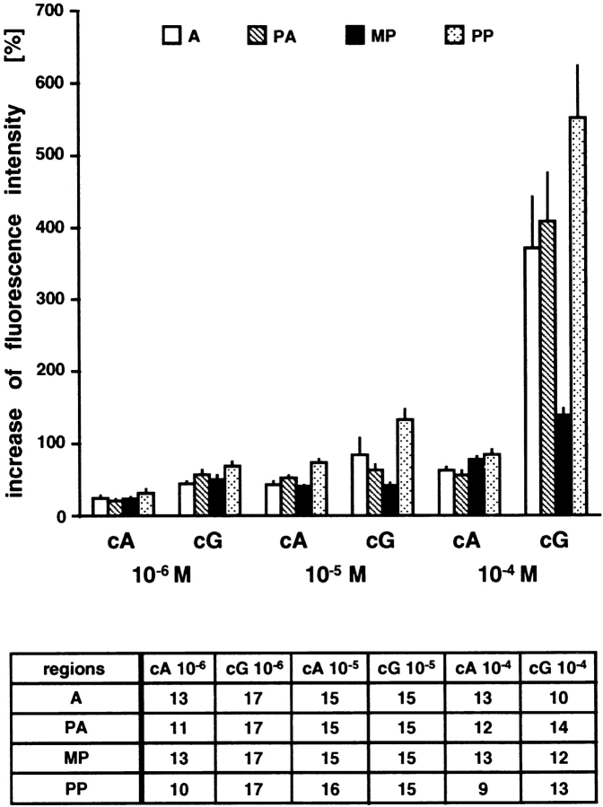
Cyclic nucleotide-gated (CNG) channels are key elements of cGMP- and cAMP-signaling pathways in vertebrate photoreceptor cells and in olfactory sensory neurons, respectively. These channels form heterooligomeric complexes composed of at least two distinct subunits (alpha and beta). The alpha subunit of cone photoreceptors is also present in mammalian sperm. Here we identify one short and several long less abundant transcripts of beta subunits in testis. The alpha and beta subunits are expressed in a characteristic temporal and spatial pattern in sperm and precursor cells. In mature sperm, the alpha subunit is observed along the entire flagellum, whereas the short beta subunit is restricted to the principal piece of the flagellum. These findings suggest that different forms of CNG channels coexist in the flagellum. Confocal microscopy in conjunction with the Ca2+ indicator Fluo-3 shows that the CNG channels serve as a Ca2+ entry pathway that responds more sensitively to cGMP than to cAMP. Assuming that CNG channel subtypes differ in their Ca2+ permeability, dissimilar localization of alpha and beta subunits may give rise to a pattern of Ca2+ microdomains along the flagellum, thereby providing the structural basis for control of flagellar bending waves.


Figures

Figure 2
Figure 2
Nucleotide and deduced amino acid sequences of CNCβ1c/ -1d/ -1e. (a) Nucleotide and deduced amino acid sequence of CNCβ1c. The first nucleotide of the translational initiation codon has been assigned position +1. A nonsense codon preceding the initiation codon in the same reading frame is underlined. The initiating methionines of CNCβ1c and of the short testis β subunit (CNCβ1f) are boxed. The deduced amino acid sequence (in one-letter code) is shown below the nucleotide sequence. Amino acid residues are numbered beginning with the initiating methionine. Number of the last residue in each line is given on the right-hand side. Beginning with histidine 10 in CNCβ1c, the amino acid sequences of the three long β variants are identical. The transmembrane segments S1–S6, the pore region, and the cGMP-binding site are represented by lines above the sequence. Amino acid residues 32–40 that are missing in two of seven clones are written in italics. (b and c) Nucleotide and deduced amino acid sequences of the 5′ ends of CNCβ1d and CNCβ1e. The putative initiating methionines are boxed. Clone pCNCβ1d does not contain a nonsense codon upstream of the putative initiation codon; therefore, the initiation site is not certain (question mark). In pCNCβ1e, a nonsense codon preceding the initiation codon in the same reading frame is underlined. These sequence data are available from GenBank/EMBL/DDBJ under accession number AF074012 (CNCβ1c), AF074013 (CNCβ1d), AF074014 (CNCβ1e).
Figure 1
Figure 1
Cloning of β subunits and Northern blot analysis. (a) Schematic drawing of the primary structure of β subunits from bovine rod photoreceptor (CNCβ1a) and testis (CNCβ1c, CNCβ1f). CNCβ1a consists of a GARP part (aa 1–571) and a β′ part (aa 572–1394). The GARP part is almost identical to the bovine GARP (Sugimoto et al., 1991). Glu refers to the glutamic acid–rich region; the transmembrane segments 1–6, the cGMP-binding site (cGMP), and the calmodulin-binding sites (C) are depicted as boxes. P, pore region. The different 5′ ends of the long testis β subunits are represented as a black box. The COOH-terminal calmodulin-binding site does not modulate the channel activity (Grunwald et al., 1998; Weitz et al., 1998). The location of partial clone pCNCβP, and of riboprobes A and B used for Northern blot analysis is indicated. The location of primers 1–7 and primer R for 5′ RACE is shown on a larger scale below the corresponding region of CNCβ1a. (b) Northern blot analysis of poly(A)+ RNA from bovine testis (T) and retina (R). Probe A corresponding to aa 469–578 of CNCβ1a hybridized to transcripts of ∼7.4 kb and ∼4.4 kb from retina and to an ∼3.3-kb transcript from testis. Probe B corresponding to aa 909–1081 hybridized to transcripts of ∼3.3 and 2.4 kb from testis and to an ∼7.4-kb transcript from retina. Autoradiographic exposure was 3 d for testis samples and 12 h for retina samples. Integrity of poly(A)+ RNA was confirmed by Northern blot analysis of transcripts encoding actin and CNCα2 (data not shown). (c) PCR amplification of β transcripts from testis (T) and retina (R) cDNA. Blot hybridization of PCR fragments amplified with primer pairs 4/5, 4/6, and 4/7. The blot was hybridized with a DNA probe amplified from cDNA of CNCβ1a with primer pair 4/7. Positions of size markers are given on the left-hand side.
Figure 7
Figure 7
Ca2+ imaging of a bovine sperm cell. Fluorescence intensity of the calcium indicator dye, Fluo-3, before (A) and after (B) liberation of 8-pCPT-cGMP from DMNB 8-pCPT-cGMP (10 μM). Fluorescence intensities are indicated by an artificial color code. The part of the sperm flagellum that was illuminated by UV light is indicated. A, acrosomal region; PA, postacrosomal region; MP, midpiece; PP, principal piece. Bar, 7.5 μm.
Figure 3
Figure 3
Western blot analysis. Western blot of heterologously expressed long (CNCβ1c) and short (CNCβ1f) testis β subunit and of membrane proteins from bovine rod outer segments, testicular tissue, and cauda epididymal sperm. The blot was probed with polyclonal antibodies FPc 21K (40 ng/ml) and PPc 32K (100 ng/ml). ROS, membranes from bovine rod outer segments (2 μg protein each); βℓ, βs: membranes from COS-1 cells transfected with cDNA encoding CNCβ1c (βℓ: 50 μg protein each); and with cDNA encoding CNCβ1f (βs: 2 μg protein each); T, membranes from testicular tissue (30 μg protein each); S, membranes from cauda epididymal sperm (30 μg protein each). In membranes of rod outer segments, FPc 21K and PPc 32K label a less abundant ∼105-kD polypeptide in addition to the 240-kD β subunit (CNCβ1a). When heterologously expressed, both the long (CNCβ1c) and short (CNCβ1f) testis β subunit give rise to a doublet of polypeptides. The apparent molecular masses of the lower bands are ∼150 kD (CNCβ1c) and ∼76 kD (CNCβ1f). The upper bands of CNCβ1c and CNCβ1f may represent posttranslationally modified β polypeptides. Antibody FPc 21K did not label any polypeptide in sperm membranes, whereas antibody PPc 32K recognized an ∼80-kD membrane protein in sperm. Molecular size standards are shown on the left-hand side.
Figure 4
Figure 4
Immunohistochemical localization of CNG channel subunits in bovine testis. Cross-sections of seminiferous tubules were stained with antibodies specific for the α subunit (PPc 23, 3 μg/ml: a and c) and the β subunit (PPc 32K, 0.9 μg/ml: b and d) or in the absence of primary antibody (e). In c, d, and e, part of a cross-sectioned seminiferous tubule is shown. The staining pattern for both antibodies differs among individual tubules depending on the stage of spermatogenesis (a and b). The α subunit-specific antibody stained granules of late spermatids (arrows) and flagella (arrowheads). The β subunit immunoreactivity is already detectable in spermatocytes (open arrows); arrows denote staining of spermatids, and arrowheads denote staining of flagella. No staining is observed in the absence of primary antibody (e). Bars: 50 μm (a and b); 16 μm (c–e).
Figure 5
Figure 5
Immunohistochemical localization of CNG channel subunits in bovine epididymis. Epididymal cross-sections were stained with PPc 23 (a and c), PPc 32K (b and d), or in the absence of primary antibody (e) as in Fig. 4. In c, d, and e part of a cross-sectioned epididymal duct is shown. PPc 23 and PPc 32K stained sperm (arrowheads) inside the epididymal ducts. In the absence of primary antibody (e), sperm (arrowhead) are not labeled. Bar: 100 μm (a and b); 16 μm (c–e).
Figure 6
Figure 6
Immunocytochemical localization of CNG channel subunits in mature sperm. Sperm were stained with antibodies PPc 23 (1.5 μg/ml; a and c) and PPc 32K (0.3 μg/ml; b and d); (a and b) phase-contrast micrographs; (c and d) same fields in epifluorescence mode; M, midpiece; P, principal piece of the flagellum. The α subunit immunoreactivity is detectable along almost the entire flagellum (c), whereas the β subunit immunoreactivity is restricted to the proximal part of the principal piece (d). Preincubation of the primary antibodies with the respective immunogenic antigen abolished the specific staining (e and f). Bar, 20 μm.
Figure 8
Figure 8
Increase of fluorescence intensity in sperm after photolysis of caged 8-pCPT-cGMP. Sperm were incubated with 10 μM DMNB 8-pCPT-cGMP. The increase of fluorescence (mean ± SEM) in various regions (for abbreviations see Fig. 7) of sperm was determined at the following conditions (extracellular concentrations in mM): UV: 2 Ca2+, no DMNB 8-pCPT-cGMP; Ca2+: 2 Ca2+; 0 Ca2+: no Ca2+, 0.5 EGTA; Mg2+: 2 Ca2+, 15 Mg2+; Dil: 2 Ca2+, 0.025 d-cis- diltiazem; Ver: 2 Ca2+, 0.0025 verapamil; Sta: 2 Ca2+, 0.001 staurosporine. Number of analyzed regions is given in the chart.
Figure 9
Figure 9
Increase of fluorescence intensity in sperm after photolysis of caged 8-Br-cGMP and caged 8-Br-cAMP. Sperm were incubated with MCM 8-Br-cAMP (cA) or MCM 8-Br-cGMP (cG; 10−4, 10−5, 10−6 M each). The increase of fluorescence (mean ± SEM) in regions (for abbreviations see Fig. 7) of sperm was determined in the presence of 2 mM extracellular Ca2+. Number of analyzed regions is given in the chart. The differences between the fluorescence intensities obtained at 10−4 M cA and 10−4 M cG are statistically significant (P < 0.05, unpaired t test).

References

    1. Ardell MD, Aragon I, Oliveira L, Porche GE, Burke E, Pittler SJ. The β subunit of human rod photoreceptor cGMP-gated cation channel is generated from a complex transcription unit. FEBS Lett. 1996;389:213–218. - PubMed
    1. Arnoult C, Zeng Y, Florman HM. ZP3-dependent activation of sperm cation channels regulates acrosomal secretion during mammalian fertilization. JCell Biol. 1996;134:637–645. - PMC - PubMed
    1. Aviv H, Leder P. Purification of biologically active globin messenger RNA by chromatography on oligothymidylic acid-cellulose. Proc Natl Acad Sci USA. 1972;69:1408–1412. - PMC - PubMed
    1. Biel M, Zong X, Ludwig A, Sautter A, Hofmann F. Molecular cloning and expression of a modulatory subunit of the cyclic nucleotide-gated cation channel. J Biol Chem. 1996;271:6349–6355. - PubMed
    1. Bönigk W, Müller F, Middendorff R, Weyand I, Kaupp UB. Two alternatively spliced forms of the cGMP-gated channel α-subunit from cone photoreceptor are expressed in the chick pineal organ. J Neurosci. 1996;16:7458–7468. - PMC - PubMed

Publication types

MeSH terms

Associated data