. 1998 Jul 27;142(2):511-22.
doi: 10.1083/jcb.142.2.511.

A-kinase anchoring protein 100 (AKAP100) is localized in multiple subcellular compartments in the adult rat heart

Affiliations

A-kinase anchoring protein 100 (AKAP100) is localized in multiple subcellular compartments in the adult rat heart

J Yang et al. J Cell Biol. .

Abstract

Stimulation of beta-adrenergic receptors activates type I and II cyclic AMP-dependent protein kinase A, resulting in phosphorylation of various proteins in the heart. It has been proposed that PKA II compartmentalization by A-kinase-anchoring proteins (AKAPs) regulates cyclic AMP-dependent signaling in the cell. We investigated the expression and localization of AKAP100 in adult hearts. By immunoblotting, we identified AKAP100 in adult rat and human hearts, and showed that type I and II regulatory (RI and II) subunits of PKA are present in the rat heart. By immunofluorescence and confocal microscopy of rat cardiac myocytes and cryostat sections of rat left ventricle papillary muscles, we localized AKAP100 to the nucleus, sarcolemma, intercalated disc, and at the level of the Z-line. After double immunostaining of transverse cross-sections of the papillary muscles with AKAP100 plus alpha-actinin-specific antibodies or AKAP100 plus ryanodine receptor-specific antibodies, confocal images showed AKAP100 localization at the region of the transverse tubule/junctional sarcoplasmic reticulum. RI is distributed differently from RII in the myocytes. RII, but not RI, was colocalized with AKAP100 in the rat heart. Our studies suggest that AKAP100 tethers PKA II to multiple subcellular compartments for phosphorylation of different pools of substrate proteins in the heart.


Figures
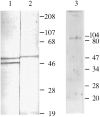
Figure 1
Figure 1
Immunoblot analysis of RI, RII, and AKAP100 in adult hearts. Soluble fraction of the rat left ventricle extracts (lanes 1 and 2, from a preparative gel) and crude protein extracts from isolated rat left ventricular myocytes (lane 3, 50 μg/lane) were separated on 10% SDS-PAGE gels by using the mini gel system (Bio-Rad Laboratories, Hercules, CA). The immunoblots were incubated with RI-specific monoclonal antibody (lane 1; 0.25 μg/ml), RII-specific goat antibodies (lane 2; 10 μg/ml) and AKAP100-specific rabbit antibodies (lane 3; 3.5 μg/ml), respectively. The protein bands were visualized by color development as described in the text (lanes 1 and 2) or by ECL (lane 3). Molecular weight markers are in kD.
Figure 2
Figure 2
Immunoblot, immunoprecipitation, and cAMP-agarose copurification analyses of AKAP100 in the rat heart. In A, human (lane 2) and rat (lane 3) left ventricle homogenates were separated on a 10% SDS-PAGE gel (100 μg/lane) followed by immunoblotting with anti-AKAP100 antibodies. Unstained molecular weight markers were loaded on lane 1. In B, rat left ventricle homogenates (0.5 ml) were incubated with 10 μg of AKAP100-specific rabbit IgGs (lane 1), 10 μg of RII-specific goat IgGs (lane 2), and 200 μl of cAMP-agarose (lane 3). The AKAP100-IgG complexes were precipitated by incubation with Protein G Sepharose beads. The precipitated proteins were solubilized from the agarose or sepharose beads with SDS-PAGE sample buffer followed by immunoblotting with anti-AKAP100 antibodies. As controls for the assay, 2 μg of rabbit IgGs and goat IgGs were loaded on lanes 4 and 5, respectively. Prestained molecular weight markers are loaded on lane 6. In C, human (lane 1) and rat (lane 2) left ventricle homogenates and human AKAP100 recombinant bacterial lysates (lane 3) were separated on a 10% SDS-PAGE gel (100 μg/lane) followed by immunoblotting with anti-AKAP100 antibodies that have been previously preabsorbed by AKAP100 recombinant bacterial lysates. As a control, 2 μg of rabbit IgGs were loaded on lane 4. In D, AKAP100 recombinant bacterial lysates (50 μg/lane) were separated on a 10% SDS-PAGE gel followed by immunoblotting with affinity-purified anti-AKAP100 IgGs as described in the text. The Hoefer (A–C) and Bio-Rad (D) SDS-PAGE systems were used for these studies. A and B represent different lanes from the same gel. The protein bands in A–C were visualized by color development, whereas the immunoblot shown in D was developed by ECL. Molecular weight markers are in kD.
Figure 3
Figure 3
Characterization of human AKAP100 and RII recombinant proteins. In A, human AKAP100 recombinant bacterial lysates were analyzed by immunblotting (lanes 1 and 2, from a preparative gel) and RII overlay (lanes 3 and 4, 100 μg/lane) assays. Lanes 1 and 2 were incubated with anti-6 His tag antibody and anti-AKAP100 antibodies, respectively. The protein bands were visualized by ECL. The RII overlay assay was performed as described in the text with (lane 4) or without (lane 3) the inhibitory peptide Ht31. The 32P-labeled RII binding protein was detected by PhosphorImaging (Molecular Dynamics, Inc.). In B, purified RII recombinant protein was analyzed by Coomassie blue staining (lane 1, 50 μg/lane) and immunoblotting (lane 2, 10 μg/lane) with anti-RII antibodies. The protein band was visualized by ECL. Lane 3 was loaded with 10 μl of 32P-labeled RII, which was detected by PhosphorImaging. Molecular weights are in kD.
Figure 4
Figure 4
Immunolocalization of AKAP100 in longitudinal sections of the rat papillary muscle. Longitudinal sections of rat papillary muscles were immunostained with AKAP100-specific rabbit antibodies and examined by confocal microscopy. Localization of the primary antibodies was detected with LRSC-conjugated donkey anti–rabbit IgG antibodies. The transverse periodic staining (T), the nuclear staining (N), and the intercalated disc staining (I) are indicated. Arrowheads label the AKAP100 staining between the transverse staining. Bar, 5 μm.
Figure 7
Figure 7
Colocalization of AKAP100 with α-actinin and RyR in longitudinal sections of cardiac myocytes. Longitudinal cryostat sections of rat papillary muscles (A–C) or freshly isolated adult rat left ventricular myocytes (D–F) were double-immunostained with either α-actinin–specific (A) and AKAP100-specific (B) antibodies or RyR-specific (D) and AKAP100-specific (E) antibodies. The anti-AKAP100 rabbit antibodies were detected with LRSC-conjugated donkey anti–rabbit IgG antibodies. Localization of the RyR- and α-actinin-specific monoclonal antibodies were visualized by using FITC-conjugated donkey anti–mouse IgG antibodies. C is the superimposed image of A and B; F combines from the images in D and E. Transverse periodic staining (T), sarcolemmal staining (S), nuclear staining (N), and intercalated disc staining (I) are indicated. In C, arrowheads indicate the longitudinal AKAP100 staining localized between the transverse staining, whereas in D and F, arrowheads label the punctate RyR staining. The nucleus without staining is labeled with an N. Bar, 5 μm.
Figure 7
Figure 7
Colocalization of AKAP100 with α-actinin and RyR in longitudinal sections of cardiac myocytes. Longitudinal cryostat sections of rat papillary muscles (A–C) or freshly isolated adult rat left ventricular myocytes (D–F) were double-immunostained with either α-actinin–specific (A) and AKAP100-specific (B) antibodies or RyR-specific (D) and AKAP100-specific (E) antibodies. The anti-AKAP100 rabbit antibodies were detected with LRSC-conjugated donkey anti–rabbit IgG antibodies. Localization of the RyR- and α-actinin-specific monoclonal antibodies were visualized by using FITC-conjugated donkey anti–mouse IgG antibodies. C is the superimposed image of A and B; F combines from the images in D and E. Transverse periodic staining (T), sarcolemmal staining (S), nuclear staining (N), and intercalated disc staining (I) are indicated. In C, arrowheads indicate the longitudinal AKAP100 staining localized between the transverse staining, whereas in D and F, arrowheads label the punctate RyR staining. The nucleus without staining is labeled with an N. Bar, 5 μm.
Figure 5
Figure 5
Colocalization of RII with AKAP100 and immunolocalization of RI in cardiac myocytes. Freshly isolated adult rat left ventricular myocytes were double-stained with RII- (A) and AKAP100-specific antibodies (B), or with RI-specific antibody (D). The anti-AKAP100 rabbit antibodies were detected with LRSC-conjugated donkey anti–rabbit IgG antibodies. Localization of RI-specific monoclonal antibody and RII-specific goat antibodies were visualized by using FITC-conjugated donkey anti–mouse IgG antibodies and FITC-conjugated donkey anti–goat IgG antibodies, respectively. C is the superimposed image of A and B. The transverse periodic staining (T), sarcolemmal staining (S), and the nuclear staining (N) are indicated. In D, the longitudinal staining pattern is labeled with arrowheads, and the nucleus without staining is labeled with an N. Bar, 5 μm.
Figure 5
Figure 5
Colocalization of RII with AKAP100 and immunolocalization of RI in cardiac myocytes. Freshly isolated adult rat left ventricular myocytes were double-stained with RII- (A) and AKAP100-specific antibodies (B), or with RI-specific antibody (D). The anti-AKAP100 rabbit antibodies were detected with LRSC-conjugated donkey anti–rabbit IgG antibodies. Localization of RI-specific monoclonal antibody and RII-specific goat antibodies were visualized by using FITC-conjugated donkey anti–mouse IgG antibodies and FITC-conjugated donkey anti–goat IgG antibodies, respectively. C is the superimposed image of A and B. The transverse periodic staining (T), sarcolemmal staining (S), and the nuclear staining (N) are indicated. In D, the longitudinal staining pattern is labeled with arrowheads, and the nucleus without staining is labeled with an N. Bar, 5 μm.
Figure 9
Figure 9
Colocalization of RII with AKAP100 in cross-sections of cardiac myocytes. Transverse cryostat sections of rat papillary muscles were double-immunostained with RII-specific (A) and AKAP100-specific (B) antibodies. The primary antibodies were detected as described in Fig. 5. The arrows label the nuclear staining (N) for RII and for AKAP100. The arrowheads indicates the similar RII and AKAP100 network staining patterns. Bar, 5 μm.
Figure 6
Figure 6
Localization of AKAP100 in the FITC-phalloidin–stained cardiac myocytes. Freshly isolated adult rat left ventricular myocytes were incubated with FITC-phalloidin (A) followed by immunostaining with AKAP100-specific rabbit antibodies (B). Localization of the anti-AKAP100 antibodies was detected with LRSC-conjugated donkey anti–rabbit IgG antibodies. C is the superimposed image of A and B. In A, the location of the Z-line at the center of phalloidin staining is indicated (Z), and the nucleus without staining is labeled with an N. In A and C, transverse periodic staining (T), sarcolemmal staining (S), and nuclear staining (N) are shown. Bar, 5 μm.
Figure 6
Figure 6
Localization of AKAP100 in the FITC-phalloidin–stained cardiac myocytes. Freshly isolated adult rat left ventricular myocytes were incubated with FITC-phalloidin (A) followed by immunostaining with AKAP100-specific rabbit antibodies (B). Localization of the anti-AKAP100 antibodies was detected with LRSC-conjugated donkey anti–rabbit IgG antibodies. C is the superimposed image of A and B. In A, the location of the Z-line at the center of phalloidin staining is indicated (Z), and the nucleus without staining is labeled with an N. In A and C, transverse periodic staining (T), sarcolemmal staining (S), and nuclear staining (N) are shown. Bar, 5 μm.
Figure 8
Figure 8
Colocalization of AKAP100 with α-actinin and RyR in cross-sections of cardiac myocytes. Cross-sections of the rat papillary muscle were double-immunostained with either α-actinin–specific (A) and AKAP100-specific (B) antibodies or RyR-specific (D) and AKAP100-specific (E) antibodies. The primary antibodies were detected as described in Fig. 5. C is the superimposed image of A and B; F is combined from the images in D and E. The myocytes are labeled as in Fig. 5. In B, arrowheads indicate the network pattern of the AKAP100 staining, which is not seen in the α-actinin staining (A). In C, AKAP100 staining localized in the boundary of the α-actinin staining is indicated by arrowheads. In D and E, arrowheads indicate the similar RyR and AKAP100 network staining. In F, arrowheads indicate the punctate RyR staining that does not overlap with the AKAP100 staining. Bar, 5 μm.
Figure 8
Figure 8
Colocalization of AKAP100 with α-actinin and RyR in cross-sections of cardiac myocytes. Cross-sections of the rat papillary muscle were double-immunostained with either α-actinin–specific (A) and AKAP100-specific (B) antibodies or RyR-specific (D) and AKAP100-specific (E) antibodies. The primary antibodies were detected as described in Fig. 5. C is the superimposed image of A and B; F is combined from the images in D and E. The myocytes are labeled as in Fig. 5. In B, arrowheads indicate the network pattern of the AKAP100 staining, which is not seen in the α-actinin staining (A). In C, AKAP100 staining localized in the boundary of the α-actinin staining is indicated by arrowheads. In D and E, arrowheads indicate the similar RyR and AKAP100 network staining. In F, arrowheads indicate the punctate RyR staining that does not overlap with the AKAP100 staining. Bar, 5 μm.

Similar articles

Cited by

References

    1. Beall JA, Mitchell GF. Identification of a particular antigen from a parasite cDNA library using antibodies affinity purified from selected portions of Western blot. J Immunol Methods. 1986;86:217–223. - PubMed
    1. Bregman DB, Bhattacharyya N, Rubin CS. High affinity binding protein for the regulatory subunit of cAMP-dependent protein kinase II-B. J Biol Chem. 1989;264:4648–4656. - PubMed
    1. Burton KA, Johnson BD, Hausken ZE, Westenbroek RE, Idzerda RL, Scheuer T, Scott JD, Catterall WA, McKnight GS. Type II regulatory subunits are not required for the anchoring-dependent modulation of Ca21 channel activity by cAMP-dependent protein kinase. Proc Natl Acad Sci USA. 1997;94:11067–11072. - PMC - PubMed
    1. Carl SL, Felix K, Caswell AH, Brandt NR, Ball WJ, Jr, Vaghy PL, Meissner G, Ferguson DG. Immunolocalization of sarcolemmal dihydropyridine receptor and sarcoplasmic reticular triadin and ryanodine receptor in rabbit ventricle and atrium. J Cell Biol. 1995;129:673–682. - PMC - PubMed
    1. Carr DW, Hausken ZE, Fraser IDC, Stofko-Hahn RE, Scott JD. Association of the type II cAMP-dependent protein kinase with a human thyroid RII-anchoring protein. J Biol Chem. 1992;267:13376–13382. - PubMed

Publication types

MeSH terms