. 1998 Jul 27;142(2):545-55.
doi: 10.1083/jcb.142.2.545.

Coordination of an array of signaling proteins through homo- and heteromeric interactions between PDZ domains and target proteins

Affiliations

Coordination of an array of signaling proteins through homo- and heteromeric interactions between PDZ domains and target proteins

X Z Xu et al. J Cell Biol. .

Abstract

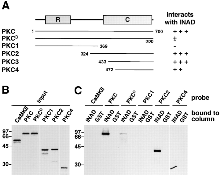
The rapid activation and feedback regulation of many G protein signaling cascades raises the possibility that the critical signaling proteins may be tightly coupled. Previous studies show that the PDZ domain containing protein INAD, which functions in Drosophila vision, coordinates a signaling complex by binding directly to the light-sensitive ion channel, TRP, and to phospholipase C (PLC). The INAD signaling complex also includes rhodopsin, protein kinase C (PKC), and calmodulin, though it is not known whether these proteins bind to INAD. In the current work, we show that rhodopsin, calmodulin, and PKC associate with the signaling complex by direct binding to INAD. We also found that a second ion channel, TRPL, bound to INAD. Thus, most of the proteins involved directly in phototransduction appear to bind to INAD. Furthermore, we found that INAD formed homopolymers and the homomultimerization occurred through two PDZ domains. Thus, we propose that the INAD supramolecular complex is a higher order signaling web consisting of an extended network of INAD molecules through which a G protein-coupled cascade is tethered.


Figures

Figure 3
Figure 3
INAD interacted with opsin, TRPL, and PKC via either PDZ3L or PDZ4. (A) Schematic indicating the PDZ domains to which the opsin, TRPL, PKC, and PLC interacted in a coimmunoprecipitation assay after expressing the proteins in 293T cells. Those INAD fragments that coimmunoprecipitated with the opsin, TRPL, PKC, or a PLC fragment encoding the last 123 amino acids were indicated with a + whereas those that did not were indicated with a −. N/D, experiments that were not done. (B) Representative results from A indicating that TRPL coimmunoprecipitated with PDZ3L and PDZ4. A plasmid encoding the COOH-terminal end of TRPL (residues 676–1,124) was cotransfected into 293T cells with a second construct encoding INAD or portions of INAD (panel A, left) fused to a MYC tag. TRPL was immunoprecipitated from cell lysates with anti-TRPL antibodies and a Western blot of the immune complexes and total cell lysates (Input) were probed with anti-MYC antibodies. (C) Representative results summarized in A indicating that PKC bound to either PDZ3L or PDZ4. A plasmid encoding FLAG-tagged PKC (pPKC-F) was cotransfected into 293T cells along with a second construct encoding full-length or fragments of INAD fused to MYC tags. Immunoprecipitations were performed with anti-MYC antibodies and Western blots of the immune complexes and total cell lysates were probed with anti-FLAG antibodies. (D) TRPC3 and Shaker B did not interact with PDZ3-4 of INAD in 293T cells. pPDZ3.4-M (PDZ3-4 of INAD with a Myc tag) was coexpressed with either pTRPC3-F or pShB in 293T cells. Cell lysates were immunoprecipitated with anti-MYC or anti-FLAG antibodies and the Western blots were probed with anti-FLAG, anti-MYC, or anti-ShB1 antibodies. (E) Representative results showing that PLC binds to PDZ1 and not other domains such as PDZ3 and PDZ4. A plasmid encoding MYC-tagged PLC (pPLC-M) was cotransfected into 293T cells along with a second construct the various INAD forms (indicated in panel A) fused to MYC tags. Immunoprecipitations were performed with anti-MYC antibodies and Western blots of the immune complexes and total cell lysates were probed with anti-FLAG antibodies. (F) Schematic of INAD–PDZ domains and calmodulin-binding domain. The arbitrary boundaries of the five PDZ domains are indicated in amino acids.
Figure 3
Figure 3
INAD interacted with opsin, TRPL, and PKC via either PDZ3L or PDZ4. (A) Schematic indicating the PDZ domains to which the opsin, TRPL, PKC, and PLC interacted in a coimmunoprecipitation assay after expressing the proteins in 293T cells. Those INAD fragments that coimmunoprecipitated with the opsin, TRPL, PKC, or a PLC fragment encoding the last 123 amino acids were indicated with a + whereas those that did not were indicated with a −. N/D, experiments that were not done. (B) Representative results from A indicating that TRPL coimmunoprecipitated with PDZ3L and PDZ4. A plasmid encoding the COOH-terminal end of TRPL (residues 676–1,124) was cotransfected into 293T cells with a second construct encoding INAD or portions of INAD (panel A, left) fused to a MYC tag. TRPL was immunoprecipitated from cell lysates with anti-TRPL antibodies and a Western blot of the immune complexes and total cell lysates (Input) were probed with anti-MYC antibodies. (C) Representative results summarized in A indicating that PKC bound to either PDZ3L or PDZ4. A plasmid encoding FLAG-tagged PKC (pPKC-F) was cotransfected into 293T cells along with a second construct encoding full-length or fragments of INAD fused to MYC tags. Immunoprecipitations were performed with anti-MYC antibodies and Western blots of the immune complexes and total cell lysates were probed with anti-FLAG antibodies. (D) TRPC3 and Shaker B did not interact with PDZ3-4 of INAD in 293T cells. pPDZ3.4-M (PDZ3-4 of INAD with a Myc tag) was coexpressed with either pTRPC3-F or pShB in 293T cells. Cell lysates were immunoprecipitated with anti-MYC or anti-FLAG antibodies and the Western blots were probed with anti-FLAG, anti-MYC, or anti-ShB1 antibodies. (E) Representative results showing that PLC binds to PDZ1 and not other domains such as PDZ3 and PDZ4. A plasmid encoding MYC-tagged PLC (pPLC-M) was cotransfected into 293T cells along with a second construct the various INAD forms (indicated in panel A) fused to MYC tags. Immunoprecipitations were performed with anti-MYC antibodies and Western blots of the immune complexes and total cell lysates were probed with anti-FLAG antibodies. (F) Schematic of INAD–PDZ domains and calmodulin-binding domain. The arbitrary boundaries of the five PDZ domains are indicated in amino acids.
Figure 1
Figure 1
INAD directly interacted with rhodopsin, TRPL, PKC, and calmodulin. (A) Rhodopsin and INAD coimmunoprecipitated from fly heads. Fly head extracts were used for immunoprecipitations with anti-rhodopsin (Rh) antibodies or nonimmune serum (NIS). A Western blot of the immune complexes was probed with rabbit anti-INAD antibodies followed by 125I- labeled protein A. Horseradish peroxidase-conjugated secondary antibodies were used in subsequent experiments (B–H). (B) INAD and opsin coimmunoprecipitated from 293T cells. pINAD (encoding full-length INAD) was transfected into 293T cells or cotransfected with pRh1 (encoding full length opsin [Op]). The cell lysates were immunoprecipitated with rabbit anti-rhodopsin antibodies or NIS and the Western blot containing the immunoprecipitates and total cell lysates (Input) was probed with anti-INAD antibodies. The volume of lysates loaded in the input lanes was 20% of that used in the corresponding lanes containing the immunoprecipitates. The same ratio was followed in subsequent experiments. (C) Opsin bound to a GST–INAD fusion protein. In vitro–translated opsin proteins labeled with [35S]methionine (Op) were incubated with a GST–INAD fusion protein or GST alone bound to glutathione–Sepharose beads. SDS sample buffer was added to the beads, the eluates were fractionated by SDS-PAGE, and then the dried gel was exposed using a PhosphorImager. (D) TRPL and INAD coimmunoprecipitated from fly heads. Immunoprecipitations were performed using fly head extracts and anti-TRPL antibodies or NIS. The immune complexes were fractionated by SDS-PAGE and a Western blot was probed with anti-INAD antibodies. (E) TRPL and INAD coimmunoprecipitated from 293T cells. INAD was expressed in 293T cells or coexpressed with TRPL and immunoprecipitations were performed with anti-TRPL antibodies. The immune complexes and total cell lysates (Input) were fractionated by SDS-PAGE and the Western blot was probed with anti-INAD antibodies. (F) TRPL bound to GST– INAD. In vitro–translated COOH-terminal TRPL labeled with 35S (C-TRPL; residues 676–1,124) was incubated with a GST–INAD fusion protein immobilized on glutathione–Sepharose beads. The bound proteins were eluted with SDS sample buffer, the eluates were fractionated by SDS-PAGE, and then the dried gel was exposed using a PhosphorImager. (G) PKC and INAD coimmunoprecipitated from 293T cells. pPKC-F (PKC with a FLAG epitope tag) and pINAD-M (INAD with a MYC tag) were cotransfected into 293T cells. Immunoprecipitations were performed with anti-MYC antibodies, and a Western blot of the immunocomplexes was probed with anti-FLAG antibodies. (H) PKC bound to GST– INAD. In vitro–translated 35S-PKC was incubated with a GST– INAD fusion protein immobilized on glutathione–Sepharose beads. The bound proteins were eluted with SDS sample buffer, fractionated by SDS-PAGE, and then exposed using a PhosphorImager. (I) TRPC3 and INAD did not coimmunoprecipitate from 293T cells. pTRPC3-F (TRPC3 with a FLAG tag) and pINAD were cotransfected in 293T cells, immunoprecipitations were carried out with anti-FLAG or anti-INAD antibodies, and Western blots were probed with either anti-INAD or anti-FLAG antibodies. To demonstrate that TRPC3 and INAD were immunoprecipitated by the primary antibodies, the same Western blots were reprobed with anti-FLAG or INAD antibodies (results are shown at bottom). (J) Shaker B and INAD did not coimmunoprecipitate from 293T cells. pShB (full-length ShB1 cDNA under the cytomegalovirus promoter; gift of M. Li, Johns Hopkins University School of Medicine, Baltimore, MD) and pINAD were coexpressed in 293T cells. Immunoprecipitations were carried out with anti-INAD antibodies and the Western blot was probed with anti-ShB antibodies (gift of M. Li). Bottom panel, the same blot reprobed with anti-INAD antibodies. (K) CaMKII did not coimmunoprecipitate with INAD from 293T cells. pCKII-M (CaMKII with a Myc tag) and pINAD were cotransfected in 293T cells, immunoprecipitations were performed with anti-INAD antibodies, and then the Western blot was probed with anti-Myc antibodies. Bottom panel, the same blot reprobed with anti-INAD antibodies.
Figure 2
Figure 2
Calmodulin interacted directly with INAD. Total bacterial extracts expressing GST–INAD fusion proteins (residues included in each construct are indicated above each lane) or GST alone were fractionated by SDS-PAGE and transferred to polyvinylene difluoride. The membranes were then probed with biotin-labeled calmodulin. Left, protein size markers.
Figure 4
Figure 4
COOH-terminal fragment of PKC specifically interacted with INAD. (A) Schematic showing that COOH-terminal fragments of PKC interacted with INAD in column-binding assays. The regulatory domain (R) and catalytic domain (C) of PKC are indicated. PKCD was the full-length PKC with the last three residues substituted with aspartic acids. Polypeptides interacting or not interacting with INAD were indicated with a + or −, respectively. Based on quantification using a PhosphorImager (refer to Materials and Methods), the relative levels of the interactions were arbitrarily assigned from − to +++. The NH2- and COOH-terminal residues included in each protein fragments were indicated. (B) The input probe (with [35S]methionine) used in C are shown. CaMKII and various PKC fragments shown in A were in vitro– translated using a TNT kit (Promega Corp.), separated by SDS-PAGE, and detected after using a PhosphorImager (1-h exposure). (C) A COOH-terminal fragment of PKC interacted with INAD in the column-binding assay. 35S-labeled probes from B were incubated with a GST–INAD or GST-bound glutathione column. After several washes, the beads were eluted with SDS sample buffer, the elutes were fractionated by SDS-PAGE, and then the signals were detected using a PhosphorImager (12-h exposures).
Figure 5
Figure 5
INAD homomultimerized in vitro through PDZ3 and PDZ4. (A) INAD homomultimerized in 293T cells. MYC- and FLAG-tagged INAD (INAD-M and INAD-F) were coexpressed in 293T cells. Immunoprecipitations were performed with anti-MYC antibodies or NIS and the Western blot was probed with anti-FLAG antibodies. (B) INAD displayed homomeric interactions in a column binding assay. In vitro–translated 35S-INAD was coincubated with GST–INAD or GST immobilized on a glutathione column. The 35S-INAD was eluted with SDS sample buffer, fractionated by SDS-PAGE, and then exposed using a PhosphorImager. (C) SAP 102 did not interact with INAD. A plasmid encoding MYC-tagged SAP 102 (gift of R. Huganir, Johns Hopkins University School of Medicine) was cotransfected with pINAD in 293T cells. Cell lysates were immunoprecipitated with anti-INAD antibodies and a Western blot was probed with anti-MYC or anti-INAD antibodies. (D and E) INAD homomultimerized via either PDZ3 or PDZ4. Constructs containing MYC-tagged fragments of INAD were cotransfected in 293T cells with plasmids encoding full-length INAD (pINAD). Cell lysates were immunoprecipitated with anti-MYC antibodies and Western blots were probed with anti-INAD antibodies. Constructs that coimmunoprecipitated with pINAD were indicated with a + whereas those that did not coimmunoprecipitate were indicated with a −. E, representative results summarized in panel D). (F) SAP 102 did not interact with INAD PDZ3-PDZ4 (PDZ3.4-F). SAP 102-M and pPDZ3.4-F (PDZ3-PDZ4 with a FLAG tag) were coexpressed in 293T cells. The coimmunoprecipitation was performed with anti-FLAG antibodies and the Western blot was probed with anti-MYC or anti-FLAG antibodies. (G) PDZ3 and PDZ4 displayed homophilic and heterophilic interactions. MYC- or FLAG-tagged INAD PDZ3 or PDZ4 constructs (PDZ3L-M, PDZ3L-F, PDZ4L-M, and PDZ4L-F) were cotransfected into 293T cells as indicated. Cell lysates were immunoprecipitated with anti-MYC antibodies or nonimmune serum (NIS). The Western blots were probed with anti-FLAG antibodies. (H and I) PDZ3-PDZ4 formed polymers. INAD fragments including either PDZ1 and PDZ2 (PDZ1-2) or PDZ3 and PDZ4 (PDZ3-4) were translated in vitro with [35S]methionine and fractionated by sucrose gradient centrifugation. Both PDZ1-2 and PDZ3-4 were loaded onto the same gradient. Collected fractions were resolved by SDS-PAGE and processed for autoradiography shown in I. The protein bands in panel I were quantified with a PhosphorImager and corresponding readings were plotted versus fraction numbers in H. Fraction numbers as well as those fractions that contained the peak levels of the marker proteins included in the gradient were indicated.
Figure 6
Figure 6
PDZ–PDZ interactions did not preclude PDZ–target interactions. (A) Models for the PDZ–PDZ and PDZ–target interaction: PDZ–PDZ interaction precludes binding of targets, PDZ–target binding prevents PDZ-PDZ interaction, PDZ–PDZ and PDZ–target binding can occur simultaneously. (B) TRPL, PDZ3, and PDZ3L formed a ternary complex. COOH-terminal TRPL, MYC-tagged PDZ3 (PDZ3-M) and FLAG-tagged PDZ3 long-form (PDZ3L-F) were expressed in duplicate or triplicate in 293T cells as indicated. Cell lysates were immunoprecipitated with anti-TRPL or anti-MYC antibodies and the Western blots were probed with either anti-MYC (left two panels) or anti-FLAG antibodies (right two panels). (C) PKC, PDZ3, and PDZ3L formed a ternary complex. FLAG-tagged PKC (PKC-F), MYC-tagged PDZ3 (PDZ3-M), MYC-tagged PDZ3 long-form (PDZ3L-M) and FLAG-tagged PDZ3 long-form (PDZ3L-F) were doubly- or triply-expressed in 293T cells as indicated. Cell lysates were immunoprecipitated with anti-MYC antibodies or nonimmune serum and the Western blots were probed with anti-FLAG antibodies. The third panel in B and the fourth panel in C are duplicates. (D) Homomultimerization and target binding were mediated through overlapping but different regions in PDZ3. Plasmids encoding various portions of PDZ3 and linker regions (MYC-tagged) were coexpressed with pPKC-F or pINAD in 293T cells. Coimmunoprecipitations were carried out using anti-MYC antibodies. +, PDZ3 fragments that interacted with PKC or INAD in the assay.
Figure 7
Figure 7
A schematic model of the phototransduction signalplex. INAD homomultimerizes through PDZ3 and PDZ4. Rhodopsin, PKC, and TRPL bind to either PDZ3 or PDZ4. Calmodulin binds to the linker region between PDZ3 and PDZ4. Binding of TRP to PDZ3 (Shieh and Zhu, 1996) and PLC to PDZ1 and PDZ5 is based on work described in separate reports (Tsunoda et al., 1997; van Huizen et al., 1998). The individual PDZ domains (1–5) in each INAD monomer are indicated by the ovals.

Similar articles

Cited by

References

    1. Arnon A, Cook B, Gillo B, Montell C, Selinger Z, Minke B. Calmodulin regulation of light adaptation and store-operated dark current in Drosophilaphotoreceptors. Proc Natl Acad Sci USA. 1997a;94:5894–5899. - PMC - PubMed
    1. Arnon A, Cook B, Montell C, Selinger Z, Minke B. Calmodulin regulation of calcium stores in phototransduction of Drosophila. . Science. 1997b;275:1119–1121. - PubMed
    1. Bloomquist BT, Shortridge RD, Schneuwly S, Perdew M, Montell C, Steller H, Rubin G, Pak WL. Isolation of a putative phospholipase C gene of Drosophila, norpA, and its role in phototransduction. Cell. 1988;54:723–733. - PubMed
    1. Brakeman PR, Lanahan AA, Obrien R, Roche K, Barners CA, Huganir RL, Worley PF. Homer: a protein that selectively binds metabotropic glutamate receptors. Nature. 1997;386:284–288. - PubMed
    1. Brenman JE, Chao DS, Gee SH, McGee AW, Craven SE, Santillano DR, Wu Z, Huang F, Xia H, Peters MF, et al. Interaction of nitric oxide synthase with the postsynaptic density protein PSD-95 and α1-syntrophin mediated by PDZ domains. Cell. 1996;84:757–767. - PubMed

Publication types

MeSH terms