. 1998 Jul 27;142(2):573-86.
doi: 10.1083/jcb.142.2.573.

Integrin-mediated signals regulated by members of the rho family of GTPases

Affiliations

Integrin-mediated signals regulated by members of the rho family of GTPases

E A Clark et al. J Cell Biol. .

Abstract

The organization of the actin cytoskeleton can be regulated by soluble factors that trigger signal transduction events involving the Rho family of GTPases. Since adhesive interactions are also capable of organizing the actin-based cytoskeleton, we examined the role of Cdc42-, Rac-, and Rho-dependent signaling pathways in regulating the cytoskeleton during integrin-mediated adhesion and cell spreading using dominant-inhibitory mutants of these GTPases. When Rat1 cells initially adhere to the extracellular matrix protein fibronectin, punctate focal complexes form at the cell periphery. Concomitant with focal complex formation, we observed some phosphorylation of the focal adhesion kinase (FAK) and Src, which occurred independently of Rho family GTPases. However, subsequent phosphorylation of FAK and paxillin occurs in a Rho-dependent manner. Moreover, we found Rho dependence of the assembly of large focal adhesions from which actin stress fibers radiate. Initial adhesion to fibronectin also stimulates membrane ruffling; we show that this ruffling is independent of Rho but is dependent on both Cdc42 and Rac. Furthermore, we observed that Cdc42 controls the integrin-dependent activation of extracellular signal-regulated kinase 2 and of Akt, a kinase whose activity has been demonstrated to be dependent on phosphatidylinositol (PI) 3-kinase. Since Rac-dependent membrane ruffling can be stimulated by PI 3-kinase, it appears that Cdc42, PI 3-kinase, and Rac lie on a distinct pathway that regulates adhesion-induced membrane ruffling. In contrast to the differential regulation of integrin-mediated signaling by Cdc42, Rac, and Rho, we observed that all three GTPases regulate cell spreading, an event that may indirectly control cellular architecture. Therefore, several separable signaling pathways regulated by different members of the Rho family of GTPases converge to control adhesion-dependent changes in the organization of the cytoskeleton, changes that regulate cell morphology and behavior.


Figures
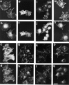
Figure 1
Figure 1
Assembly of focal adhesions and stress fibers during adhesion and spreading of Rat1 fibroblasts on fibronectin. Serum-starved Rat1 fibroblasts (a–d) or cells expressing dominant-inhibitory Cdc42 (e–h), Rac1 (I–L), or RhoA (m–q) were trypsinized, washed, and then replated on fibronectin-coated coverslips for 15 (a, b, e, f, i, j, m, and n) or 60 (c, d, g, h, k, l, p, and q) min and stained for vinculin (b, d, f, h, j, l, n, and q) and for F-actin (a, c, e, g, i, k, m, and p). Bar, 10 μm.
Figure 2
Figure 2
PDGF and serum induce the assembly of membrane ruffles and stress fibers in serum-starved adherent Rat1 cells. Parental (a–c), dominant-inhibitory Rac- (d), or Rho-expressing (e) Rat1 cells were plated on fibronectin-coated coverslips in serum-free media for 48 h. The cells were then left untreated (a) or stimulated with PDGF-BB (b and d; 3 ng/ml) or FBS (1%, c and e) for 10 min at 37°C. The cells were then washed, fixed, permeabilized, and then F-actin present in stress fibers was stained with phalloidin. Bar, 10 μm.
Figure 3
Figure 3
Expression of dominant- inhibitory mutants of Rho-family members Cdc42, Rac1, and RhoA in Rat1 cells. Rat1 fibroblasts (lane 1) or cells expressing a Myc epitope–tagged N17Cdc42 (lane 2), N17Rac1 (lane 3), or N19RhoA (lane 4) were grown for 48 h in the absence of 2 ng/ml tetracycline and the expression of these proteins was examined by immunoblotting with an antibody (9E10) to the Myc epitope.
Figure 4
Figure 4
Rho, Rac, and Cdc42 are required for Rat1 cells to spread on fibronectin. Serum-starved parental (Rat1) or dominant-inhibitory Cdc42- (N17Cdc42), Rac1- (N17Rac), or RhoA-expressing (N19Rho) Rat1 fibroblasts were trypsinized, washed, and then replated on fibronectin- or polylysine-coated wells for 20 min. The cells were then fixed and stained with crystal violet. Random fields of cells were photographed and the percent cells spread was determined. Error bars represent the standard deviation in triplicate samples. Similar results were obtained in three separate experiments. (Students' t-test: N17Cdc42, P < 0.01; N17Rac, P < 0.02; N19Rho, P < 0.1)
Figure 5
Figure 5
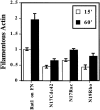
Quantitation of F-actin content in Rat1 cells adhering to fibronectin. Serum-starved parental (Rat1) or dominant-inhibitory Cdc42- (N17Cdc42), Rac1- (N17Rac), or RhoA-expressing (N19Rho) Rat1 fibroblasts were trypsinized, washed, and then replated on fibronectin-coated wells for 15 or 60 min. The cells were then fixed and phalloidin-stained, and the amount of phalloidin bound quantitated as described in Materials and Methods. The relative F-actin content was determined by dividing the amount of phalloidin bound in fibronectin-adherent cells by the amount of phalloidin bound in fibronectin-adherent Rat1 cells at 15 min. Error bars represent the standard deviation in triplicate samples. Similar results were obtained in three separate experiments.
Figure 6
Figure 6
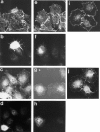
The role of Cdc42, Rac, and Rho in the assembly of focal adhesions and stress fibers in Rat1 fibroblasts. Rat1 cells transfected with 10 μg of the pCMV plasmid encoding Myc-N17Cdc42 (a–d) or Myc-N17Rac1 (e–h) or Rat1 cells treated with C3 transferase (i and j) as described in Materials and Methods were trypsinized, washed, and then replated on fibronectin-coated coverslips for 60 min and stained for F-actin (a, e, and i), β1 integrin (c and g), vinculin (j), or the Myc epitope tag (b, d, f, and h).
Figure 7
Figure 7
A role for Rho in integrin-mediated tyrosine phosphorylation of FAK. In A and B, serum-starved parental (Rat1) or dominant-inhibitory Cdc42- (N17Cdc42), Rac1- (N17Rac), or RhoA-expressing (N19Rho) Rat1 fibroblasts were trypsinized, washed, and then replated on fibronectin-coated wells for 0, 10, 30, or 90 min. In C, Rat1 cells treated with C3 transferase (+C3) as described in Materials and Methods were used and plated for 0, 30, or 90 min. The cells were then lysed, and 50 μg of the lysates were immunoprecipitated with a polyclonal antiserum to FAK, subjected to electrophoresis, and then immunoblotted with an anti-phosphotyrosine antibody (A and C) or an anti-FAK antibody (B). The results presented here are representative of eight separate experiments.
Figure 8
Figure 8
A role for Rho and Cdc42 in integrin-mediated tyrosine phosphorylation of paxillin. In A and B, serum-starved parental (Rat1) or dominant-inhibitory Cdc42- (N17Cdc42), Rac1- (N17Rac), or RhoA-expressing (N19Rho) Rat1 fibroblasts were trypsinized, washed, and then replated on fibronectin-coated wells for 0, 10, 30, or 90 min. In C, untreated Rat1 cells (−) or Rat1 cells treated with C3 transferase (+C3) as described in Materials and Methods were used and plated for 0 or 30 min. The cells were then lysed, and 100 μg of the lysates were immunoprecipitated with a mAb to paxillin, subjected to electrophoresis, and then immunoblotted with an anti-phosphotyrosine antibody (A and C) or the anti-paxillin antibody (B).
Figure 9
Figure 9
A role for Rho and Cdc42 in integrin-mediated tyrosine phosphorylation of Erk2. In A, serum-starved parental (Rat1) or dominant-inhibitory Cdc42- (N17Cdc42), Rac1- (N17Rac), or RhoA-expressing (N19Rho) Rat1 fibroblasts were trypsinized, washed, and then replated on fibronectin-coated wells for 0, 10, 30, or 90 min. In B, Rat1 or N17Cdc42-expressing cells were replated on fibronectin for 0, 15, or 60 min, or treated in suspension with 10 ng/ml PDGF for 5 min at room temperature. In C, Rat1 cells treated with C3 transferase (+C3) as described in Materials and Methods were used and plated for 0, 15, or 60 min. The cells were then lysed, and 15 μg of the lysates were subjected to electrophoresis and immunoblotted with a pan-Erk antibody. In D, Rat1 cells were transfected with 1 μg of the pSVL plasmid encoding HA-ERK alone or in combination with 10 μg of pCMV incoding Myc-N17Cdc42. The cells were serum-starved, trypsinized, washed, and then replated on fibronectin-coated dishes for 0, 15, or 60 min. HA-ERK was immunoprecipitated from cell lysates with the 12CA5 antibody and subjected to an in vitro kinase assay using myelin basic protein (MBP) as the substrate. HA-ERK present in the cell lysates (40 μg) was detected by Western blotting with the 12CA5 antibody. The specific activity of HA-ERK is expressed relative to the specific activity of the kinase in cells transfected with HA-ERK alone and held in suspension.
Figure 10
Figure 10
Integrin-mediated tyrosine phosphorylation of Src is independent of Cdc42, Rac, and Rho. Serum-starved parental (Rat1) or dominant-inhibitory Cdc42- (N17Cdc42), Rac1- (N17Rac), or RhoA-expressing (N19Rho) Rat1 fibroblasts were trypsinized, washed, and then replated on fibronectin-coated wells for 0, 10, 30, or 90 min. The cells were then lysed, and 15 μg of the lysates were subjected to electrophoresis and immunoblotted with an antibody specific for Src phosphotyrosine-416 (Y416P) (A) or for total Src protein (B).
Figure 11
Figure 11
Integrin-mediated activation of Akt is Cdc42-dependent. Serum-starved parental (Rat1) or dominant-inhibitory Cdc42- (N17Cdc42), Rac1- (N17Rac), or RhoA-expressing (N19Rho) Rat1 fibroblasts were trypsinized, washed, and then replated on fibronectin-coated wells for 0, 15, or 60 min. Akt kinase activity was assayed using histone H2B as a substrate as described in Materials and Methods. This autoradiogram is representative of results from three separate experiments. In B, Rat1 cells transfected with 1 μg of the pCMV-6 plasmid encoding HA-Akt either alone or in combination with 10 μg of pCMV-encoding Myc-N17Cdc42 were serum starved, trypsinized, washed, and then replated on fibronectin-coated dishes for 0, 15, or 60 min. HA-Akt was immunoprecipitated with an antibody (12CA5) to the HA epitope and subjected to an in vitro kinase assay using histone H2B as the substrate. The samples were then electrophoresed and transferred to a nitrocellulose membrane for autoradiography (H2B) and quantitated on a PhosphorImager. The membrane was then probed with 12CA5 to determine the levels of HA-Akt present in the immunoprecipitate. The specific activity of HA-Akt (activity divided by protein levels) is expressed relative to the specific activity of the kinase in cells transfected with HA-Akt alone and held in suspension.
Figure 12
Figure 12
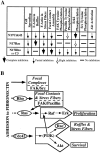
Summary of the different roles of Rho-family GTPases, Cdc42, Rac, and Rho in regulating cell morphology and integrin-mediated signaling events. The ability of N17Cdc42, N17Rac, N19Rho, or C3 transferase to inhibit completely (>90%, large arrow), partially inhibit (40–70%, small arrow), inhibit at a single time point (white arrow), or not significantly inhibit (<10%, dash) integrin-mediated morphological changes and signaling events that occur upon adhesion to fibronectin are summarized in A. B shows a model of the different roles played by Rho family GTPases in integrin-mediated signaling events that regulate cytoskeletal organization. Cell adhesion to the extracellular matrix induces the aggregation of small clusters of integrin receptors that leads to cytoskeletal organization via several pathways. Initial activation of FAK and Src, and the assembly of focal complexes are independent of Rho family members. A Rho-dependent pathway regulates the full activation of FAK, phosphorylation of paxillin, and assembly of focal adhesions. A third pathway from clustered integrins activates Akt (probably through PI 3-kinase) in a Cdc42-dependent pathway. Both Cdc42 and Rac regulate actin organization required for membrane ruffles. Adhesion-dependent Erk phosphorylation is contingent upon the activity of Cdc42 and Rho, and it requires a properly organized cytoskeleton. Rho, Cdc42, and Rac all regulate the organization of F-actin in stress fibers, although it is unlikely that they do so through a single, linear “cascade” since some events, such as FAK phosphorylation, are regulated by only a single Rho family member. Therefore, the most plausible explanation is that Rho family members regulate adhesion-initiated stress fiber formation via convergent pathways.

References

    1. Aepfelbacher M, Essler M, Huber E, Czech A, Weber PC. Rho is a negative regulator of human monocyte spreading. J Immunol. 1996;157:5070–5075. - PubMed
    1. Amano M, Ito M, Kimura K, Fukata Y, Chihara K, Nakano T, Matsuura Y, Kaibuchi K. Phosphorylation and activation of myosin by Rho-associated kinase (Rho-kinase) J Biol Chem. 1996;271:20246–20249. - PubMed
    1. Amano M, Chihara K, Kimura K, Fukata Y, Nakamura N, Matsuura Y, Kaibuchi K. Formation of actin stress fibers and focal adhesion enhanced by Rho-kinase. Science. 1997;275:1308–1311. - PubMed
    1. Barry ST, Critchley DR. The RhoA-dependent assembly of focal adhesions in Swiss 3T3 cells is associated with increased tyrosine phosphorylation and the recruitment of both pp125FAK and protein kinase C-δ to focal adhesions. J Cell Sci. 1994;107:2033–2045. - PubMed
    1. Barry ST, Flinn HM, Humphries MJ, Critchley DR, Ridley AJ. Requirement for Rho in integrin signalling. Cell Adhes Commun. 1997;4:387–398. - PubMed

Publication types