. 1998 Aug 15;511 ( Pt 1)(Pt 1):159-69.
doi: 10.1111/j.1469-7793.1998.159bi.x.

Functions of large conductance Ca2+-activated (BKCa), delayed rectifier (KV) and background K+ channels in the control of membrane potential in rabbit renal arcuate artery

Affiliations

Functions of large conductance Ca2+-activated (BKCa), delayed rectifier (KV) and background K+ channels in the control of membrane potential in rabbit renal arcuate artery

H M Prior et al. J Physiol. .

Abstract

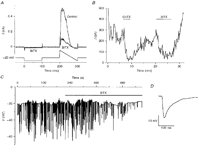
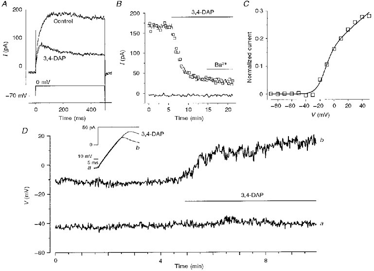
1. The types of K+ channel which determine the membrane potential of arcuate artery smooth muscle cells were investigated by patch-clamp recording from isolated cells and lumenal diameter measurements from intact pressurized renal arcuate arteries. 2. Single cells had a mean resting potential of -38 mV and were depolarized by 130 mM K+ but not by the Cl- channel blocker 4,4'-diisothiocyanatostilbene-2, 2'-disulphonic acid (DIDS). 3. Iberiotoxin did not affect the resting potential but inhibited spontaneous transient hyperpolarizations. Iberiotoxin or 1 mM tetraethylammonium (TEA+) constricted intact arteries. 3,4-Diaminopyridine (3,4-DAP)-sensitive delayed rectifier K+ (KV) channel current was elicited by depolarization but 3,4-DAP did not affect the resting potential or induce constriction in the intact artery. 4. A voltage-independent K+ current was inhibited by >= 0.1 mM barium (Ba2+) and unaffected by iberiotoxin, glibenclamide, apamin, 3,4-DAP and ouabain. In six out of ten cells, 1 mM Ba2+ depolarized the resting potential, while in the other cells the potential was resistant to all of the K+ channel blockers and ouabain. Ba2+ (0.1-1 mM) constricted the intact artery, but 10 microM Ba2+, 1 microM glibenclamide or 100 nM apamin had no effect. 5. The data suggest that resting potential is determined by background K+ channels, one type being Ba2+ sensitive and voltage independent, and another type being poorly defined due to its resistance to any inhibitor. Large conductance Ca2+-activated K+ (BKCa) and KV channels do not determine the resting potential but have separate functions to underlie transient Ca2+-induced hyperpolarizations and to protect against depolarization past about -30 mV.


Figures

Figure 1
Figure 1. Resting potential of smooth muscle cells isolated from arcuate artery
A, resting potentials for 60 separate cells. Recordings were in current clamp using the amphotericin perforated-patch whole-cell method. Resting potential values were allocated to 10 mV bins and the number of cells (N) in each bin is given as a column. The smooth curve is a fitted single Gaussian distribution with a mid-point at -33 mV (mean, -38 mV). B, membrane potential recording where the horizontal bars indicate bath application of 0.1 and 1 mm DIDS, and an increase in the extracellular K+ concentration from 5 to 130 mm.
Figure 4
Figure 4. Depolarization induced by millimolar Ba2+ or high K+
All experiments described were in current clamp using amphotericin B to permeabilize the cell-attached patch, and the 5 mm K+ bath solution was used. A, superimposed on the same plot are the resting potential (thick trace and right-hand ordinate) and the input conductance of the cell (thin trace and left-hand ordinate) measured from the amplitude of the electrotonic potential elicited by a hyperpolarizing current pulse. Ba2+ (1 mm) was bath applied. B, summary histogram for the effects of various treatments on membrane potential. Mean (±s.e.m.) depolarization (no hyperpolarizations occurred) for the resting potential (×) and the potential at the end of the depolarizing current pulse when rectification was observed (□). IbTX, 100 nm iberiotoxin alone (8 cells) and 100 nm charybdotoxin (n= 2) or 100 nm iberiotoxin (n= 1) in the presence of 1 mm Ba2+; Ba2+, 1 mm BaCl2 alone (10 cells); 3,4-DAP, 1 mm 3,4-DAP in the presence of 100 nm iberiotoxin and 1 mm Ba2+, which had no effect on their own (3 cells); Cocktail: 1 mm 3,4-DAP, 10 μM Ba2+, 1 μM glibenclamide, 100 nm apamin and 10 μM ouabain (5 cells); DIDS, 1 mm DIDS (4 cells); 130 mm K+, change from 5 to 130 mm K+ in the bath solution (9 cells). *P < 0.05 (two-tailed test) and †P < 0.05 (one-tailed test), significantly different from zero
Figure 2
Figure 2. Large conductance Ca2+-activated (BKCa) K+ channels
A, conventional whole-cell voltage-clamp recording using a holding potential of -20 mV. A square voltage step to -60 mV and a ramp change in voltage from +80 to -60 mV were applied every 10 s. Examples of current traces are shown for control and in the presence of bath-applied 100 nm iberiotoxin (IbTX). The 60 mm K+ bath solution and the 0.1 mm EGTA pipette solution were used. B, plot of current amplitude against time for current sampled at +60 mV during the ramp change in voltage described, and for the experiment, in A. Bath application of 100 nm charybdotoxin (ChTX) and 100 nm iberiotoxin is indicated by the horizontal bars. C, a current-clamp recording in which the 5 mm K+ bath solution was used and amphotericin B was used to permeabilize the cell-attached patch. This cell exhibited spontaneous transient hyperpolarizations and the peak amplitude of each transient hyperpolarization is shown as a vertical bar superimposed on the resting membrane potential. Iberiotoxin (100 nm) was bath applied as indicated by the horizontal bar. D, an example of one of the spontaneous transient hyperpolarizations (from C) on a fast time base.
Figure 3
Figure 3. Aminopyridine-sensitive delayed rectifier (KV) channels
A, conventional whole-cell voltage-clamp recording showing current in response to a square depolarizing step to 0 mV, applied every 15 s from a holding potential of -70 mV. The 5 mm K+ bath solution (including 0.1 mm Cd2+) and the 10 mm EGTA pipette solution were used. A control current trace and one in the presence of 1 mm 3,4-DAP are shown. B, a time-series plot for the experiment in A giving current amplitude at the end of the step to 0 mV (□) and current elicited by stepping from -70 to -90 mV for 0.5 s (continuous record). 3,4-DAP and 1 mm Ba2+ were bath applied. C, voltage dependence of 3,4-DAP-sensitive current determined by applying square voltage-clamp steps from -70 mV. Peak current amplitudes under control conditions and in 3,4-DAP were normalized relative to the maximum control current amplitude for each cell and averaged for 4 cells. 3,4-DAP-sensitive current was obtained by subtracting the 3,4-DAP-resistant values from the control values (□). The smooth curve is: grel(V - Vrev)/1 + exp[(V - Vm)/dV]}, where the mid-point of the Boltzmann function (Vm) is -14.9 mV and the slope (dV) is 6.5 mV, Vrev is the reversal potential, and grel is the relative conductance. D, current-clamp recording of resting potential (a) and potential at the end of the square pulse of depolarizing current (b). 3,4-DAP (1 mm) was bath applied in the presence of 1 mm Ba2+ and 100 nm iberiotoxin, which had no effect. Examples of potential during the current pulse are shown schematically in the inset.
Figure 5
Figure 5. Voltage-independent Ba2+-sensitive K+ current
A, conventional whole-cell voltage-clamp recording using a holding potential of -20 mV, 60 mm K+ bath solution and 0.1 mm EGTA pipette solution (K+ equilibrium potential, -20 mV). Plot of current amplitude during a square voltage step to -60 mV. Horizontal bars indicate bath application of 10 mm Ba2+, 100 nm iberiotoxin, 100 nm charybdotoxin, 10 μM quinine, and 1, 2 and 4 mm TEA+. Throughout the experiment the bath solution contained 1 mm 3,4-DAP, 1 μM glibenclamide (Glib), 100 nm apamin and 10 μM ouabain. B shows, for the same experiment described in A, a plot of current during a 100 ms ramp change in voltage from 0 to -60 mV, which was applied every 10 s and 100 ms after each square step to -60 mV. Control current and current in the presence of bath-applied 10 mm Ba2+ are shown. The inset plot is the difference between the two currents. C, an experiment using the same solutions as in A and B but for a different cell. Example current traces are shown in response to a 1 s step to -60 mV, applied every 10 s from -20 mV. Current under control conditions and in the presence of 0.1 mm Ba2+ is shown. D, as for C but for another cell which had an inward rectifier K+ current.
Figure 6
Figure 6. Responses of intact pressurized arcuate arteries to various K+ channel inhibitors
Columns are mean percentage values and error bars are s.e.m. Numbers in parentheses are the numbers of arteries tested. *P < 0.05 (two-tailed test) and †P < 0.05 (one-tailed test), significantly different from zero. TEA+, 1 mm tetraethylammonium; 3,4-DAP, 1 mm 3,4-DAP; Apamin, 100 nm apamin; IbTX, iberiotoxin (0.1 or 1 μM); Glib, 1 μM glibenclamide; Cocktail: 1 mm 3,4-DAP, 10 μM Ba2+, 1 μM glibenclamide, 100 nm apamin and 100 nm IbTX. When iberiotoxin was applied in isolation it was added directly to the bath solution with a reduced perfusion rate of 2 ml min−1.

Similar articles

Cited by

References

    1. Aalkjær C, Mulvany MJ, Nyborg NCB. Atrial natriuretic factor causes specific relaxation of rat renal arcuate arteries. British Journal of Pharmacology. 1985;86:447–453. - PMC - PubMed
    1. Archer SL, Huang JMC, Reeve HL, Hampl V, Tolarová S, Michelakis E, Weir EK. Differential distribution of electrophysiologically distinct myocytes in conduit and resistance arteries determines their response to nitric oxide and hypoxia. Circulation Research. 1996;78:431–442. - PubMed
    1. Beech DJ, Bolton TB. Properties of the cromakalim-induced potassium conductance in smooth muscle cells isolated from the rabbit portal vein. British Journal of Pharmacology. 1989;98:851–864. - PMC - PubMed
    1. Benham CD, Bolton TB. Spontaneous transient outward currents in single visceral and vascular smooth muscle cells of the rabbit. The Journal of Physiology. 1986;381:385–406. - PMC - PubMed
    1. Bolton TB, Imaizumi Y. Spontaneous transient outward currents in smooth muscle cells. Cell Calcium. 1996;20:141–152. - PubMed

Publication types

MeSH terms

LinkOut - more resources