. 1998 Aug 15;511 ( Pt 1)(Pt 1):265-72.
doi: 10.1111/j.1469-7793.1998.265bi.x.

Delayed production of adenosine underlies temporal modulation of swimming in frog embryo

Affiliations

Delayed production of adenosine underlies temporal modulation of swimming in frog embryo

N Dale. J Physiol. .

Abstract

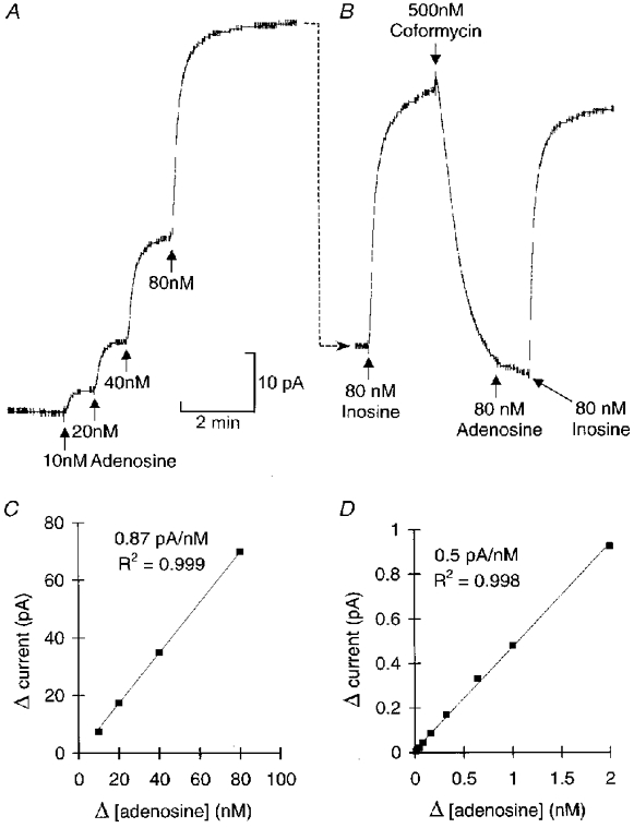
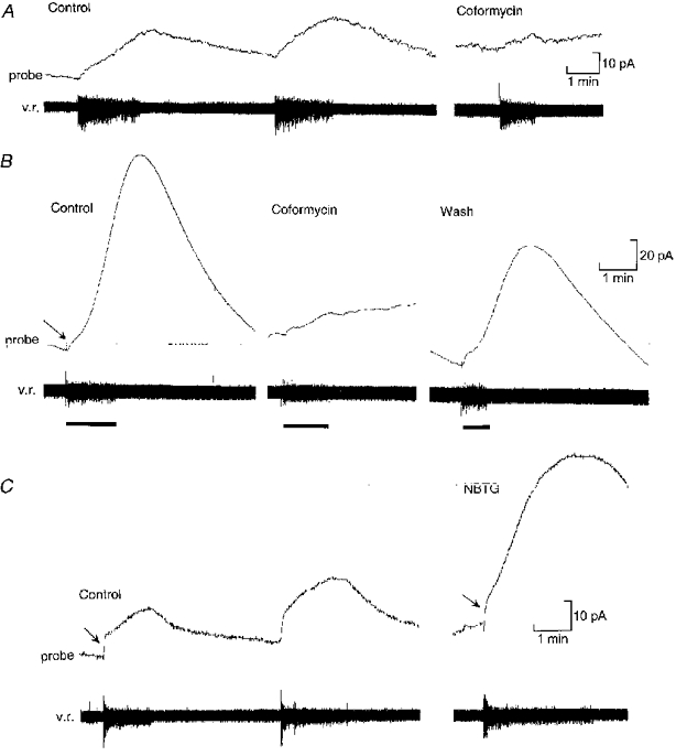
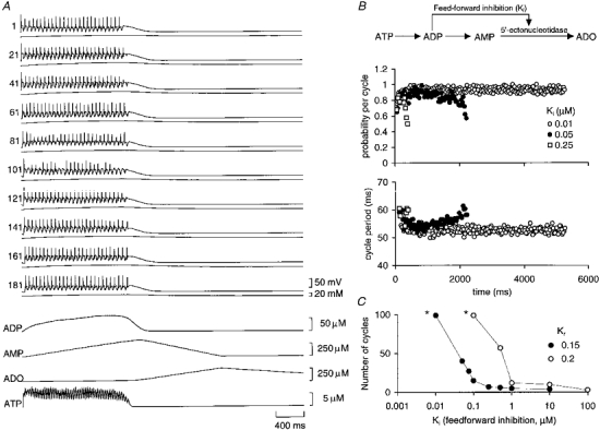
1. To investigate the dynamics of adenosine production in the spinal cord during motor activity, and its possible contribution to the temporal modulation of motor patterns, a sensor sensitive to adenosine at concentrations as low as 10 nM was devised. 2. When pressed against the outside of the spinal cord, the sensor detected slow changes in the levels of adenosine during fictive swimming that ranged from 10 to 650 nM. In four embryos where particularly large signals were recorded due to favourable probe placement, the adenosine levels continued to rise for up to a minute following cessation of activity before slowly returning to baseline. In the remaining thirteen embryos, levels of adenosine started to return slowly to baseline almost immediately after activity had stopped. 3. Inhibitors of adenosine uptake increased the magnitude of the signal recorded and slowed the recovery following cessation of activity. 4. A realistic computational model of the spinal circuitry was combined with models of extracellular breakdown of ATP to adenosine. ATP and adenosine inhibited, as in the real embryo, the voltage-gated K+ and Ca2+ currents, respectively. The model reproduced the temporal run-down of motor activity seen in the real embryo suggesting that synaptic release of ATP together with its extracellular breakdown to adenosine is sufficient to exert time-dependent control over motor pattern generation. 5. The computational analysis also suggested that the delay in the rise of adenosine levels is likely to result from feed-forward inhibition of the 5'-ectonucleotidase in the spinal cord. This inhibition is a key determinant of the rate of run-down.


Figures

Figure 1
Figure 1. In vitro calibration and characterization of the adenosine sensor
A, successive amounts of adenosine were added to the bath at each arrow to raise the concentration of adenosine in the bath by the amount indicated under each arrow. The speed of response by the probe is determined in large by mixing in the bath. The change in probe current resulting from each application of adenosine is plotted in C This shows that the probe responds in a linear fashion. B, in the same experiment 80 nm inosine was added (immediately after the 80 nm adenosine). The response to inosine (substrate for the second enzyme in the cascade) was about 25 % bigger than the response to the same amount of adenosine, indicating some loss of efficiency in the probe. Coformycin (500 nm), a specific blocker of adenosine deaminase, was added. This rapidly reduced the probe current (due to the continued presence of adenosine in the bath) and greatly attenuated the response to subsequent addition of adenosine. However, the response to inosine was unaffected. Thus coformycin only disables the first part of the cascade but leaves the rest intact making it a good test for the specificity of the device. D, after the coformycin had been washed out, the sensitivity of the probe to adenosine recovered (although it was still slightly lower than in C). This calibration shows that the response to adenosine was linear from 10 nm to 2 μm.
Figure 2
Figure 2
The sensor probe can detect adenosine released from the spinal cord during fictive locomotion A, production of adenosine during two consecutive episodes of swimming monitored by a ventral root recording (v.r.). Note the slow rise in the probe current (equivalent to a change in concentration of about 60 nm) and the slow decay after the end of the swimming episode. The record on the right shows that application of 50 nm coformycin blocks most of the probe current, indicating that the signal is largely due to the release of adenosine. B, example (from another preparation) where favourable placement of the probe relative to the spinal cord resulted in a massive signal equivalent to a change in adenosine concentration of about 370 nm. A fast component to the probe current (arrow) can be seen at the beginning of the swimming activity. The probe signal continued to rise for about 50 s after the end of the swimming episode. Application of 50 nm coformycin blocked the probe current, but left a small fast component. The large slowly developing component of the probe current was thus specifically due to the release of adenosine. C, blockers of adenosine uptake greatly enhance the release of adenosine from the spinal cord. In the control (left) the probe current involved both fast (arrow) and slow components, the slow component being equivalent to a rise in adenosine concentration of around 64 nm. After application of 1 μM NBTG (right) to block adenosine uptake, the fast component (arrow) was unchanged, but the slow component was greatly increased in amplitude and rate of rise (equivalent to a change of about 150 nm).
Figure 3
Figure 3. Computational model incorporating release of ATP and its conversion to adenosine reproduces run-down of the motor pattern
A, the activity of every 20th neuron from a simulation of 200 neurons (neurons 1-100 and 101-200 are on opposing sides of the simulated circuit). The trace underlying the membrane potential record for each neuron is the intracellular concentration of Na+. Underneath the neural records are traces that show the release of ATP, the build-up of ADP and AMP, and the production of adenosine (ADO). The feed-forward inhibition was set at 0.075 μM. Note how the peak adenosine production was greatly delayed relative to swimming activity. B, schema showing the break-down of ATP to adenosine and feed-forward inhibition of the 5′-ectonucleotidase by ADP (top). Run-down of the motor pattern can be seen in a decay in the probability of firing (middle) and a lengthening in the cycle period of activity (bottom). Data are shown for three strengths of feed-forward inhibition, showing a case with essentially no run-down (Ki= 0.01 μM) and two other cases where the feed-forward inhibition was weaker and allowed much more rapid run-down. C, the duration of motor activity in the model depends strongly on the strength of feed-forward inhibition. Once Ki reached a certain strength, episode length was steeply dependent upon its magnitude. Data are shown for two rates of ATP release (Kr). Note that run-down proceeds more slowly for all strengths of Ki at the faster rate of ATP release. The asterisks indicate that the number of cycles counted for these data points was terminated by the length of the simulation and not by the occurrence of run-down in the model.

Similar articles

Cited by

References

    1. Agarwal RP, Parks RE. Adenosine deaminase from human erythrocytes. Methods in Enzymology. 1978;51:502–507. - PubMed
    1. Arshavsky YI, Orlovsky GN, Panchin YV, Roberts A, Soffe SR. Neuronal control of swimming locomotion: analysis of the pteropod mollusc Clione and embryos of the amphibian Xenopus. Trends in Neurosciences. 1993;16:227–232. 10.1016/0166-2236(93)90161-E. - DOI - PubMed
    1. Berners MOM, Boutelle MG, Fillenz M. Online measurement of brain glutamate with an enzyme/polymer coated tubular electrode. Analytical Chemistry. 1994;66:2017–2021. - PubMed
    1. Clarke JDW, Roberts A. Interneurones in the Xenopus embryo spinal cord: sensory excitation and activity during swimming. The Journal of Physiology. 1984;354:345–362. - PMC - PubMed
    1. Dale N. Kinetic characterization of the voltage-gated currents possessed by Xenopus embryo spinal neurons. The Journal of Physiology. 1995a;489:473–488. - PMC - PubMed

Publication types

LinkOut - more resources