Comparative Study
. 1998 Aug;180(15):3741-9.
doi: 10.1128/JB.180.15.3741-3749.1998.

The regulated outer membrane protein Omp21 from Comamonas acidovorans is identified as a member of a new family of eight-stranded beta-sheet proteins by its sequence and properties

Affiliations
Comparative Study

The regulated outer membrane protein Omp21 from Comamonas acidovorans is identified as a member of a new family of eight-stranded beta-sheet proteins by its sequence and properties

C Baldermann et al. J Bacteriol. 1998 Aug.

Abstract

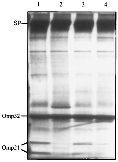
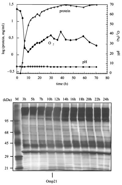
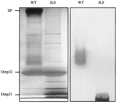
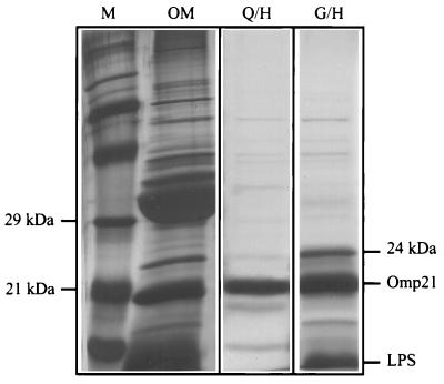
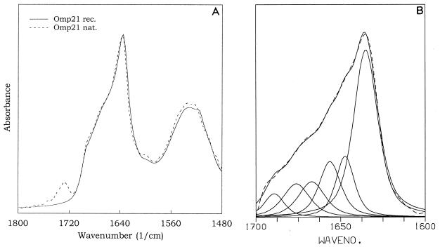
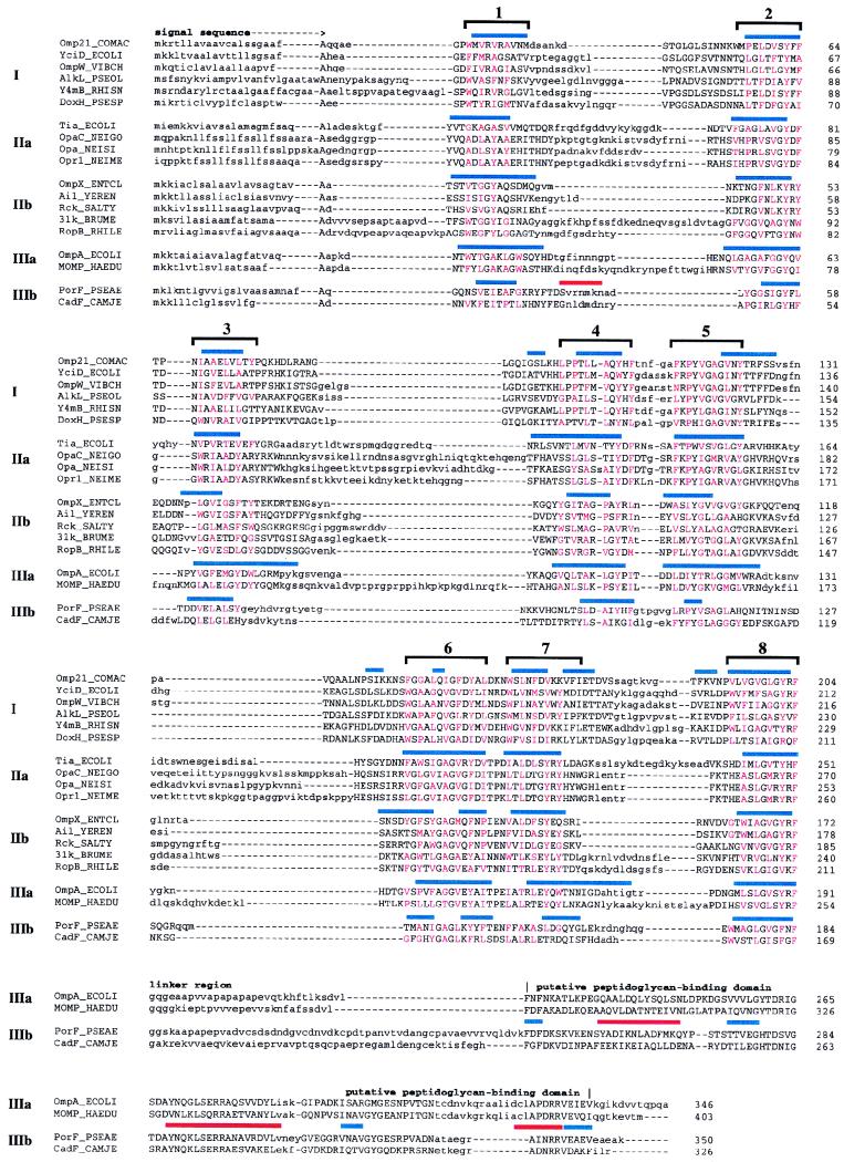
Omp21, a minor outer membrane protein of the soil bacterium Comamonas acidovorans, was purified from a spontaneous mutant lacking a surface layer and long-chain lipopolysaccharide. Omp21 synthesis is enhanced by oxygen depletion, and the protein has a variable electrophoretic mobility in sodium dodecyl sulfate-polyacrylamide gel electrophoresis due to its heat-modifiable behavior. The structural gene omp21 encodes a precursor of 204 amino acids with a putative signal peptide of 21 amino acids. Mature Omp21 is a typical outer membrane protein with a high content of beta structure as determined by infrared spectroscopy. Sequence comparisons show that it belongs to a new outer membrane protein family, characterized by eight amphipathic beta strands, which includes virulence proteins, such as the neisserial opacity proteins, Salmonella typhimurium Rck, and Yersinia enterocolitica Ail, as well as the major outer membrane proteins OmpA from Escherichia coli and OprF from Pseudomonas aeruginosa.


Figures

FIG. 1
FIG. 1
SDS-PAGE of the outer membrane proteins from C. acidovorans grown in batch cultures under different aeration conditions. The gel shows the protein patterns of cells grown in complex medium at low (lane 1) and high (lane 2) aeration and cells grown in mineral medium at low (lane 3) and high (lane 4) aeration. Omp21, running at 21 and 24 kDa, is expressed in cultures with limited oxygen. SP: regular surface protein; Omp32: porin.
FIG. 2
FIG. 2
Induction of Omp21 expression in cells grown in mineral medium at high aeration in the fermentor under controlled pH, malic acid, and temperature conditions. Top: total cell protein (μ = 0.53 h−1) and relative oxygen concentration (100% denotes 8.5 mg of O2/liter). Bottom: outer membrane protein patterns in SDS-PAGE from samples taken at different times of growth. Omp21 appears after 10 h at a relative O2 concentration of about 20% and is present in all samples beyond 24 h.
FIG. 3
FIG. 3
SDS-PAGE of outer membrane preparations from C. acidovorans type strain (WT) and mutant strain JL0. The gels were stained for protein (left panel) and for LPS (right panel). Mutant cells (JL0) are deficient in the surface layer protein (SP) and exhibit short-chain LPS only.
FIG. 4
FIG. 4
SDS-PAGE of Omp21 preparations from mutant strain JL0. Left panel: marker proteins (M) and C8En-extracted outer membrane proteins (OM). Middle panel: Omp21 purified by ionic exchange (Q) and hydroxyapatite chromatography (H). This preparation contains a particularly low level of LPS and almost no conformers at 23 and 24 kDa. Right panel: Omp21 purified by gel filtration (G) and hydroxyapatite chromatography (H). Note the additional conformer at 24 kDa. The protein was silver stained.
FIG. 5
FIG. 5
Nucleotide sequence and derived amino acid sequence of Omp21 from C. acidovorans. The putative ribosome-binding site (AAGA) is printed in boldface. The characteristic rho-independent terminator region is indicated by dotted lines. Translation stop codons are marked by asterisks. Amino acid residues +1 to +21 represent a typical bacterial signal sequence. Underlined amino acid sequences, corresponding to 51% of the mature protein, were determined by amino acid sequence analysis (J1 to J3, W1, and W2 indicate the identical peptides from the various protein bands separated in SDS-PAGE [see the text]). Dashed lines with a plus every 10 nucleotides are provided for orientation.
FIG. 6
FIG. 6
Attenuated total reflection FTIR spectra of Omp21. (A) Spectra of recombinant Omp21 (rec.) and native Omp21 (nat.) in association with LPS. The spectra are displayed in the amide I and amide II regions centered around 1,640 and 1,530 cm−1. The absorption at 1,730 cm−1 originates from carbonyl-ester vibrations of fatty acids bound in LPS molecules that do not contribute to the amide I band absorptions used for secondary-structure quantification. (B) Band form analysis between 1,700 and 1,600 cm−1 reveals a number of vibrations (solid lines) whose superposition defines the spectrum (dotted line). The strong absorption at 1,635 cm−1 and the band at 1,693 cm−1 are indicative of antiparallel β-sheet structure.
FIG. 7
FIG. 7
Alignment and secondary-structure prediction for Omp21 and homologous proteins. Representative proteins from each group in Table 1 are shown. For sequences in group III, the start of the linker region leading to the putative peptidoglycan-binding domain is denoted. Sequence regions that could not be aligned within any of the five groups are shown in lowercase letters. Red capitals mark the hydrophobic amino acids located in the conserved amphipathic sequence stretches. Bars above each aligned group indicate β strands (blue) and α helices (red) predicted by the PHD program (groups I, IIb, and IIIb) or by the method of Gromiha and Ponnuswamy (groups IIa and IIIa). Brackets above complete alignments emphasize the predicted eight-stranded β sheet of this family of outer membrane proteins.

References

    1. Altschul S F, Madden T L, Schaffer A A, Zhang J, Zhang Z, Miller W, Lipman D J. Gapped BLAST and PSI-BLAST: a new generation of protein database search programs. Nucleic Acids Res. 1997;25:3389–3402. - PMC - PubMed
    1. Applegate M, Juhn G, Liechty M, Moore M, Hozier J. Use of DNA purified in situ from cells embedded in agarose plugs for the molecular analysis of tk−/− mutants recovered in the L5178Y tk+/− 3.7.2C mutagen assay system. Mutat Res. 1990;245:55–59. - PubMed
    1. Baldermann, C., and H. Engelhardt. Unpublished data.
    1. Barondess J J, Beckwith J. A bacterial virulence determinant encoded by lysogenic coliphage lambda. Nature. 1990;346:871–874. - PubMed
    1. Bhat K S, Gibbs C P, Barrera O, Morrison S G, Jahnig F, Stern A, Kupsch E M, Meyer T F, Swanson J. The opacity proteins of Neisseria gonorrhoeae strain MS11 are encoded by a family of 11 complete genes. Mol Microbiol. 1991;5:1889–1901. - PubMed

Publication types

MeSH terms

Substances

Associated data

LinkOut - more resources