. 1998 Aug;180(15):3785-92.
doi: 10.1128/JB.180.15.3785-3792.1998.

Biosynthesis of Di-myo-inositol-1,1'-phosphate, a novel osmolyte in hyperthermophilic archaea

Affiliations

Biosynthesis of Di-myo-inositol-1,1'-phosphate, a novel osmolyte in hyperthermophilic archaea

L Chen et al. J Bacteriol. 1998 Aug.

Abstract

Biosynthesis of di-myo-inositol-1,1'-phosphate (DIP) is proposed to occur with myo-inositol and myo-inositol 1-phosphate (I-1-P) used as precursors. Activation of the I-1-P with CTP and condensation of the resultant CDP-inositol (CDP-I) with myo-inositol then generates DIP. The sole known biosynthetic pathway of inositol in all organisms is the conversion of D-glucose-6-phosphate to myo-inositol. This conversion requires two key enzymes: L-I-1-P synthase and I-1-P phosphatase. Enzymatic assays using 31P nuclear magnetic resonance spectroscopy as well as a colorimetric assay for inorganic phosphate have confirmed the occurrence of L-I-1-P synthase and a moderately specific I-1-P phosphatase. The enzymatic reaction that couples CDP-I with myo-inositol to generate DIP has also been detected in Methanococcus igneus. 13C labeling studies with [2,3-13C]pyruvate and [3-13C]pyruvate were used to examine this pathway in M. igneus. Label distribution in DIP was consistent with inositol units formed from glucose-6-phosphate, but the label in the glucose moiety was scrambled via transketolase and transaldolase activities of the pentose phosphate pathway.


Figures
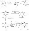
FIG. 1
FIG. 1
Proposed biosynthetic pathway for DIP showing the four key enzymatic activities. Based on similar transformations in other organisms, cofactors are indicated for several of the steps.
FIG. 2
FIG. 2
1H-coupled 31P NMR spectrum (202.3 MHz) of an I-1-P synthase assay mixture containing 5 mM G-6-P, 1 mM NAD+, 50 mM Tris acetate (pH 8.2), and 100 μl of 60 to 75% ammonium sulfate protein extract that had been incubated at 85°C for 4 h. Note that only I-1-P is a doublet in the coupled spectrum.
FIG. 3
FIG. 3
1H-decoupled 31P spectra (202.3 MHz) for analysis of specific I-1-P phosphatase activities from incubations (55°C, 16 h) of mixtures containing 6 mM MgCl2, 50 mM Tris acetate (pH 8.0), and the following: 2 mM I-1-P and the 0 to 44% fraction (A), 2 mM I-1-P and the 44 to 85% fraction (B), 2 mM G-6-P and the 0 to 44% fraction (C), and 2 mM G-6-P and the 44 to 85% fraction (D).
FIG. 4
FIG. 4
31P spectrum (121.4 MHz) for analysis of DIP synthase activity. Fifty microliters of CDP-I stock solution (30 mM), 300 μl of dialyzed cell extract, and 5 mM myo-inositol were incubated at 65°C for 2 h. The insets show expansions of the 3- to 5-ppm region, the phosphate monoester region, and the DIP region at ∼−1 ppm. Each phosphorus resonance in the spectrum is labeled.
FIG. 5
FIG. 5
1H-1H TOCSY NMR spectrum of DIP partially purified from the DIP synthase assay mixture. The vertical dotted line identifies all six DIP inositol protons in the ring spin system.
FIG. 6
FIG. 6
Predicted 13C labeling for DIP using [2,3-13C]pyruvate: ∗, label from C-3 of pyruvate; ▵, label from C-2 of pyruvate.
FIG. 7
FIG. 7
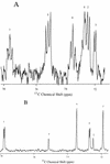
1H-decoupled 13C-NMR spectra of DIP obtained from labeling of M. igneus cultures with [2,3-13C]pyruvate (A) and [3-13C]pyruvate (B). Identities of the different DIP carbons are indicated by numbers above the resonances. Relative enrichment of DIP carbons in panel B is monitored by the integrated intensities.

Similar articles

Cited by

References

    1. Barnett J E G, Brice R E, Corina D L. Colorimetric determination of inositol monophosphates as an assay for d-glucose-6-phosphate-1L-myo-inositol 1-phosphate cyclase. Biochem J. 1970;119:183–186. - PMC - PubMed
    1. Chen I W, Charalampous F C. Biochemical studies on inositol. VIII. Purification and properties of the enzyme system which converts glucose-6-phosphate to inositol. J Biol Chem. 1965;240:3507–3512. - PubMed
    1. Chen L, Roberts M F. Cloning and expression of the inositol monophosphatase gene from Methanococcus jannaschii and characterization of the enzyme. Appl Environ Microbiol. 1998;64:2609–2615. - PMC - PubMed
    1. Ciulla R A. Response of methanogens to osmotic stress: in vitro and in vivo NMR studies. Ph.D. dissertation. Boston, Mass: Department of Chemistry, Boston College; 1995.
    1. Ciulla R A, Burggraf S, Stetter K O, Roberts M F. Occurrence and role of di-myo-inositol-1,1′-phosphate in Methanococcus igneus. Appl Environ Microbiol. 1994;60:3660–3664. - PMC - PubMed

Publication types

MeSH terms

LinkOut - more resources