

Dentilisin activity affects the organization of the outer sheath of Treponema denticola
- PMID: 9683480
- PMCID: PMC107367
- DOI: 10.1128/JB.180.15.3837-3844.1998
Dentilisin activity affects the organization of the outer sheath of Treponema denticola
Abstract
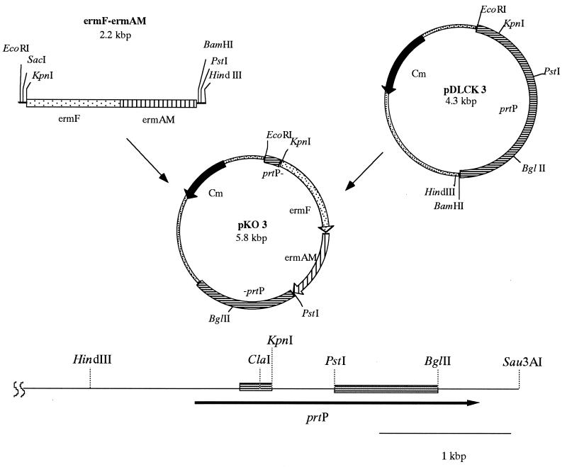
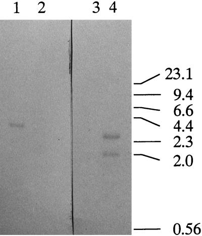
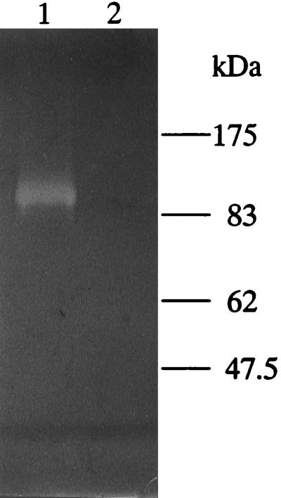
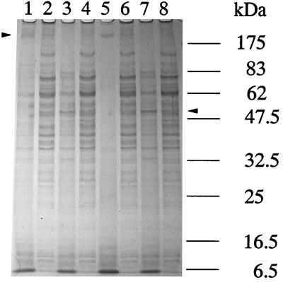
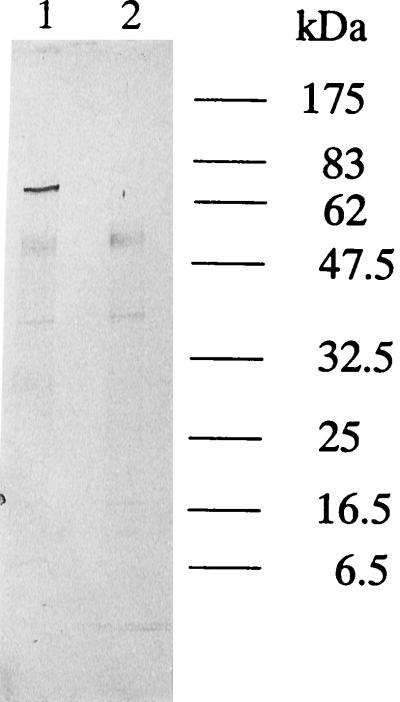
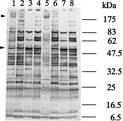
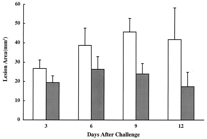
Prolyl-phenylalanine-specific serine protease (dentilisin) is a major extracellular protease produced by Treponema denticola. The gene, prtP, coding for the protease was recently cloned and sequenced (K. Ishihara, T. Miura, H. K. Kuramitsu, and K. Okuda, Infect. Immun. 64:5178-5186, 1996). In order to determine the role of this protease in the physiology and virulence of T. denticola, a dentilisin-deficient mutant, K1, was constructed following electroporation with a prtP-inactivated DNA fragment. No chymotrypsin-like protease activity was detected in the dentilisin-deficient mutant. In addition, the high-molecular-mass oligomeric protein characteristic of the outer sheath of the organism decreased in the mutant. Furthermore, the hydrophobicity of the mutant was decreased, and coaggregation of the mutant with Fusobacterium nucleatum was enhanced compared to that of the wild-type organism. The results obtained with a mouse abscess model system indicated that the virulence of the mutant was attenuated relative to that of the wild-type organism. These results suggest that dentilisin activity plays a major role in the structural organization of the outer sheath of T. denticola. The loss of dentilsin activity and the structural change in the outer sheath affect the pathogenicity of T. denticola.
Figures

Similar articles
-
A 43-kDa protein of Treponema denticola is essential for dentilisin activity.FEMS Microbiol Lett. 2004 Mar 19;232(2):181-8. doi: 10.1016/S0378-1097(04)00067-9. FEMS Microbiol Lett. 2004. PMID: 15033237
-
Binding of Porphyromonas gingivalis fimbriae to Treponema denticola dentilisin.FEMS Microbiol Lett. 2003 Sep 26;226(2):267-71. doi: 10.1016/S0378-1097(03)00615-3. FEMS Microbiol Lett. 2003. PMID: 14553921
-
Insertional inactivation of the prtP gene of Treponema denticola confirms dentilisin's disruption of epithelial junctions.J Mol Microbiol Biotechnol. 2000 Oct;2(4):581-6. J Mol Microbiol Biotechnol. 2000. PMID: 11075935
-
Approaches to Understanding Mechanisms of Dentilisin Protease Complex Expression in Treponema denticola.Front Cell Infect Microbiol. 2021 May 18;11:668287. doi: 10.3389/fcimb.2021.668287. eCollection 2021. Front Cell Infect Microbiol. 2021. PMID: 34084756 Free PMC article. Review.
-
Molecular pathogenesis of the cell surface proteins and lipids from Treponema denticola.FEMS Microbiol Lett. 1999 Dec 15;181(2):199-204. doi: 10.1111/j.1574-6968.1999.tb08844.x. FEMS Microbiol Lett. 1999. PMID: 10585538 Review.
Cited by
-
The major outer sheath protein (Msp) of Treponema denticola has a bipartite domain architecture and exists as periplasmic and outer membrane-spanning conformers.J Bacteriol. 2013 May;195(9):2060-71. doi: 10.1128/JB.00078-13. Epub 2013 Mar 1. J Bacteriol. 2013. PMID: 23457251 Free PMC article.
-
Investigation of the potential regulator proteins associated with the expression of major surface protein and dentilisin in Treponema denticola.J Oral Microbiol. 2020 Oct 11;12(1):1829404. doi: 10.1080/20002297.2020.1829404. J Oral Microbiol. 2020. PMID: 33149843 Free PMC article.
-
Two paralogous families of a two-gene subtilisin operon are widely distributed in oral treponemes.J Bacteriol. 2003 Dec;185(23):6860-9. doi: 10.1128/JB.185.23.6860-6869.2003. J Bacteriol. 2003. PMID: 14617650 Free PMC article.
-
Virulence factors of the oral spirochete Treponema denticola.J Dent Res. 2011 Jun;90(6):691-703. doi: 10.1177/0022034510385242. Epub 2010 Oct 12. J Dent Res. 2011. PMID: 20940357 Free PMC article. Review.
-
Detection of Treponema denticola in atherosclerotic lesions.J Clin Microbiol. 2001 Mar;39(3):1114-7. doi: 10.1128/JCM.39.3.1114-1117.2001. J Clin Microbiol. 2001. PMID: 11230436 Free PMC article.
References
-
- Boyer H W, Roulland-Dussoix D. A complementation analysis of the restriction and modification of DNA in Escherichia coli. J Mol Biol. 1969;41:459–472. - PubMed
-
- Chan E C S, Soiboo R, Keng T, Psarra N, Hurley R, Cheng S L, Iugovaz I. Treponema denticola (ex Brumpt 1925) sp. nov. nom. rev. and identification of new spirochete isolates from periodontal pockets. Int J Syst Bacteriol. 1993;43:196–203. - PubMed
-
- Choi B-K, Nattermann H, Grund S, Haider W, Gröbel U B. Spirochetes from digital dermatitis lesions in cattle are closely related to treponemes associated with human periodontitis. Int J Syst Bacteriol. 1997;47:175–181. - PubMed
Publication types
MeSH terms
Substances
LinkOut - more resources
Full Text Sources
Molecular Biology Databases
