. 1998 Aug;180(15):3954-66.
doi: 10.1128/JB.180.15.3954-3966.1998.

Genetic analysis of dioxin dioxygenase of Sphingomonas sp. Strain RW1: catabolic genes dispersed on the genome

Affiliations

Genetic analysis of dioxin dioxygenase of Sphingomonas sp. Strain RW1: catabolic genes dispersed on the genome

J Armengaud et al. J Bacteriol. 1998 Aug.

Abstract

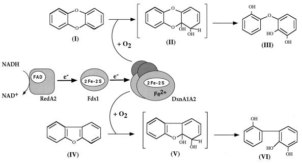
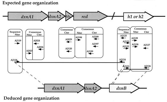
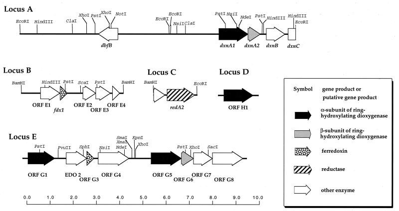
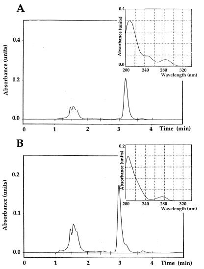
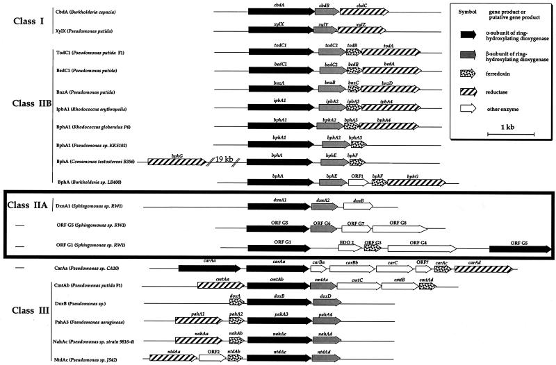
The dioxin dioxygenase of Sphingomonas sp. strain RW1 activates dibenzo-p-dioxin and dibenzofuran for further metabolism by introducing two atoms of oxygen at a pair of vicinal carbon atoms, one of which is involved in one of the bridges between the two aromatic rings, i.e., an angular dioxygenation. The dxnA1 and dxnA2 cistrons encoding this dioxygenase have been cloned and shown to be located just upstream of a hydrolase gene which specifies an enzyme involved in the subsequent step of the dibenzofuran biodegradative pathway. Genes encoding the electron supply system of the dioxygenase are not clustered with the dioxygenase gene but rather are located on two other distinct and separate genome segments. Moreover, whereas expression of dxnA1A2 is modulated according to the available carbon source, expression of the dbfB gene encoding the ring cleavage enzyme of the dibenzofuran pathway, which is located in the neighborhood of dxnA1A2 but oriented in the opposite direction, is constitutive. The scattering of genes for the component proteins of dioxin dioxygenase system around the genome of Sphingomonas sp. strain RW1, and the differential expression of dioxin pathway genes, is unusual and contrasts with the typical genetic organization of catabolic pathways where component cistrons tend to be clustered in multicistronic transcriptional units. The sequences of the alpha and beta subunits of the dioxin dioxygenase exhibit only weak similarity to other three component dioxygenases, but some motifs such as the Fe(II) binding site and the [2Fe-2S] cluster ligands are conserved. Dioxin dioxygenase activity in Escherichia coli cells containing the cloned dxnA1A2 gene was achieved only through coexpression of the cognate electron supply system from RW1. Under these conditions, exclusively angular dioxygenation of dibenzofuran and dibenzo-p-dioxin was obtained. The dioxin dioxygenase was not active in E. coli cells coexpressing a class IIB electron supply system. In the course of the isolation of the dxnA1 and dxnA2 cistrons, a number of other catabolic genes dispersed over different genome segments were identified, which may indicate greater catabolic potential than was previously suspected. This finding is consistent with the catabolic versatility of members of the genus Sphingomonas, which is becoming increasingly evident, and may indicate a less well evolved and regulated but more dynamic genetic organization in this organism than is the case for better-studied pathways in organisms such as Pseudomonas species.


Figures

FIG. 1
FIG. 1
The dioxin dioxygenase and its electron supply system. The reaction carried out by the multicomponent ring-hydroxylating dioxin dioxygenase and its electron transfer chain are shown. A flavoprotein reductase, RedA2, accepts electrons from NADH and transfers them via the ferredoxin Fdx1 to the terminal oxygenase. The reduced terminal oxygenase catalyzes the angular oxidation of dibenzo-p-dioxin and dibenzofuran. Chemical designations: (I), dibenzo-p-dioxin; (II), 4,4a-dihydroxy-dihydro-dibenzo-p-dioxin; (III), 2,2′,3-THD-ether; (IV), dibenzofuran; (V), 4,4a-dihydroxy-dihydro-dibenzofuran; (VI), 2,2′,3-THB. The two unstable compounds which spontaneously transform to other products are indicated in brackets.
FIG. 2
FIG. 2
PCR-based strategy developed to clone the dxnA1 and dxnA2 genes. From different alignments of related proteins, C-terminal (Cter) and N-terminal (Nter) consensus sequences were defined as indicated by the central boxes. Degenerate forward and reverse primers, indicated by small arrows, were then designed from these consensus sequences and from the N-terminal sequence of each purified polypeptide determined by Edman degradation. The two PCR products obtained from the multiple combinations of primers (indicated with dashed lines) resulted in determination of the gene organization of the dxn locus.
FIG. 3
FIG. 3
Genetic organizations of loci encompassing the genes described in this study. The positions and orientations of the different ORFs detected within the loci described in the text are shown by full arrows. The scale bar at the bottom indicates gene sizes in kilobase pairs.
FIG. 4
FIG. 4
Phylogenetic tree obtained from alignment of the four α-subunit sequences with related proteins. The sequences of 16 α subunits of ring-hydroxylating oxygenases and the four sequences reported in this study were compiled by using the GeneWorks software (version 2.5N) from IntelliGenetics; the multiple alignment analysis was performed with the Phylip package programs. The phylogenetic unrooted tree was drawn by using TreeView. The horizontal bar indicates the percent divergence (distance). The numbers on some of the branches refer to the confidence (percent) estimated by bootstrap analysis (100 replications). The proteins are labeled by trivial abbreviations. Their accession codes in the SwissProt or GenBank databases and their origins are D86080 for aniline dioxygenase from Acinetobacter sp. strain YAA (AtdA-YAA), D90884 for dioxygenase from E. coli K-12 (BedC1-K12), L046642 for benzene dioxygenase from Pseudomonas putida ML2 (BedC1-ML2), M76990 for benzoate oxygenase from Acinetobacter calcoaceticus ADP1 (BenA-ADP1), X80041 for biphenyl dioxygenase from Rhodococcus globerulus P6 (BphA1-P6), P37333 for biphenyl dioxygenase from Burkholderia sp. strain LB400 (BphA-LB400), U47637 for biphenyl dioxygenase from Comamonas testosteroni B-356 (BphA-B356), M83949 for naphthalene 1,2-dioxygenase from P. putida G7 (NahAc-G7), U49496 for naphthalene dioxygenase from Pseudomonas sp. strain 9816 (NahAc-9816), U49504 for 2-nitrotoluene dioxygenase from Pseudomonas sp. strain JS42 (NtdAc-JS42), U62430 for ORF 2-like dioxygenase from Burkholderia sp. strain DNT (ORF 2-DNT), AB004059 for polycyclic aromatic hydrocarbon dioxygenase from P. putida OUS82 (PahAc-OUS82), U78099 for chlorobenzene dioxygenase from Burkholderia sp. strain PS12 (TecA1-PS12), U11420 for 2,4,5-trichlorophenoxyacetic acid oxygenase from Pseudomonas cepacia ACC1100 (TftA-ACC1100), J04996 for toluene dioxygenase from P. putida F1 (TodC1-F1), U51165 for dioxygenase from Cycloclasticus oligotrophus RB1 (XylC1-RB1), and P23099 for toluate 1,2-dioxygenase from P. putida ML2 (XylX-mt2).
FIG. 5
FIG. 5
Conserved sequences that characterize the Rieske-type [2Fe-2S] cluster and Fe(II) binding sites in α subunits. Well-conserved fingerprint sequence regions in the N-terminal part of the α subunits of ring-hydroxylating dioxygenases are aligned. Shaded characters represent conserved residues; arrows indicate the amino acids involved in binding the Rieske-type [2Fe-2S] cluster (A) and the mononuclear iron atom (B). The corresponding consensus sequences for the entire family of this type of protein are shown. Accession numbers in the GenBank database and origins of the large α subunits are P37333 for biphenyl dioxygenase from Burkholderia sp. strain LB400 (BphA-LB400), D17319 for biphenyl dioxygenase from Pseudomonas sp. strain KKS102 (BphA-KKS102), U78099 for chlorobenzene dioxygenase from Burkholderia sp. strain PS12 (TecA1-PS12), and J04996 for toluene dioxygenase from Pseudomonas putida F1 (TodC1-F1).
FIG. 6
FIG. 6
Immunodetection of DxnA1 in Sphingomonas sp. strain RW1. Cells harvested from liquid cultures of Sphingomonas sp. strain RW1 and E. coli(pAJ127) grown under different conditions were lysed in sodium dodecyl sulfate-containing buffer, and their proteins were subjected to electrophoresis on 10% sodium dodecyl sulfate–glycine polyacrylamide gels and immunostained after transfer to nitrocellulose membranes. One membrane (A) was stained by an alkaline phosphatase-based reaction, whereas the other (B) was stained by a chemiluminescence-based reaction. (A) Samples of Sphingomonas sp. strain RW1 grown in M9 medium with 2 mM dibenzofuran as the sole carbon source (lane 1), LB medium (lane 2), M9 medium with 2 mM salicylate (lane 3), and M9 medium with 2 mM acetate (lane 4); (B) samples of E. coli DH5α(pAJ127) grown in LB medium (lane 5) and Sphingomonas sp. strain RW1 grown in M9 medium with 2 mM dibenzofuran as the sole carbon source (lane 1). The arrowhead shows the position of the 48-kDa large subunit of the dioxin dioxygenase.
FIG. 7
FIG. 7
HPLC analysis of product formation by E. coli DH5α(pAJ127)(pAJ130). Conversion of dibenzofuran (A) and dibenzo-p-dioxin (B) by E. coli DH5α(pAJ127)(pAJ130) was analyzed by HPLC as detailed in Materials and Methods. The chromatograms were recorded at a wavelength of 210 nm and correspond to samples taken after 3 h of incubation. Products eluting between 1.0 and 2.0 min were also formed by E. coli control strains with or without substrates and therefore are not produced by the dioxin dioxygenase activity. The spectra of the specific products formed by the dioxin dioxygenase activity, 2,2′,3-THB eluting at 3.2 min and 2,2′,3-THD-ether eluting at 3.0 min, are shown in the insets.
FIG. 8
FIG. 8
Comparative molecular organization of three-component dioxygenases. The organization of each of the genes encoding the different polypeptides of representative hydroxylating enzymes is shown. The general name of each cluster, the name of the microorganism containing the enzymes, and the class (according to Batie et al., [4]) to which the enzymes belong are indicated at the left. Gene names are given above the ORFs, and arrows indicate the direction of transcription. Each cluster was aligned with the others, taking the gene specifying the α subunit dioxygenase as a reference. Genes are drawn according to size and relative position. Accession numbers in the DDBJ/EMBL/GenBank database of the corresponding sequences are X79076 for cbdA (dibenzothiophene dioxygenase) from Pseudomonas sp., M64747 for xylX (toluate dioxygenase) from Pseudomonas putida, J04996 for todC1 (toluene dioxygenase) from P. putida F1, L04642 for bedC1 (benzene dioxygenase) from P. putida ML2, M17904 for bnzA (benzene dioxygenase) from P. putida, U24277 for ipbA1 (isopropylbenzene dioxygenase) from Rhodococcus erythropolis BD2, X80041 for bphA1 (biphenyl dioxygenase) from Rhodococcus globerulus P6, D17319 for bphA1 (biphenyl dioxygenase) from Pseudomonas sp. strain KKS102, U47637 for bphA (biphenyl dioxygenase) from Comamonas testosteroni B-356, M86348 for bphA (biphenyl dioxygenase) from Burkholderia sp. strain LB400, D89064 for carAa (carbazole dioxygenase) from Pseudomonas sp. strain CA10, U24215 for cmtAa (p-cumate dioxygenase) from P. putida F1, M60405 for doxB (dibenzothiophene dioxygenase) from Pseudomonas sp., D84146 for pahA3 (naphthalene dioxygenase) from Pseudomonas aeruginosa PaK1, M83949 for nahAc (naphthalene dioxygenase) from P. putida NCIB 9816-4, and U49504 for ntdAc (2-nitrotoluene dioxygenase) from Pseudomonas sp. strain JS42.

References

    1. Armengaud J, Timmis K N. Molecular characterization of Fdx1, a putidaredoxin-type [2Fe-2S] ferredoxin able to transfer electrons to the dioxin dioxygenase of Sphingomonas sp. RW1. Eur J Biochem. 1997;247:833–842. - PubMed
    1. Armengaud J, Timmis K N. The reductase RedA2 of the multicomponent dioxin dioxygenase system of Sphingomonas sp. RW1 is related to class-I cytochrome P450-type reductases. Eur J Biochem. 1998;253:437–444. - PubMed
    1. Asturias J A, Diaz E, Timmis K N. The evolutionary relationship of biphenyl dioxygenase from gram-positive Rhodococcus globerulus P6 to multicomponent dioxygenases from gram-negative bacteria. Gene. 1995;156:11–18. - PubMed
    1. Batie C J, Ballou D P, Correll C C. Phthalate dioxygenase reductase and related flavin-iron-sulfur containing electron transferases. In: Müller F, editor. Chemistry and biochemistry of flavoenzymes. Boca Raton, Fla: CRC Press; 1992. pp. 543–566.
    1. Beil S, Happe B, Timmis K N, Pieper D H. Genetic and biochemical characterization of the broad spectrum chlorobenzene dioxygenase from Burkholderia sp. strain PS12: dechlorination of 1,2,4,5-tetrachlorobenzene. Eur J Biochem. 1997;247:190–199. - PubMed

Publication types

MeSH terms

Associated data

LinkOut - more resources