. 1998 Aug 3;188(3):421-9.
doi: 10.1084/jem.188.3.421.

Lyn, Jak2, and Raf-1 kinases are critical for the antiapoptotic effect of interleukin 5, whereas only Raf-1 kinase is essential for eosinophil activation and degranulation

Affiliations

Lyn, Jak2, and Raf-1 kinases are critical for the antiapoptotic effect of interleukin 5, whereas only Raf-1 kinase is essential for eosinophil activation and degranulation

K Pazdrak et al. J Exp Med. .

Abstract

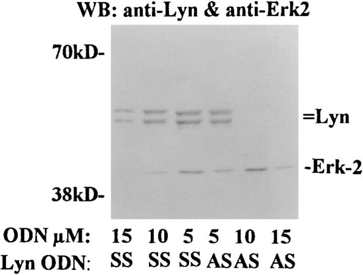
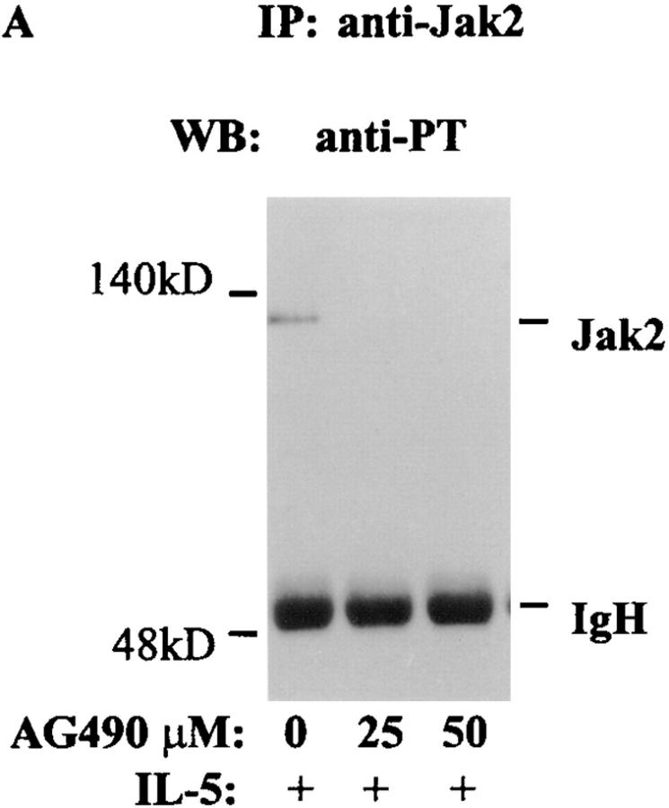
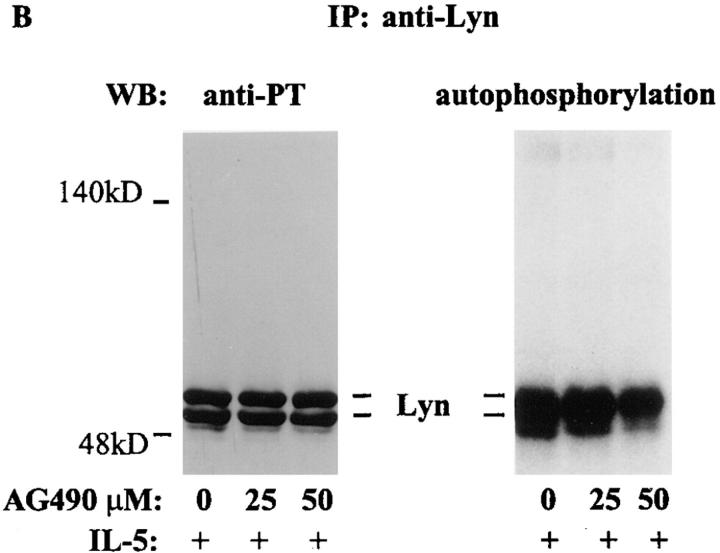
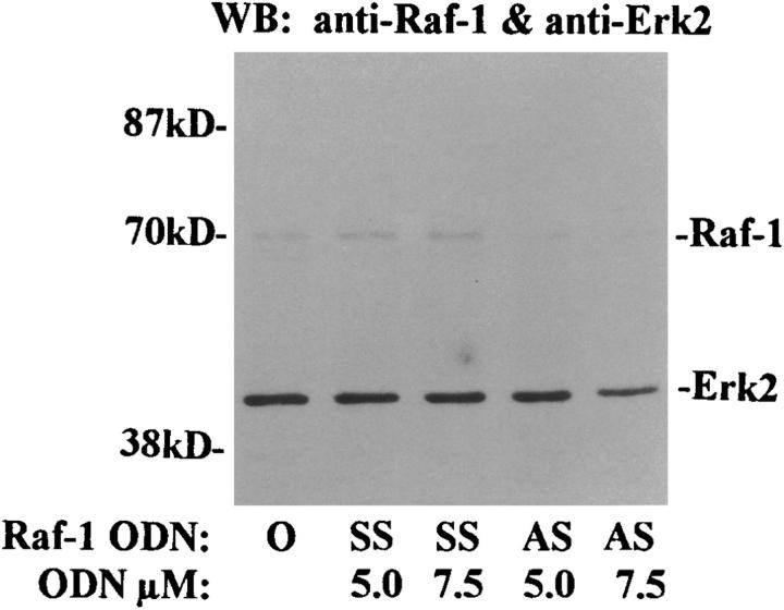
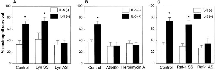
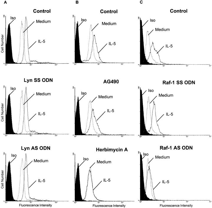
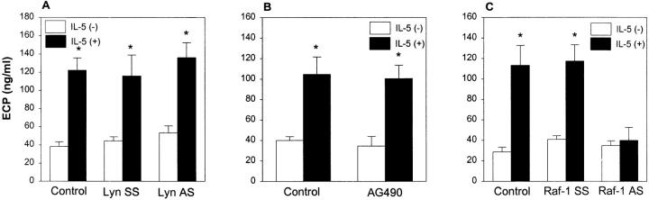
Interleukin (IL)-5 has been shown to activate many signaling molecules in eosinophils, but their functional relevance remains unknown. We have examined the functional relevance of Lyn, Jak2, and Raf-1 kinases in eosinophil survival, upregulation of adhesion molecules and degranulation. To this goal we used Lyn and Raf-1 antisense (AS) oligodeoxynucleotides (ODN) to inhibit the expression of these proteins and tyrphostin AG490 to specifically block the activation of Jak2. We have demonstrated that all three kinases are important for IL-5- induced suppression of eosinophil apoptosis. However, Lyn and Jak2 tyrosine kinases are not important for the upregulation of CD11b and the secretion of eosinophil cationic protein. In contrast, Raf-1 kinase is critical for both these functions. This is the first identification of specific signaling molecules responsible for three important functions of eosinophils. We have established a central role for Raf-1 kinase in regulating eosinophil survival, expression of beta2 integrins and degranulation. Further, there appears to be a dissociation between two receptor-associated tyrosine kinases, i.e., Lyn and Jak2, and the activation of Raf-1 kinase. The delineation of the functional relevance of signaling molecules will help design therapeutic approaches targeting specific eosinophil function.


Figures

Figure 1
Figure 1
The effect of Lyn AS ODN on Lyn and ERK-2 kinases expression. Purified eosinophils were incubated with Lyn antisense (AS ) or sense (SS ) ODN for 6 h. The cells were then washed and lysed. The lysates were electrophoresed, transferred to Hybond membrane, and then Western blotted with anti-Lyn and anti–ERK-2 antibodies. The blot was developed with the enhanced chemiluminescence system. Lyn AS ODN at 10 μM concentration blocked the expression of Lyn but not ERK-2 kinase (n = 3). At 15 μM concentration both AS and SS ODN inhibited the expression of the kinases.
Figure 2
Figure 2
The effect of AG490 on IL-5–induced tyrosine phosphorylation of Jak2 and Lyn and autophosphorylation of Lyn kinase. Eosinophils were pretreated with AG490 for 6 h and then stimulated with IL-5. The cells were lysed and immunoprecipitated with antibodies against p53/56 Lyn or Jak2 kinases. The immunoprecipitates were immunoblotted with an antiphosphotyrosine antibody. Further, the anti-Lyn immunoprecipitate was used in an autophosphorylation assay. IL-5–induced tyrosine phosphorylation of Jak2 (A) and p53/56 Lyn (B, left) kinases. AG490 at 25 and 50 μM concentrations inhibited tyrosine phosphorylation of Jak2 but not Lyn kinase (n = 3). AG490 did not affect autophosphorylation of Lyn kinase at 25 μM but had some inhibitory effects at 50 μM concentration (B, right).
Figure 2
Figure 2
The effect of AG490 on IL-5–induced tyrosine phosphorylation of Jak2 and Lyn and autophosphorylation of Lyn kinase. Eosinophils were pretreated with AG490 for 6 h and then stimulated with IL-5. The cells were lysed and immunoprecipitated with antibodies against p53/56 Lyn or Jak2 kinases. The immunoprecipitates were immunoblotted with an antiphosphotyrosine antibody. Further, the anti-Lyn immunoprecipitate was used in an autophosphorylation assay. IL-5–induced tyrosine phosphorylation of Jak2 (A) and p53/56 Lyn (B, left) kinases. AG490 at 25 and 50 μM concentrations inhibited tyrosine phosphorylation of Jak2 but not Lyn kinase (n = 3). AG490 did not affect autophosphorylation of Lyn kinase at 25 μM but had some inhibitory effects at 50 μM concentration (B, right).
Figure 3
Figure 3
The effect of Raf-1 antisense ODN on the expression of Raf-1 and ERK-2 kinases (A) and phosphorylation of ERK-2 (B). (A) Purified eosinophils were incubated with medium (O), Raf-1 AS (AS), or SS ODN (SS ) for 6 h. The cells were then washed and lysed. The lysates were electrophoresed and immunoblotted with anti– Raf-1 and anti–ERK-2 antibodies. Raf-1 AS ODN at 5 and 7.5 μM concentrations blocked the expression of Raf-1 but not ERK-2 kinase (n = 3). The SS ODN did not affect the expression of the kinases. (B) Cells were pretreated with medium (O) or 7.5 μM concentration of Raf-1 AS (AS ) and SS ODN (SS ) for 6 h and then stimulated with (+) or without (−) IL-5. The cells were lysed, immunoprecipitated with an anti-ERK2 antibody, and immunoblotted with an antiphosphotyrosine antibody. IL-5–induced tyrosine phosphorylation of ERK-2 kinase. Raf-1 AS but not SS ODN inhibited the phosphorylation of ERK-2 kinase (left). The membrane was then stripped and reprobed with the anti–ERK-2 antibody (right), which showed an equal amount of ERK-2 protein in each lane.
Figure 3
Figure 3
The effect of Raf-1 antisense ODN on the expression of Raf-1 and ERK-2 kinases (A) and phosphorylation of ERK-2 (B). (A) Purified eosinophils were incubated with medium (O), Raf-1 AS (AS), or SS ODN (SS ) for 6 h. The cells were then washed and lysed. The lysates were electrophoresed and immunoblotted with anti– Raf-1 and anti–ERK-2 antibodies. Raf-1 AS ODN at 5 and 7.5 μM concentrations blocked the expression of Raf-1 but not ERK-2 kinase (n = 3). The SS ODN did not affect the expression of the kinases. (B) Cells were pretreated with medium (O) or 7.5 μM concentration of Raf-1 AS (AS ) and SS ODN (SS ) for 6 h and then stimulated with (+) or without (−) IL-5. The cells were lysed, immunoprecipitated with an anti-ERK2 antibody, and immunoblotted with an antiphosphotyrosine antibody. IL-5–induced tyrosine phosphorylation of ERK-2 kinase. Raf-1 AS but not SS ODN inhibited the phosphorylation of ERK-2 kinase (left). The membrane was then stripped and reprobed with the anti–ERK-2 antibody (right), which showed an equal amount of ERK-2 protein in each lane.
Figure 4
Figure 4
The effect of Lyn AS ODN (A), AG490 (B) and Raf-1 AS ODN (C ) on survival-prolonging activity of IL-5 on eosinophils. (A) Eosinophils were pretreated with 10 μM concentration of Lyn AS and SS ODN or medium (Control ) for 6 h. The cells were then stimulated with (+) or without (−) IL-5 (10−10 M) for 1.5 h. Eosinophil viability was assessed 32 h later. The control and Lyn SS-treated cells showed significant prolongation of eosinophil survival (n = 5, *P <0.01, paired t test) after IL-5 stimulation. This survival prolonging effect of IL-5 was blocked by pretreatment of cells with Lyn AS ODN. (B) Purified eosinophils were pretreated with buffer (Control ), AG490 (25 μM) or Herbimycin A (2 μM) for 6 h. The cells were then stimulated with (+) or without (−) IL-5 for 1.5 h. Eosinophil viability was assessed 32 h later. The IL-5–induced prolongation of eosinophil survival was completely abrogated by both AG490 and Herbimycin A (n = 5, *P <0.01). (C ) Eosinophils were pretreated with 7.5 μM concentration of Raf-1 AS, SS ODN, or medium (Control ) for 6 h. The cells were then stimulated with (+) or without (−) IL-5 (10−10 M) for 1.5 h. Eosinophil viability was assessed 32 h later. The control and SS ODN-treated cells showed significant prolongation of eosinophil survival (n = 5, *P <0.01, paired t test) after IL-5 stimulation. Raf-1 AS ODN completely blocked this survival prolonging effect of IL-5.
Figure 5
Figure 5
The effect of Lyn AS ODN (A), AG490 (B), and Raf-1 AS ODN (C ) on CD11b expression. (A). Eosinophils were pretreated with medium (Control ), Lyn SS, or AS ODN and then stimulated with medium or IL-5. Then the cells were stained with an anti-CD11b FITC-conjugated mAb or isotype control mAb and analyzed by a FACScan®. In control experiments, IL-5 upregulated the expression of CD11b. Neither AS nor SS Lyn ODN had any effect on eosinophil CD11b expression (n = 3). (B). Jak2 kinase and CD11b expression. Eosinophils were pretreated with medium (Control ), AG490 (25 μM) or Herbimycin A (2 μM) and then stimulated with medium or IL-5. Then the cells were stained with the anti-CD11b FITC-conjugated mAb or isotype control mAb. In control experiments, IL-5 upregulated the expression of CD11b. The Jak2 inhibitor AG490 did not block the upregulation of CD11b, but a broad spectrum tyrosine kinase inhibitor Herbimycin A did (n = 3). (C ) Eosinophils were pretreated with medium (Control ), Raf-1 SS or AS ODN and then stimulated with medium or IL-5. Then the cells were stained with the anti-CD11b FITC-conjugated mAb or isotype control mAb. In control and Raf-1 SS ODN experiments, IL-5 upregulated the expression of CD11b. Raf-1 AS ODN blocked the upregulation of CD11b by IL-5 (n = 3).
Figure 6
Figure 6
The effect of Lyn AS ODN (A), AG490 (B), and Raf-1 AS ODN (C ) on ECP release. (A) Eosinophils were pretreated with medium (Control ), Lyn SS or AS ODN and, then, primed with (+) or without (−) IL-5. The cells were then challenged with a suboptimal concentration of PMA (0.1 ng/ml) which by itself did not induce ECP release. The IL-5–priming of cells caused significant secretion of ECP from eosinophils (*P <0.01, n = 4). Neither AS nor SS Lyn ODN affected ECP release. (B). Eosinophils were pretreated with medium (Control ) or AG490 (25 μM) and then primed with (+) or without (−) IL-5 as described above. The cells were then challenged with a suboptimal concentration of PMA (0.1 ng/ml). The IL-5–priming of cells caused significant secretion of ECP from eosinophils (*P <0.05, n = 4). Pretreatment of cells with AG490 did not block ECP release. (C ) Eosinophils were pretreated with medium (Control), Raf-1 SS or AS ODN and, then, primed with (+) or without (−) IL-5. The cells were then challenged with PMA (0.1 ng/ml). The IL-5–priming of cells caused significant secretion of ECP from control and SS ODN-treated eosinophils (*P <0.05, n = 4). The Raf-1 AS ODN completely prevented ECP release.

References

    1. Corrigan CJ, Kay AB. T cells and eosinophils in the pathogenesis of asthma. Immunol Today. 1992;13:501–503. - PubMed
    1. Owen, W.F., Jr., and K.F. Austen. 1994. Cytokine regulation of eosinophil-mediated inflammatory reactions by modulation of eosinophil programmed cell death and subsequent priming for augmented function. In Eosinophils In Allergy and Inflammation. G.J. Gleich and A.B. Kay, editors. Marcel Decker, New York. 239–253.
    1. Resnick MB, Weller PF. Mechanisms of eosinophil recruitment. Am J Res Cell Mol Biol. 1993;8:349–355. - PubMed
    1. Yamaguchi Y, Tsuda T, Suda J, Eguchi M, Miura Y, Harada N, Tominaga A, Takatsu K. Purified interleukin-5 supports the terminal differentiation and proliferation of murine eosinophilic precursors. J Exp Med. 1988;167:43–56. - PMC - PubMed
    1. Walsh GM, Hartnell A, Wardlaw AJ, Kurihara K, Sanderson CJ, Kay AB. IL-5 enhances the in vitro adhesion of human eosinophils, but not neutrophils, in a leukocyte integrin (CD11/CD18)-dependent manner. Immunology. 1990;71:258–265. - PMC - PubMed

Publication types

MeSH terms