. 1998 Aug 3;188(3):527-37.
doi: 10.1084/jem.188.3.527.

Rac-1 regulates nuclear factor of activated T cells (NFAT) C1 nuclear translocation in response to Fcepsilon receptor type 1 stimulation of mast cells

Affiliations

Rac-1 regulates nuclear factor of activated T cells (NFAT) C1 nuclear translocation in response to Fcepsilon receptor type 1 stimulation of mast cells

H Turner et al. J Exp Med. .

Abstract

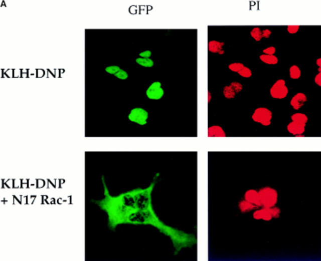
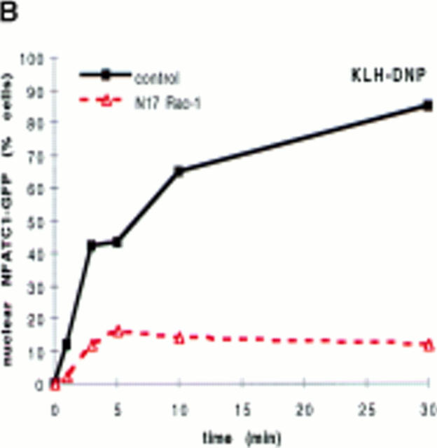
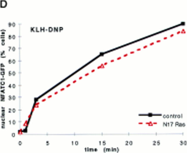
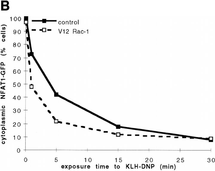
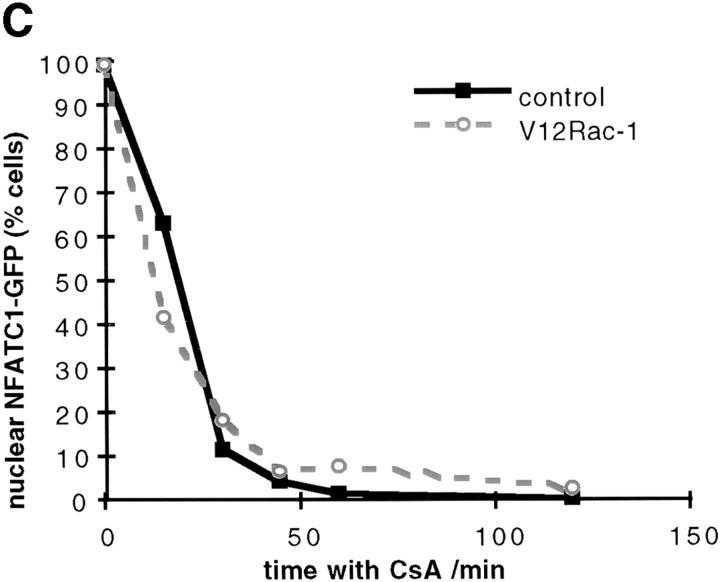
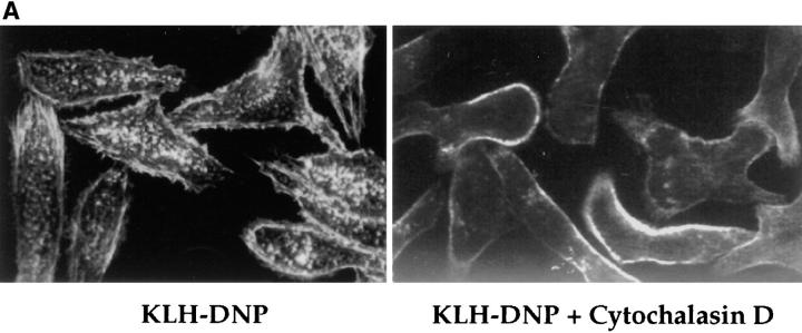
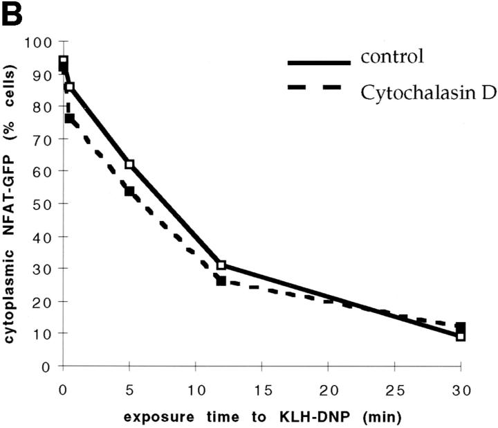
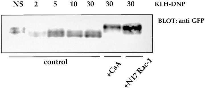
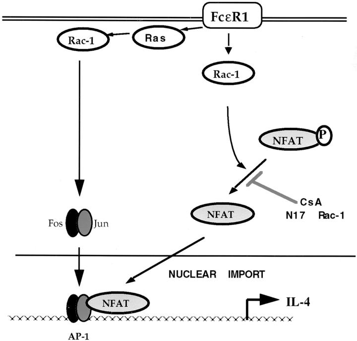
Transcription factors of the nuclear factor of activated T cells (NFAT) family play a key role in antigen receptor-mediated responses in lymphocytes by controlling induction of a wide variety of cytokine genes. The GTPases Ras and Rac-1 have essential functions in regulation of NFAT transcriptional activity in the mast cell system, where Fcepsilon receptor type 1 (FcepsilonR1) ligation results in induction of multiple NFAT target genes. This report examines the precise biochemical basis for the Rac-1 dependency of FcepsilonR1 activation of NFAT in mast cells. We are able to place Rac-1 in two positions in the signaling network that regulates the assembly and activation of NFAT transcriptional complexes in lymphocytes. First, we show that activity of Rac-1 is required for FcepsilonR1-mediated NFATC1 dephosphorylation and nuclear import. Regulation of NFAT localization by the FcepsilonR1 is a Rac-dependent but Ras-independent process. This novel signaling role for Rac-1 is distinct from its established regulation of the actin cytoskeleton. Our data also reveal a second GTPase signaling pathway regulating NFAT transcriptional activity, in which Rac-1 mediates a Ras signal. These data illustrate that the GTPase Rac-1 should now be considered as a component of the therapeutically important pathways controlling NFATC1 subcellular localization. They also reveal that GTPases may serve multiple functions in cellular responses to antigen receptor ligation.


Figures

Figure 1
Figure 1
(A) NFATC1-GFP marker construct. Schematically shown here, the full-length NFATC1 molecule was fused to the GFP reporter by cloning into the pEGFP-1 vector (Clontech). TAD, Transactivation domain. HR, Homology region. SRR, Serine-rich region. SP, Ser/Pro box. (B) NFATC1-GFP is cytosolic in resting RBL2H3 mast cells. RBL2H3 cells were transfected with 8 μg NFATC1-GFP and recovered for 6 h on glass coverslips in complete medium at 37°C. Cells were costained with PI, fixed, and mounted as described. (C and D) FcεR1 cross-linking induces NFATC1-GFP nuclear import in RBL2H3 mast cells. RBL2H3 cells were transfected with 8 μg NFATC1-GFP and recovered for 6 h on glass coverslips in complete medium at 37°C. Cells were IgE primed and stimulated with 500 ng/ml KLH-DNP for 30 min (C) or for the indicated times (D). Cells were either costained with PI, fixed, and mounted as described (C), or cells were scored for predominant localization of GFP (D). (E) FcεR1 induction of NFATC1-GFP nuclear import is CN regulated and CsA sensitive. RBL2H3 cells were transfected with 8 μg NFATC1-GFP reporter alone or in combination with 20 μg activated CN plasmid (CNM) and recovered for 6 h on glass coverslips in complete medium at 37°C. Cells were primed and stimulated with 500 ng/ml KLH-DNP for 30 min in the absence or presence of 50 nM CsA. NS, Not stimulated. Cells were fixed, mounted, and scored for predominant localization of GFP as described.
Figure 1
Figure 1
(A) NFATC1-GFP marker construct. Schematically shown here, the full-length NFATC1 molecule was fused to the GFP reporter by cloning into the pEGFP-1 vector (Clontech). TAD, Transactivation domain. HR, Homology region. SRR, Serine-rich region. SP, Ser/Pro box. (B) NFATC1-GFP is cytosolic in resting RBL2H3 mast cells. RBL2H3 cells were transfected with 8 μg NFATC1-GFP and recovered for 6 h on glass coverslips in complete medium at 37°C. Cells were costained with PI, fixed, and mounted as described. (C and D) FcεR1 cross-linking induces NFATC1-GFP nuclear import in RBL2H3 mast cells. RBL2H3 cells were transfected with 8 μg NFATC1-GFP and recovered for 6 h on glass coverslips in complete medium at 37°C. Cells were IgE primed and stimulated with 500 ng/ml KLH-DNP for 30 min (C) or for the indicated times (D). Cells were either costained with PI, fixed, and mounted as described (C), or cells were scored for predominant localization of GFP (D). (E) FcεR1 induction of NFATC1-GFP nuclear import is CN regulated and CsA sensitive. RBL2H3 cells were transfected with 8 μg NFATC1-GFP reporter alone or in combination with 20 μg activated CN plasmid (CNM) and recovered for 6 h on glass coverslips in complete medium at 37°C. Cells were primed and stimulated with 500 ng/ml KLH-DNP for 30 min in the absence or presence of 50 nM CsA. NS, Not stimulated. Cells were fixed, mounted, and scored for predominant localization of GFP as described.
Figure 2
Figure 2
(A and B) N17Rac-1 prevents FcεR1-induced NFATC1-GFP nuclear import. RBL2H3 mast cells were transfected with 8 μg NFATC1-GFP reporter alone or in combination with 20 μg pEF-N17Rac-1. Cells were recovered for 6 h on glass coverslips before IgE priming and stimulation with 500 ng/ml KLH-DNP for 30 min (A) or for the indicated times (B). Cells were fixed and mounted, and localization of GFP was visualized as described in Materials and Methods (A) or scored for predominant localization of NFATC1-GFP (B). (C) Ionomycin induction of NFATC1-GFP nuclear accumulation is unaffected by N17Rac-1. RBL2H3 cells were transfected with 8 μg NFATC1-GFP alone or in combination with 20 μg pEF-N17Rac-1. Cells were seeded onto glass coverslips, recovered for 6 h, and stimulated for the indicated times using 500 ng/ml Ionomycin or vehicle control. Cells were fixed, mounted, and scored for predominant localization of GFP as described. (D) Dominant inhibition of Ras does not affect NFATC1-GFP nuclear import. RBL2H3 cells were transfected with 8 μg NFATC1-GFP alone or in combination with 20 μg pRSV-N17 Ras. Cells were seeded onto glass coverslips, recovered for 6 h, and stimulated for the indicated times using 500 ng/ml KLH-DNP. Cells were fixed, mounted, and scored for predominant localization of GFP as described.
Figure 2
Figure 2
(A and B) N17Rac-1 prevents FcεR1-induced NFATC1-GFP nuclear import. RBL2H3 mast cells were transfected with 8 μg NFATC1-GFP reporter alone or in combination with 20 μg pEF-N17Rac-1. Cells were recovered for 6 h on glass coverslips before IgE priming and stimulation with 500 ng/ml KLH-DNP for 30 min (A) or for the indicated times (B). Cells were fixed and mounted, and localization of GFP was visualized as described in Materials and Methods (A) or scored for predominant localization of NFATC1-GFP (B). (C) Ionomycin induction of NFATC1-GFP nuclear accumulation is unaffected by N17Rac-1. RBL2H3 cells were transfected with 8 μg NFATC1-GFP alone or in combination with 20 μg pEF-N17Rac-1. Cells were seeded onto glass coverslips, recovered for 6 h, and stimulated for the indicated times using 500 ng/ml Ionomycin or vehicle control. Cells were fixed, mounted, and scored for predominant localization of GFP as described. (D) Dominant inhibition of Ras does not affect NFATC1-GFP nuclear import. RBL2H3 cells were transfected with 8 μg NFATC1-GFP alone or in combination with 20 μg pRSV-N17 Ras. Cells were seeded onto glass coverslips, recovered for 6 h, and stimulated for the indicated times using 500 ng/ml KLH-DNP. Cells were fixed, mounted, and scored for predominant localization of GFP as described.
Figure 3
Figure 3
(A and B) Effect of V12Rac-1 upon NFATC1-GFP nuclear accumulation in RBL2H3 mast cells. RBL2H3 cells were transfected with 8 μg NFATC1-GFP reporter alone or in combination with 20 μg pEF-V12Rac-1. Cells were recovered for 6 h on glass coverslips before IgE priming and stimulation for 30 min with the indicated concentrations of KLH-DNP (A) or stimulation with 500 ng/ml KLH-DNP for the indicated times (B). Cells were fixed, mounted, and scored for predominant localization of GFP as described. (C) Effect of V12Rac-1 on NFATC1-GFP nuclear export in RBL2H3 mast cells. RBL2H3 cells were transfected with 8 μg NFATC1-GFP reporter alone or in combination with 20 μg pEF-V12Rac-1. Cells were recovered for 6 h on glass coverslips before stimulation for 30 min with 500 ng/ml Ionomycin to induce nuclear uptake of NFATC1-GFP. The medium was then exchanged twice with warm DMEM/10% FCS before addition of 50 nM CsA to prevent further import. At the indicated time points after CsA addition, cells were fixed, mounted, and scored for predominant localization of GFP as described.
Figure 3
Figure 3
(A and B) Effect of V12Rac-1 upon NFATC1-GFP nuclear accumulation in RBL2H3 mast cells. RBL2H3 cells were transfected with 8 μg NFATC1-GFP reporter alone or in combination with 20 μg pEF-V12Rac-1. Cells were recovered for 6 h on glass coverslips before IgE priming and stimulation for 30 min with the indicated concentrations of KLH-DNP (A) or stimulation with 500 ng/ml KLH-DNP for the indicated times (B). Cells were fixed, mounted, and scored for predominant localization of GFP as described. (C) Effect of V12Rac-1 on NFATC1-GFP nuclear export in RBL2H3 mast cells. RBL2H3 cells were transfected with 8 μg NFATC1-GFP reporter alone or in combination with 20 μg pEF-V12Rac-1. Cells were recovered for 6 h on glass coverslips before stimulation for 30 min with 500 ng/ml Ionomycin to induce nuclear uptake of NFATC1-GFP. The medium was then exchanged twice with warm DMEM/10% FCS before addition of 50 nM CsA to prevent further import. At the indicated time points after CsA addition, cells were fixed, mounted, and scored for predominant localization of GFP as described.
Figure 4
Figure 4
Cytochalasin D does not affect NFATC1-GFP nuclear import. (A) RBL2H3 mast cells were seeded onto glass coverslips. Cells were IgE primed and stimulated with 500 ng/ml KLH-DNP for 30 min in the absence (left) or presence (right) of 500 nM Cytochalasin D. Cells were fixed and permeabilized with Triton X-100, and polymerized actin was visualized using 0.25 μg/ml Rhodamine-Phalloidin. (B) RBL2H3 cells were transfected with 8 μg NFATC1-GFP reporter. Cells were recovered for 6 h on glass coverslips before IgE priming and stimulation for the indicated times with 500 ng/ml KLH-DNP in the absence or presence of 500 nM Cytochalasin D.
Figure 4
Figure 4
Cytochalasin D does not affect NFATC1-GFP nuclear import. (A) RBL2H3 mast cells were seeded onto glass coverslips. Cells were IgE primed and stimulated with 500 ng/ml KLH-DNP for 30 min in the absence (left) or presence (right) of 500 nM Cytochalasin D. Cells were fixed and permeabilized with Triton X-100, and polymerized actin was visualized using 0.25 μg/ml Rhodamine-Phalloidin. (B) RBL2H3 cells were transfected with 8 μg NFATC1-GFP reporter. Cells were recovered for 6 h on glass coverslips before IgE priming and stimulation for the indicated times with 500 ng/ml KLH-DNP in the absence or presence of 500 nM Cytochalasin D.
Figure 5
Figure 5
FcεR1-mediated NFATC1-GFP dephosphorylation is inhibited by N17Rac-1 or CsA. 107 RBL2H3 cells per point were transfected with 10 μg NFATC1-GFP alone or in combination with 20 μg pEF-N17Rac-1 and recovered for 6 h in the absence or presence of 50 nM CsA, before IgE priming and stimulation for the indicated times (in minutes) with 500 ng/ml KLH-DNP. NS, Not stimulated. Cells were harvested and lysed for Western blot analysis in a buffer containing 1% NP-40, 50 mM Hepes, pH 7.4, 10 mM iodoacetamide, 1 mM PMSF, and 0.42 M NaCl. Proteins were acetone precipitated and resolved by 6% SDS-PAGE before anti-GFP Western blot analysis using 1 μg/ml anti-GFP.
Figure 6
Figure 6
Activated Rac-1 can rescue the dominant inhibition of NFAT/AP-1 transcriptional activity by N17Ras. 107 RBL2H3 cells per condition were transfected with 15 μg IL-4 NFAT/AP-1 CAT reporter alone or in combination with the indicated regulatory plasmids (A) 15 μg pEF-N17Rac-1, 15 μg RSV-N17 Ras; (B) 15 μg pEF-V12Rac-1, and 15 μg pEF-V12 Ras. Cells were recovered for 6 h, then stimulated using the indicated doses of KLH-DNP for 16 h. Cells were harvested and assayed for CAT activity as described. Results are representative of three experiments.
Figure 6
Figure 6
Activated Rac-1 can rescue the dominant inhibition of NFAT/AP-1 transcriptional activity by N17Ras. 107 RBL2H3 cells per condition were transfected with 15 μg IL-4 NFAT/AP-1 CAT reporter alone or in combination with the indicated regulatory plasmids (A) 15 μg pEF-N17Rac-1, 15 μg RSV-N17 Ras; (B) 15 μg pEF-V12Rac-1, and 15 μg pEF-V12 Ras. Cells were recovered for 6 h, then stimulated using the indicated doses of KLH-DNP for 16 h. Cells were harvested and assayed for CAT activity as described. Results are representative of three experiments.
Figure 7
Figure 7
Model of NFAT/AP-1 complex regulation by Ras family GTPases in mast cells. Ras and Rac-1 are required for transcriptional activity of the NFAT/AP-1 complex. Ras signals do not regulate the subcellular localization of NFAT protein. Rac-1 regulates NFAT phosphorylation status and, hence, nuclear import. The effect of dominant inhibition of Ras upon NFAT/AP-1 transcriptional activity can be rescued by activated Rac-1, indicating that there is a Ras-dependent Rac-1 signaling pathway acting upon the NFAT/AP-1 complex in addition to the Ras-independent effect of Rac-1 upon NFAT subcellular localization. This latter pathway is postulated to target the AP-1 binding partner of NFAT.

Similar articles

Cited by

References

    1. Daeron M. Fc receptor biology. Annu Rev Immunol. 1997;15:203–234. - PubMed
    1. Jouvin MH, Numerof RP, Kinet JP. Signal transduction through the conserved motifs of the high affinity IgE receptor Fc epsilon RI. Semin Immunol. 1995;7:29–35. - PubMed
    1. Rao A, Luo C, Hogan P. Transcription factors of the NFAT family: regulation and function. Annu Rev Immunol. 1997;15:707–748. - PubMed
    1. Turner H, Cantrell DA. Distinct Ras effector pathways are involved in FcεR1 regulation of the transcriptional activity of Elk-1 and NFAT in mast cells. J Exp Med. 1997;185:43–57. - PMC - PubMed
    1. Boguski MS, McCormick F. Proteins regulating Ras and its relatives. Nature. 1993;366:643–654. - PubMed

Publication types

MeSH terms