. 1998 Aug 3;188(3):539-48.
doi: 10.1084/jem.188.3.539.

Transendothelial migration of megakaryocytes in response to stromal cell-derived factor 1 (SDF-1) enhances platelet formation

Affiliations

Transendothelial migration of megakaryocytes in response to stromal cell-derived factor 1 (SDF-1) enhances platelet formation

T Hamada et al. J Exp Med. .

Abstract

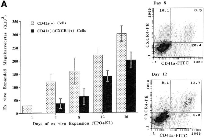
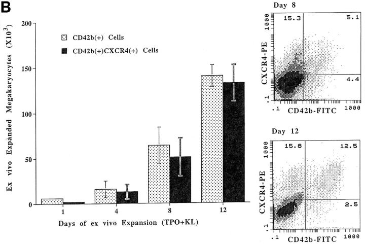
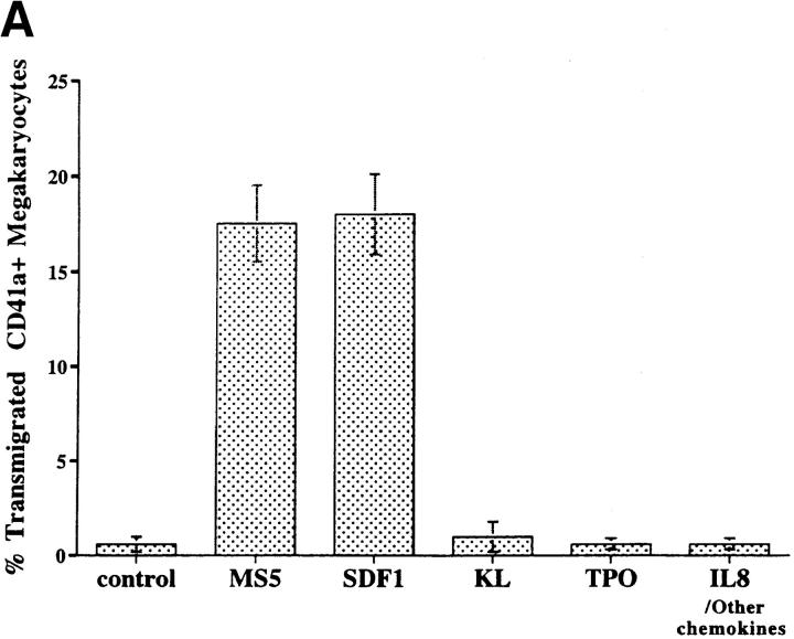
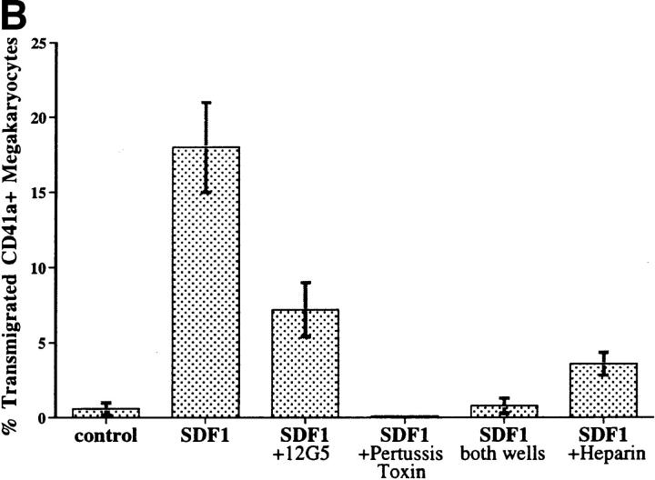
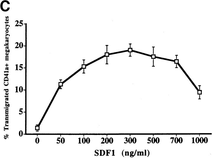
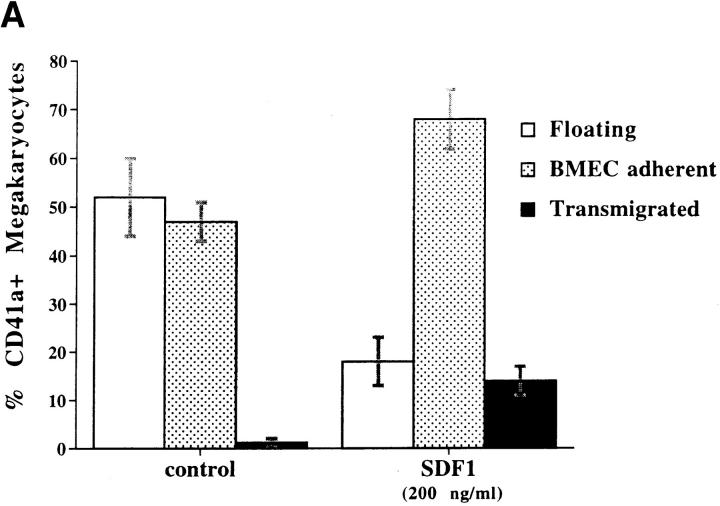
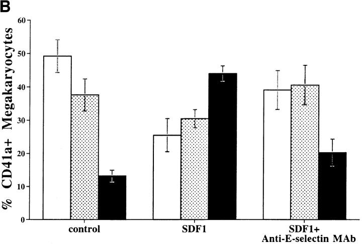
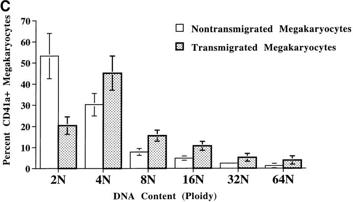
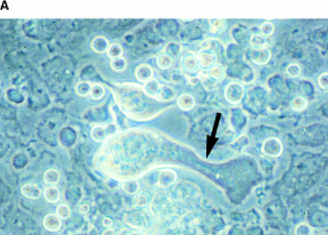
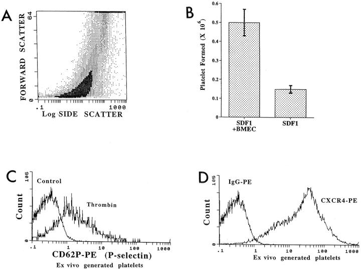
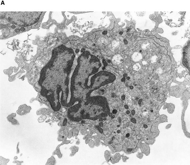
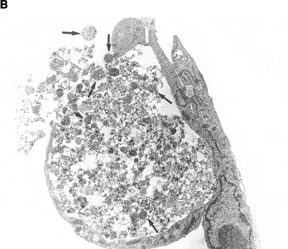
Although thrombopoietin has been shown to promote megakaryocyte (MK) proliferation and maturation, the exact mechanism and site of platelet formation are not well defined. Studies have shown that MKs may transmigrate through bone marrow endothelial cells (BMEC), and release platelets within the sinusoidal space or lung capillaries. In search for chemotactic factor(s) that may mediate transmigration of MKs, we have discovered that mature polyploid MKs express the G protein-coupled chemokine receptor CXCR4 (Fusin, LESTR). Therefore, we explored the possibility that stromal cell-derived factor 1 (SDF-1), the ligand for CXCR4, may also induce transendothelial migration of mature MKs. SDF-1, but not other CXC or CC chemokines, was able to mediate MK migration (ED50 = 125 pmol/liter). The MK chemotaxis induced by SDF-1 was inhibited by the CXCR4-specific mAb (12G5) and by pertussis toxin, demonstrating that signaling via the G protein-coupled receptor CXCR4 was necessary for migration. SDF-1 also induced MKs to migrate through confluent monolayers of BMEC by increasing the affinity of MKs for BMEC. Activation of BMEC with interleukin 1beta resulted in a threefold increase in the migration of MKs in response to SDF-1. Neutralizing mAb to the endothelial-specific adhesion molecule E-selectin blocked the migration of MKs by 50%, suggesting that cellular interaction of MKs with BMEC is critical for the migration of MKs. Light microscopy and ploidy determination of transmigrated MKs demonstrated predominance of polyploid MKs. Virtually all platelets generated in the lower chamber also expressed CXCR4. Platelets formed in the lower chamber were functional and expressed P-selectin (CD62P) in response to thrombin stimulation. Electron microscopy of the cells that transmigrated through the BMEC monolayers in response to SDF-1 demonstrated the presence of intact polyploid MKs as well as MKs in the process of platelet formation. These results suggest that SDF-1 is a potent chemotactic factor for mature MKs. Expression of CXCR4 may be the critical cellular signal for transmigration of MKs and platelet formation.


Figures

Figure 1
Figure 1
CXCR4 is expressed on mature MKs. CD34+ cells isolated from umbilical CB were ex vivo expanded into MKs with TPO and KL. Every 4 d, an aliquot of the expanding MKs was removed, and the number of CD41a+CXCR4+ or CD42b+CXCR4+ cells was quantified with two-color flow cytometry (right). (A) Although on days 1–4 of expansion there are very few CD41a+ cells that express CXCR4, by day 8, 40% of the expanding CD41a+ cells expressed CXCR4. On days 12–16 of expansion, 75% of CD41a+ cells express CXCR4 (n = 5, P <0.05). The CD41a population is comprised of immature MKs, immature uncommitted cells, or myeloid progenitor cells. (B) During MK expansion, a small percentage of expanding MKs express the maturation marker CD42b. Virtually all maturing CD42b+ MKs express CXCR4. Although on day 8 there are very few mature CD42b+CXCR4+ cells, by day 12 a large number of expanding MKs are CD42b+CXCR4+ (n = 5, P <0.01). The majority of the CD42b population is immature CD41a+ MKs and myeloid progenitor cells.
Figure 1
Figure 1
CXCR4 is expressed on mature MKs. CD34+ cells isolated from umbilical CB were ex vivo expanded into MKs with TPO and KL. Every 4 d, an aliquot of the expanding MKs was removed, and the number of CD41a+CXCR4+ or CD42b+CXCR4+ cells was quantified with two-color flow cytometry (right). (A) Although on days 1–4 of expansion there are very few CD41a+ cells that express CXCR4, by day 8, 40% of the expanding CD41a+ cells expressed CXCR4. On days 12–16 of expansion, 75% of CD41a+ cells express CXCR4 (n = 5, P <0.05). The CD41a population is comprised of immature MKs, immature uncommitted cells, or myeloid progenitor cells. (B) During MK expansion, a small percentage of expanding MKs express the maturation marker CD42b. Virtually all maturing CD42b+ MKs express CXCR4. Although on day 8 there are very few mature CD42b+CXCR4+ cells, by day 12 a large number of expanding MKs are CD42b+CXCR4+ (n = 5, P <0.01). The majority of the CD42b population is immature CD41a+ MKs and myeloid progenitor cells.
Figure 2
Figure 2
SDF-1 induces migration of MK cells. (A) SDF-1 (200 ng/ml) or conditioned medium from the MS5 stromal cell line, which is known to contain SDF-1, induced migration of 15–20% of MKs. Replacement of SDF-1 with TPO (100 ng/ml) and KL (100 ng/ml) failed to induce migration of MKs (n = 4, P <0.05). Replacement of SDF-1 with other known chemokines and cytokines such as VEGF or bFGF at 100 ng/ml, IL-11 or IL-6 at 100–200 ng/ml, IL-8 (200 ng/ml), or other chemokines at 100–200 ng/ml, including the CXC (IL-8, NAP-2, IP10, and MIG) or CC chemokines (MIP-1α, MIP-3α [LARC, Exodus-1], MIP-3β [ELC], RANTES, MCP-1, -3, and -5, 6Ckine/Exodus-2/secondary lymphoid tissue chemokine [SLC], thymus-expressed chemokine [TECK], and PF4), also failed to induce migration of MKs (n = 3, P <0.01 for all experiments). (B) Addition of SDF-1 (200 ng/ml) to the lower chamber of the 5-μm transwell plates resulted in the migration from the upper chamber of 20% of ex vivo–expanded MKs (day 12). Incubation of cells with pertussis toxin (2.5 μg/ml) resulted in complete blockage of migration of MKs. Addition of SDF-1 to the upper and the lower chamber of transwells to disrupt the gradient resulted in complete abrogation of MK migration. Addition of heparin or mAb to CXCR4 (12G5, 40 μg/ ml) to the transmigration medium resulted in partial inhibition of migration of MKs (n = 4, P <0.05 for both heparin and 12G5). (C) SDF-1 at various concentrations (50–1,000 ng/ml) was placed in the lower chamber of the transwell plates with MKs expanded for 14 d, and the number of CD41a+ cells migrating through the 5-μm transwells was quantified by two-color flow cytometry. The ED50 for MKs is ∼125 pmol/liter (n = 3).
Figure 2
Figure 2
SDF-1 induces migration of MK cells. (A) SDF-1 (200 ng/ml) or conditioned medium from the MS5 stromal cell line, which is known to contain SDF-1, induced migration of 15–20% of MKs. Replacement of SDF-1 with TPO (100 ng/ml) and KL (100 ng/ml) failed to induce migration of MKs (n = 4, P <0.05). Replacement of SDF-1 with other known chemokines and cytokines such as VEGF or bFGF at 100 ng/ml, IL-11 or IL-6 at 100–200 ng/ml, IL-8 (200 ng/ml), or other chemokines at 100–200 ng/ml, including the CXC (IL-8, NAP-2, IP10, and MIG) or CC chemokines (MIP-1α, MIP-3α [LARC, Exodus-1], MIP-3β [ELC], RANTES, MCP-1, -3, and -5, 6Ckine/Exodus-2/secondary lymphoid tissue chemokine [SLC], thymus-expressed chemokine [TECK], and PF4), also failed to induce migration of MKs (n = 3, P <0.01 for all experiments). (B) Addition of SDF-1 (200 ng/ml) to the lower chamber of the 5-μm transwell plates resulted in the migration from the upper chamber of 20% of ex vivo–expanded MKs (day 12). Incubation of cells with pertussis toxin (2.5 μg/ml) resulted in complete blockage of migration of MKs. Addition of SDF-1 to the upper and the lower chamber of transwells to disrupt the gradient resulted in complete abrogation of MK migration. Addition of heparin or mAb to CXCR4 (12G5, 40 μg/ ml) to the transmigration medium resulted in partial inhibition of migration of MKs (n = 4, P <0.05 for both heparin and 12G5). (C) SDF-1 at various concentrations (50–1,000 ng/ml) was placed in the lower chamber of the transwell plates with MKs expanded for 14 d, and the number of CD41a+ cells migrating through the 5-μm transwells was quantified by two-color flow cytometry. The ED50 for MKs is ∼125 pmol/liter (n = 3).
Figure 3
Figure 3
SDF-1 induces transmigration of MKs through BMEC monolayers. (A) Ex vivo–expanded MKs (day 12) were incubated in the upper chamber of the transwell plates coated with confluent monolayers of resting inactivated BMEC. Introduction of SDF-1 (200 ng/ml) in the lower chamber induced increased migration of MKs. In addition, SDF-1 increased the affinity of MKs for BMEC monolayers. (B) Ex vivo–expanded MKs were incubated in the upper chamber of 5-μm transwell plates coated with IL-1β–treated BMEC monolayers, in the presence and absence of neutralizing mAbs to E-selectin, VCAM-1, and ICAM-1. IL-1β treatment of BMEC results in a threefold increase in adhesion and a fourfold increase in transmigration of MKs in response to SDF-1. mAb to E-selectin but not to ICAM-1 or VCAM-1 resulted in significant inhibition of transmigration of MKs through IL-1β–activated BMEC monolayers.
Figure 3
Figure 3
SDF-1 induces transmigration of MKs through BMEC monolayers. (A) Ex vivo–expanded MKs (day 12) were incubated in the upper chamber of the transwell plates coated with confluent monolayers of resting inactivated BMEC. Introduction of SDF-1 (200 ng/ml) in the lower chamber induced increased migration of MKs. In addition, SDF-1 increased the affinity of MKs for BMEC monolayers. (B) Ex vivo–expanded MKs were incubated in the upper chamber of 5-μm transwell plates coated with IL-1β–treated BMEC monolayers, in the presence and absence of neutralizing mAbs to E-selectin, VCAM-1, and ICAM-1. IL-1β treatment of BMEC results in a threefold increase in adhesion and a fourfold increase in transmigration of MKs in response to SDF-1. mAb to E-selectin but not to ICAM-1 or VCAM-1 resulted in significant inhibition of transmigration of MKs through IL-1β–activated BMEC monolayers.
Figure 4
Figure 4
Ploidy analysis of migrated MKs. (A) Attachment of MKs to BMEC results in profound morphological changes, including a unilateral pseudopod formation (arrow). (B) Light microscopy of Wright/Giemsa-stained transmigrated MKs in response to SDF-1 (200 ng/ml) demonstrated a predominance of polyploid MKs. (C) Ploidy analysis of migrated MKs in response to SDF-1 demonstrated a predominance of polyploid MKs (n = 4, P <0.05 for 4, 8, 16, and 32N).
Figure 4
Figure 4
Ploidy analysis of migrated MKs. (A) Attachment of MKs to BMEC results in profound morphological changes, including a unilateral pseudopod formation (arrow). (B) Light microscopy of Wright/Giemsa-stained transmigrated MKs in response to SDF-1 (200 ng/ml) demonstrated a predominance of polyploid MKs. (C) Ploidy analysis of migrated MKs in response to SDF-1 demonstrated a predominance of polyploid MKs (n = 4, P <0.05 for 4, 8, 16, and 32N).
Figure 5
Figure 5
Generation of functional platelets by migrated MKs. CD41a+ platelets generated from day 12 ex vivo–expanded MKs migrating through either bare transwells or BMEC monolayers in response to SDF-1 (200 ng/ml) were collected, fixed in 0.7% formalin, and immediately subjected to flow cytometry. (A) Forward and log side scatter of platelets generated from migrating MKs in response to SDF-1 demonstrate a characteristic scatter profile identical to that of in vivo–generated platelets. (B) CD41a+CXCR4+ platelets generated 24 h after the migration of MKs were quantified by two-color flow cytometry. Migration of MKs through BMEC in response to SDF-1 results in significantly larger numbers of platelets than that of MKs through bare transwells. (C) Platelets generated in response to SDF-1 and BMEC are functional and express P-selectin (CD62P) after thrombin stimulation. (D) Virtually all platelets generated from migrating MKs express CXCR4.
Figure 6
Figure 6
Electron microscopic analysis of MKs that have migrated through BMEC. MKs migrating through BMEC monolayers were recovered 12–24 h after the addition of SDF-1 (200 ng/ml) from the lower chamber of the transwell plates, and were analyzed by electron microscopy. (A) A large intact polyploid MK that has maintained its morphological integrity 3 h after transmigration through BMEC and 5-μm pores. Original magnification: ×4,000. (B) An MK 12 h after transmigration in the process of platelet formation. Multiple distinct platelets (arrows) are seen throughout the cytoplasm of the MK. Original magnification: ×2,500.
Figure 6
Figure 6
Electron microscopic analysis of MKs that have migrated through BMEC. MKs migrating through BMEC monolayers were recovered 12–24 h after the addition of SDF-1 (200 ng/ml) from the lower chamber of the transwell plates, and were analyzed by electron microscopy. (A) A large intact polyploid MK that has maintained its morphological integrity 3 h after transmigration through BMEC and 5-μm pores. Original magnification: ×4,000. (B) An MK 12 h after transmigration in the process of platelet formation. Multiple distinct platelets (arrows) are seen throughout the cytoplasm of the MK. Original magnification: ×2,500.

Similar articles

Cited by

References

    1. Kaushansky K, Lok S, Holly RD, Broudy VC, Lin N, Bailey MC, Forstrom JW, Buddle MM, Oort PJ, Hagen FS, et al. Promotion of megakaryocyte progenitor expansion and differentiation by the c-Mpl ligand thrombopoietin. Nature. 1994;369:568–571. - PubMed
    1. Wendling F, Maraskovsky E, Debili N, Florindo C, Teepe M, Titeux M, Methia N, Breton-Gorius J, Cosman D, Vainchenker W. cMpl ligand is a humoral regulator of megakaryocytopoiesis. Nature. 1994;369:571–574. - PubMed
    1. Zucker-Franklin D, Kaushansky K. Effect of thrombopoietin on the development of megakaryocytes and platelets: an ultrastructural analysis. Blood. 1996;88:1632–1638. - PubMed
    1. Kaushansky K, Broudy VC, Lin N, Jorgensen MJ, McCarty J, Fox N, Zucker-Franklin D, Lofton-Day C. Thrombopoietin, the Mp1 ligand, is essential for full megakaryocyte development. Proc Natl Acad Sci USA. 1995;92:3234–3238. - PMC - PubMed
    1. de Sauvage FJ, Carver-Moore K, Luoh SM, Ryan A, Dowd M, Eaton DL, Moore MW. Physiological regulation of early and late stages of megakaryocytopoiesis by thrombopoietin. J Exp Med. 1996;183:651–656. - PMC - PubMed

Publication types