. 1998 Aug 3;188(3):549-59.
doi: 10.1084/jem.188.3.549.

The Vav-Rac1 pathway in cytotoxic lymphocytes regulates the generation of cell-mediated killing

Affiliations

The Vav-Rac1 pathway in cytotoxic lymphocytes regulates the generation of cell-mediated killing

D D Billadeau et al. J Exp Med. .

Abstract

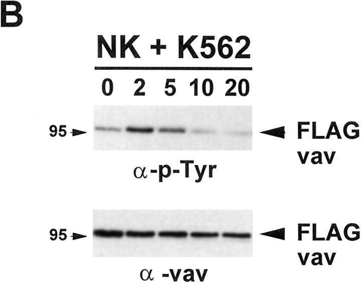
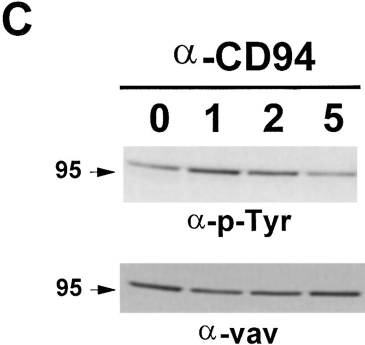
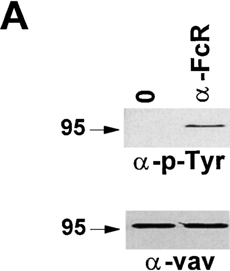
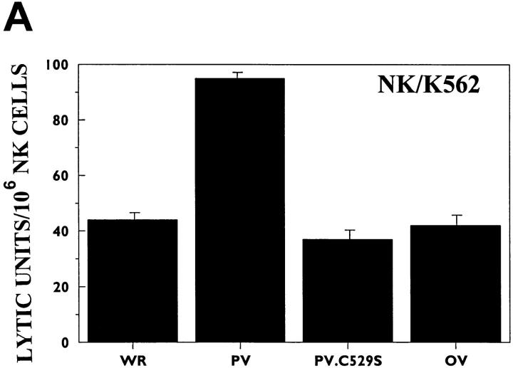
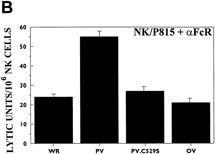
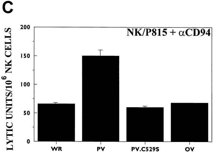
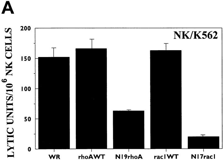
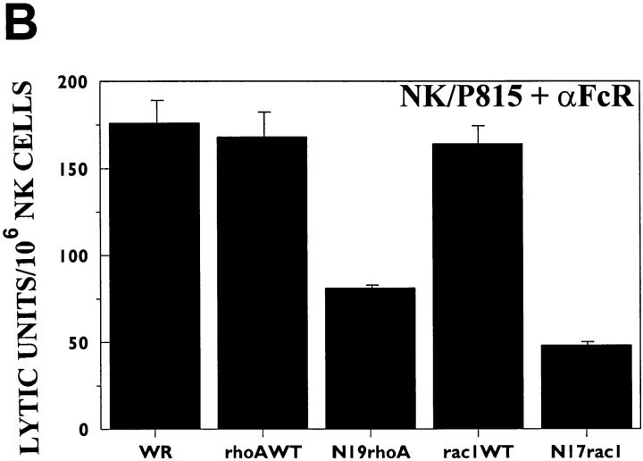
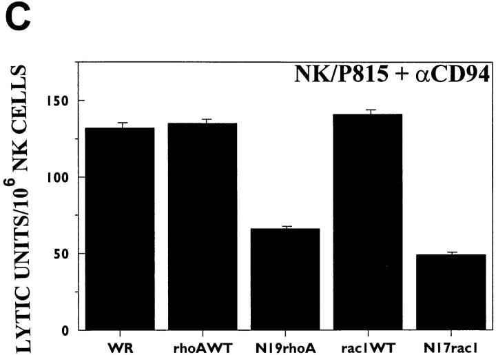
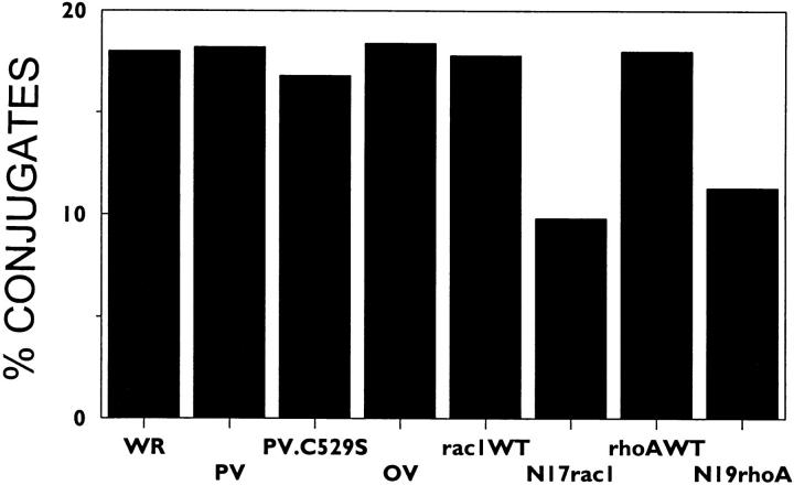
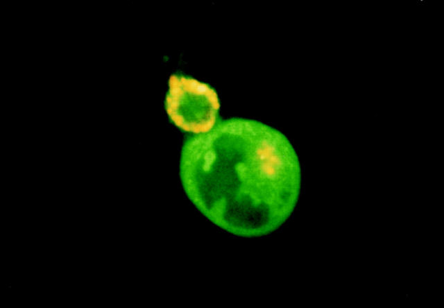
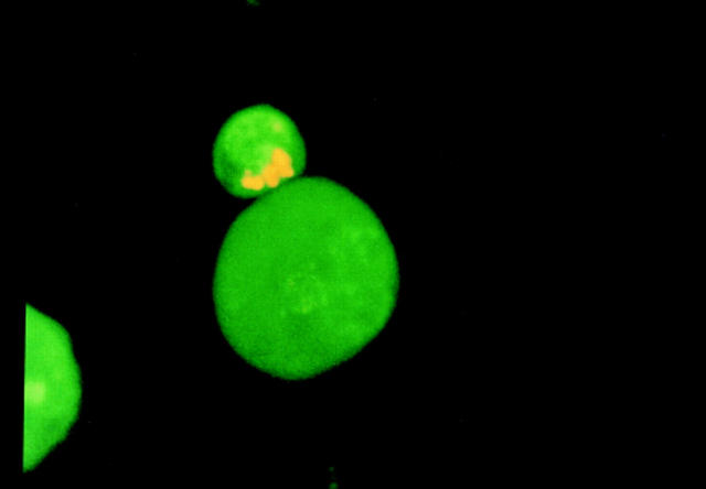
The Rac1 guanine nucleotide exchange factor, Vav, is activated in hematopoietic cells in response to a large variety of stimuli. The downstream signaling events derived from Vav have been primarily characterized as leading to transcription or transformation. However, we report here that Vav and Rac1 in natural killer (NK) cells regulate the development of cell-mediated killing. There is a rapid increase in Vav tyrosine phosphorylation during the development of antibody-dependent cellular cytotoxicity and natural killing. In addition, overexpression of Vav, but not of a mutant lacking exchange factor activity, enhances both forms of killing by NK cells. Furthermore, dominant-negative Rac1 inhibits the development of NK cell-mediated cytotoxicity by two mechanisms: (a) conjugate formation between NK cells and target cells is decreased; and (b) those NK cells that do form conjugates have decreased ability to polarize their granules toward the target cell. Therefore, our results suggest that in addition to participating in the regulation of transcription, Vav and Rac1 are pivotal regulators of adhesion, granule exocytosis, and cellular cytotoxicity.


Figures

Figure 1
Figure 1
Vav is tyrosine phosphorylated during natural killing and after KAR cross-linking. (A) Vav was immunoprecipitated from 5 × 10⁶ NK cells after 1 min of FcR cross-linking with anti-FcR 3G8 mAb (α-FcR). (B) NK cells were infected with vaccinia virus expressing the FLAG-Vav construct. Using anti-FLAG mAb, viral expressed FLAG-Vav was immunoprecipitated from 5 × 10⁶ NK cells which had been incubated at 37°C for the indicated times (in minutes) with 2.5 × 10⁶ K562 cells. (C) Vav was immunoprecipitated from 5 × 10⁶ NK cells after cross-linking of the activating CD94–NKG2 complex for the indicated times (in minutes) at 37°C. In all cases, the immunoprecipitates were resolved by SDS-PAGE, transferred to a nylon membrane, and probed with either antiphosphotyrosine mAb (upper panels, α-p-Tyr) or anti-Vav polyclonal rabbit sera (lower panels, α-vav). This is a representative example from three separate experiments.
Figure 1
Figure 1
Vav is tyrosine phosphorylated during natural killing and after KAR cross-linking. (A) Vav was immunoprecipitated from 5 × 10⁶ NK cells after 1 min of FcR cross-linking with anti-FcR 3G8 mAb (α-FcR). (B) NK cells were infected with vaccinia virus expressing the FLAG-Vav construct. Using anti-FLAG mAb, viral expressed FLAG-Vav was immunoprecipitated from 5 × 10⁶ NK cells which had been incubated at 37°C for the indicated times (in minutes) with 2.5 × 10⁶ K562 cells. (C) Vav was immunoprecipitated from 5 × 10⁶ NK cells after cross-linking of the activating CD94–NKG2 complex for the indicated times (in minutes) at 37°C. In all cases, the immunoprecipitates were resolved by SDS-PAGE, transferred to a nylon membrane, and probed with either antiphosphotyrosine mAb (upper panels, α-p-Tyr) or anti-Vav polyclonal rabbit sera (lower panels, α-vav). This is a representative example from three separate experiments.
Figure 2
Figure 2
Vav is involved in cell-mediated cytotoxicity. NK clones were infected with the indicated recombinant vaccinia virus. Infected NK clones were incubated with 51Cr-labeled K562 (A), P815 cells coated with 0.15 μg/ml of the anti-FcR mAb 3G8 (B), or P815 cells coated with 0.15 μg/ml of the anti-CD94 mAb HP-3B1 (C). This is a representative example of eight separate experiments. Data are expressed as lytic units/106 cells ± 1 SD.
Figure 2
Figure 2
Vav is involved in cell-mediated cytotoxicity. NK clones were infected with the indicated recombinant vaccinia virus. Infected NK clones were incubated with 51Cr-labeled K562 (A), P815 cells coated with 0.15 μg/ml of the anti-FcR mAb 3G8 (B), or P815 cells coated with 0.15 μg/ml of the anti-CD94 mAb HP-3B1 (C). This is a representative example of eight separate experiments. Data are expressed as lytic units/106 cells ± 1 SD.
Figure 3
Figure 3
Rac1 regulates cell-mediated cytotoxicity. NK clones were infected with the indicated recombinant vaccinia virus. Infected NK clones were incubated with 51Cr-labeled K562 (A), P815 cells coated with 0.15 μg/ml of the anti-FcR mAb 3G8 (B), or P815 cells coated with 0.15 μg/ml of the anti-CD94 mAb HP-3B1 (C). This is a representative example of eight separate experiments. Data are expressed as lytic units/106 cells ± 1 SD.
Figure 3
Figure 3
Rac1 regulates cell-mediated cytotoxicity. NK clones were infected with the indicated recombinant vaccinia virus. Infected NK clones were incubated with 51Cr-labeled K562 (A), P815 cells coated with 0.15 μg/ml of the anti-FcR mAb 3G8 (B), or P815 cells coated with 0.15 μg/ml of the anti-CD94 mAb HP-3B1 (C). This is a representative example of eight separate experiments. Data are expressed as lytic units/106 cells ± 1 SD.
Figure 4
Figure 4
Dominant-negative Rac1 inhibits conjugate formation. NK clones infected with the indicated recombinant vaccinia virus were stained intracellularly with sulfofluorescein (green fluorescence), and then incubated for 10 min at 37°C with K562 target cells which had been stained intracellularly with hydroethidine (red fluorescence). Using flow cytometry, the percentage of NK cells forming conjugates were scored based on the simultaneous emission of both green and red fluorescence. 104 events were analyzed per sample. The data presented are a representative example of five different experiments.
Figure 5
Figure 5
Visualization of acridine orange–stained granules in NK cells by fluorescence microscopy. Acridine orange–labeled NK clones were incubated with K562 target cells as described in Materials and Methods. After this incubation, the cells were placed on a slide, and the fluorescent granules were observed using fluorescence microscopy. (Top) Representative example of a conjugate in which the cytolytic granules of the NK cell have not polarized with respect to the K562 target cell. (Bottom) Conjugate in which the granules in the NK cell have been polarized toward the K562 target cell. Scale: 0.1 μM/1 pixel; picture width = 512 pixels).
Figure 5
Figure 5
Visualization of acridine orange–stained granules in NK cells by fluorescence microscopy. Acridine orange–labeled NK clones were incubated with K562 target cells as described in Materials and Methods. After this incubation, the cells were placed on a slide, and the fluorescent granules were observed using fluorescence microscopy. (Top) Representative example of a conjugate in which the cytolytic granules of the NK cell have not polarized with respect to the K562 target cell. (Bottom) Conjugate in which the granules in the NK cell have been polarized toward the K562 target cell. Scale: 0.1 μM/1 pixel; picture width = 512 pixels).

Similar articles

Cited by

References

    1. Katzav S, Martin-Zanca D, Barbacid M. Vav, a novel human oncogene derived from a locus ubiquitously expressed in hematopoietic cells. EMBO (Eur Mol Biol Organ) J. 1989;8:2283–2290. - PMC - PubMed
    1. Katzav S, Cleveland JL, Heslop HE, Pulido D. Loss of the amino-terminal helix-loop-helix domain of the vav proto-oncogene activates its transforming potential. Mol Cell Biol. 1991;11:1912–1920. - PMC - PubMed
    1. Bustelo XR. The VAV family of signal transduction molecules. Crit Rev Oncog. 1996;7:65–88. - PubMed
    1. Collins T, Deckert M, Altman A. Views on Vav. Immunol Today. 1997;18:221–225. - PubMed
    1. Bustelo XR, Ledbetter JA, Barbacid M. Product of the vavproto-oncogene defines a new class of tyrosine protein kinase substrates. Nature. 1992;356:68–71. - PubMed

Publication types

MeSH terms