. 1998 Aug 4;95(16):9129-33.
doi: 10.1073/pnas.95.16.9129.

Small heat shock protein of Methanococcus jannaschii, a hyperthermophile

Affiliations

Small heat shock protein of Methanococcus jannaschii, a hyperthermophile

R Kim et al. Proc Natl Acad Sci U S A. .

Abstract

Small heat shock proteins (sHSPs) belong to a family of 12- to 43-kDa proteins that are ubiquitous and are conserved in amino acid sequence among all organisms. A sHSP homologue of Methanococcus jannaschii, a hyperthermophilic Archaeon, forms a homogeneous multimer comprised of 24 monomers with a molecular mass of 400 kDa in contrast to other sHSPs that show heterogeneous oligomeric complexes. Electron microscopy analysis revealed a spherically shaped oligomeric structure approximately 15-20 nm in diameter. The protein confers thermal protection of other proteins in vitro as found in other sHSPs. Escherichia coli cell extracts containing the protein were protected from heat-denatured precipitation when heated up to 100 degreesC, whereas extracts from cells not expressing the protein were heat-sensitive at 60 degreesC. Similar results were obtained when purified sHSP protein was added to an E. coli cell lysate. The protein also prevented the aggregation of two purified proteins: single-chain monellin (SCM) at 80 degreesC and citrate synthase at 40 degreesC.


Figures
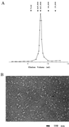
Figure 1
Figure 1
SEC and electron microscopy (EM) of Mj HSP16.5. (A) SEC was performed by using a Tosohaas TSK G4000SW column as described in Materials and Methods. Purified Mj HSP16.5 elutes as a single peak of molecular mass ≈440 kDa. The column was standardized with the following markers as indicated above the figure: carbonic anhydrase (29 kDa); albumin (66 kDa); apoferritin (443 kDa); thyroglobulin (699 kDa). (B) Negative stain EM image of Mj HSP16.5 (1 μM) appears as small particles ≈15–20 nm in diameter. The arrow shows a particle displaying a hole. (Bar = 100 nm.)
Figure 2
Figure 2
Thermal stability of an E. coli crude extract expressing Mj HSP16.5. Extract of soluble E. coli proteins from induced BL21(DE3)/pSJS1240 and BL21(DE3)/pSJS1240 cells expressing Mj HSP16.5 were prepared as described in Materials and Methods. Aliquots of cell extract (4 mg/ml) were heated for 20 min at various temperatures, as indicated. After being allowed to cool to room temperature, samples were centrifuged at 10,000 × g for 5 min. The supernatants (10 μl) were analyzed by SDS/PAGE. Lanes: 1–7, BL21(DE3)/pSJS1240 cell extract heated at 20°, 60°, 70°, 80°, 90°, 100°, and 110°C, respectively; 8, molecular mass markers in kilodaltons; 9–15, transformed BL21(DE3)/pSJS1240 cell extract expressing Mj HSP16.5 heated at 20°, 60°, 70°, 80°, 90°, 100°, and 110°C, respectively.
Figure 3
Figure 3
Thermal protection of E. coli cell extract by purified Mj HSP16.5. Cell extract (CE) from E. coli BL21(DE3)/pSJS1240 (2 mg/ml) was incubated with Mj HSP16.5 or cytochrome C at 2:1 or 1:1 (wt/wt) ratio at 80°C for 20 min. Samples were centrifuged, and the pellets were resuspended in the original volume. Ten microliters of each sample were run on a 15% SDS/PAGE gel. Lanes: 1, molecular mass markers in kilodaltons; 2, cell extract (CE), unheated (U); 3–4, CE, heated (H) 80°C, 20 min: insoluble pellet (I), supernatant (S); 5, CE:Mj HSP16.5 (2:1 wt/wt ratio), U; 6–7, CE:Mj HSP16.5 (2:1 wt/wt ratio), H 80°C, 20 min: I, S; 8, CE:Mj HSP16.5 (1:1 wt/wt ratio), U; 9–10, CE:Mj HSP16.5 (1:1 wt/wt ratio), H 80°C, 20 min: I, S; 11, CE:cytochrome C (1:1 wt/wt ratio), U; 12–13, CE:cytochrome C (1:1 wt/wt ratio), H 80°C, 20 min: I, S; 14, 1 μg Mj HSP16.5; 15, 5 μg cytochrome C.
Figure 4
Figure 4
Thermal protection of SCM by purified Mj HSP16.5. SCM (90 μM) was incubated at 80°C for 20 min in the presence of Mj HSP16.5, lysozyme, or Mj 577 protein. Samples were centrifuged, and the pellets were resuspended in the original volume. Ten microliters of each sample was run on a 15% SDS/PAGE gel. Lanes: 1, molecular mass markers in kilodaltons; 2, SCM:Mj HSP16.5 (1:1 molar ratio), unheated (U); 3–4, SCM:Mj HSP16.5 (1:1 molar ratio), heated (H): supernatant (S), insoluble pellet (I); 5, SCM U; 6–7, SCM, H: S, I; 8, Mj HSP16.5, U; 9–10, Mj HSP16.5, H: S, I; 11, SCM:lysozyme (1:1 molar ratio), U; 12–13, SCM:lysozyme (1:1 molar ratio), H: S, I; 14–15, SCM:Mj 577 protein (1:1 molar ratio) H: S, I.
Figure 5
Figure 5
Thermal aggregation of CS was prevented by the addition of Mj HSP16.5. CS dimer at a 5.8-μM concentration was incubated in a spectrophotometer cell thermostatted at 40°C in 50 mM potassium phosphate (pH 7), with various concentrations of Mj HSP16.5 (1: 0, 2: 29.3 μM; 3: 58.6 μM; 4: 177 μM; 5: 234 μM). The aggregation of CS was monitored by measuring apparent light scattering (A360).

Similar articles

Cited by

References

    1. Gething M J, Sambrook J. Nature (London) 1992;355:33–45. - PubMed
    1. Hendrick J P, Hartl F-U. Annu Rev Biochem. 1993;62:349–384. - PubMed
    1. Parsell D A, Lindquist S. In: The Biology of Heat Shock Proteins and Molecular Chaperones. Morimoto R I, Tissieres A, Georgopoulos C, editors. Cold Spring Harbor, NY: Cold Spring Harbor Lab. Press; 1994. pp. 457–494.
    1. Buchner J. FASEB J. 1996;10:10–19. - PubMed
    1. Caspers G-J, Leunissen J A M, de Jong W W. J Mol Evol. 1995;40:238–248. - PubMed

Publication types

Substances

LinkOut - more resources