. 1998 Aug 4;95(16):9155-60.
doi: 10.1073/pnas.95.16.9155.

Regulation of Ich-1 pre-mRNA alternative splicing and apoptosis by mammalian splicing factors

Affiliations

Regulation of Ich-1 pre-mRNA alternative splicing and apoptosis by mammalian splicing factors

Z H Jiang et al. Proc Natl Acad Sci U S A. .

Abstract

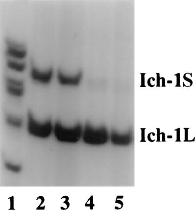
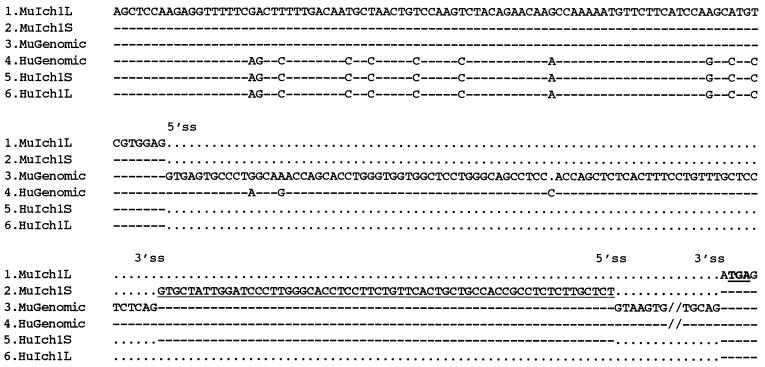
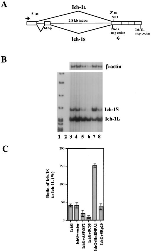
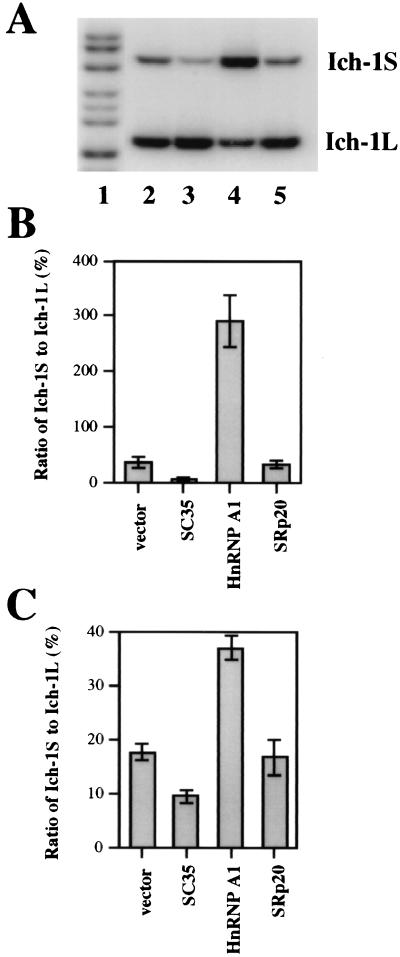
The importance of alternative splicing in regulating apoptosis has been suggested by findings of functionally antagonistic proteins generated by alternative splicing of several genes involved in apoptosis. Among these, Ich-1 (also named as caspase-2) encodes a member of the caspase family of proteases. Two forms of Ich-1 are produced as a result of alternative splicing: Ich-1L, which causes apoptosis, and Ich-1S, which prevents apoptosis. The precise nature of Ich-1 alternative splicing and its regulation remain unknown. Here, we show that the production of Ich-1L and Ich-1S transcripts results from alternative exclusion or inclusion of a 61-bp exon. Several splicing factors can regulate Ich-1 splicing. Serine-arginine-rich proteins SC35 and ASF/SF2 promote exon skipping, decreasing the ratio of Ich-1S to Ich-1L transcripts; whereas heterogeneous nuclear ribonucleoprotein A1 facilitates exon inclusion, increasing this ratio. Furthermore, in cultured cells, SC35 overexpression increases apoptosis; whereas heterogeneous nuclear ribonucleoprotein A1 overexpression decreases apoptosis. These results provide the first direct evidence that splicing factors can regulate Ich-1 alternative splicing and suggest that alternative splicing may be an important regulatory mechanism for apoptosis.


Figures

Figure 1
Figure 1
Expression of Ich-1L and Ich-1S in neural and lymphoid tissues. RT-PCR was performed with murine Ich-1 specific primers and cDNA templates reverse transcribed from Poly(A)+ RNA prepared from different murine tissues: total brain (lane 2), cerebrum (lane 3) thymus (lane 4), and spleen (lane 5). Size markers are shown in lane 1. Ich-1S transcript is longer because inclusion of the alternatively spliced exon creates a stop codon (see Fig. 2 and Fig. 3A).
Figure 2
Figure 2
Alignment of murine and human Ich-1 genomic and cDNA sequences. Sequences 1–3 are murine Ich-1L cDNA, Ich-1S cDNA and genomic DNA; sequences 4–6 are human genomic DNA, Ich-1S cDNA, and Ich-1L cDNA respectively. Identical nucleotides are indicated by “- - -” and gaps are marked by “⋅⋅⋅”. The positions of 5′ and 3′ splice sites are shown. The 61-bp exon included in the Ich-1S cDNA is underlined. A stop codon TGA in frame with Ich-1S is in bold form and the sequence of the 2.8 kb downstream exon is omitted (//).
Figure 3
Figure 3
Regulation of Ich-1 alternative splicing by SR proteins and hnRNP A1. (A) The Ich-1 minigene construct. The genomic DNA fragment containing the upstream exon, introns flanking the 61-bp exon, and the downstream exons was inserted under control of the cytomegalovirus promoter in a mammalian expression vector pcDNA3. The immediate downstream exon was fused at the SalI site to exons further downstream. The arrowheads indicate positions of specific primers used in the RT-PCR assay to detect the transfected Ich-1 minigene expression. (B and C) The Ich-1 minigene construct was co-transfected into HeLa cells with a plasmid encoding individual splicing regulators. Alternatively spliced Ich-1 products expressed from the transfected Ich-1 minigene were detected by RT-PCR with murine Ich-1 specific primers. The ratio of Ich-1S to Ich-1L was measured using a PhosphorImager. Lane 1 contains size markers. Lanes 2–8 contain RT-PCR products after transfection with pcDNA3 vector alone (lane 2), Ich-1 minigene construct (IchG) (lane 3), IchG+vector (lane 4), IchG+ASF/SF2 (lane 5), IchG+SC35 (lane 6), IchG+hnRNP A1 (lane 7), and IchG+SRp20 (lane 8). B shows a representative gel containing RT-PCR products. β-actin PCR products for the same samples were used as controls for amounts of input RNA. C shows the ratio of Ich-1S to Ich-1L as determined from three independent experiments.
Figure 4
Figure 4
Regulation of Ich-1 alternative splicing by SC35 and hnRNP A1 in 293T cells. (A and B). Effects of SC35 and hnRNP A1 on alternative splicing of the Ich-1 minigene. Panel A shows a representative gel containing RT-PCR products detected from 293T cells transfected with the Ich-1 minigene construct together with either the vector alone or a plasmid expressing splicing factor: IchG+vector (lane 2), IchG+SC35 (lane 3), IchG+hnRNP A1 (lane 4), and IchG+SRp20 (lane 5). Lane 1 contains size markers. B shows the ratio of Ich-1S to Ich-1L as determined from three independent experiments. (C) Effects of SC35 and hnRNP A1 on endogenous Ich-1 alternative splicing. The plasmids expressing individual splicing regulators were transfected into 293T cells. The alternative splicing products of the endogenous Ich-1 were detected by RT-PCR with human Ich-1 primers. Shown in C is quantitation of the ratio of Ich-1S to Ich-1L as determined from three independent experiments.
Figure 5
Figure 5
Overexpression of SC35 or hnRNP A1 modulates apoptosis. HeLa cells were transiently transfected with plasmids expressing SC35 (A and B) or hnRNP A1 (C and D) as proteins containing a myc-tag. The transfected cells were shown by immunostaining using a mAb against the myc-tag (A and C). The cells were counterstained with Hoechst dye 33258 to show the nuclear morphology (B and D). Nuclei of a fraction of SC35 overexpressing cells are fragmented and condensed (B) (as marked by arrows). The nontransfected cells showing apoptotic condensed and fragmented nuclei are marked by *. Labeled with arrowheads are several cells that overexpress SC35 or hnRNP A1 and have normal nuclear morphology. The SC35 immunostaining in the transfected healthy cells was localized to the nucleus. However, in many dying cells overexpressing SC35, the immunostaining was no longer confined to the nucleus possibly due to defects in the nuclear envelope.

Similar articles

Cited by

References

    1. Boise L H, González-García M, Postema C E, Ding L, Lindstein T, Turka L A, Mao X, Nuñez G, Thompson C B. Cell. 1993;74:597–608. - PubMed
    1. Wang L, Miura M, Bergeron L, Zhu H, Yuan J-Y. Cell. 1994;78:739–750. - PubMed
    1. Shaham S, Horvitz H R. Cell. 1996;86:201–208. - PubMed
    1. Alnemri E S, Fernandes-Alnemri T, Litwack G. J Biol Chem. 1995;270:4312–4317. - PubMed
    1. Papoff G, Cascino I, Eramo A, Starace G, Lynch D H, Ruberti G. J Immunol. 1996;156:4622–4630. - PubMed

Publication types

MeSH terms

LinkOut - more resources