. 1998 Aug 4;95(16):9167-71.
doi: 10.1073/pnas.95.16.9167.

Molecular remedy of complex I defects: rotenone-insensitive internal NADH-quinone oxidoreductase of Saccharomyces cerevisiae mitochondria restores the NADH oxidase activity of complex I-deficient mammalian cells

Affiliations

Molecular remedy of complex I defects: rotenone-insensitive internal NADH-quinone oxidoreductase of Saccharomyces cerevisiae mitochondria restores the NADH oxidase activity of complex I-deficient mammalian cells

B B Seo et al. Proc Natl Acad Sci U S A. .

Abstract

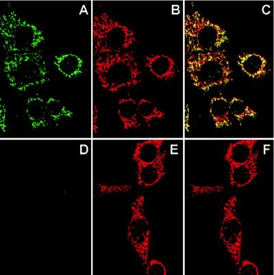
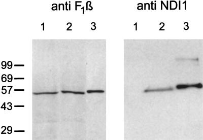
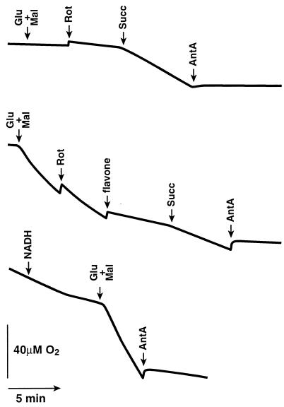
The NDI1 gene encoding rotenone-insensitive internal NADH-quinone oxidoreductase of Saccharomyces cerevisiae mitochondria was cotransfected into the complex I-deficient Chinese hamster CCL16-B2 cells. Stable NDI1-transfected cells were obtained by screening with antibiotic G418. The NDI1 gene was shown to be expressed in the transfected cells. The expressed Ndi1 enzyme was recognized to be localized to mitochondria by immunoblotting and confocal immunofluorescence microscopic analyses. Using digitonin-permeabilized cells, it was shown that the transfected cells, but not nontransfected control cells, exhibited the electron transfer activities with glutamate/malate as the respiratory substrate. The activities were inhibited by flavone, antimycin A, and KCN but not by rotenone. Added NADH did not serve as the substrate, suggesting that the expressed Ndi1 enzyme was located on the matrix side of the inner mitochondrial membranes. Furthermore, although nontransfected cells could not survive in a medium low in glucose (0.6 mM), which is a substrate of glycolysis, the NDI1-transfected cells were able to grow in the absence of added glucose. When glycolysis is slow, either at low glucose concentrations or in the presence of galactose, respiration is required for cells to survive. The mutant cells do not survive at low glucose or in galactose, but they can be rescued by Ndi1. These results indicated that the S. cerevisiae Ndi1 was expressed functionally in CCL16-B2 cells and catalyzed electron transfer from NADH in the matrix to ubiquinone-10 in the inner mitochondrial membranes. It is concluded that the NDI1 gene provides a potentially useful tool for gene therapy of mitochondrial diseases caused by complex I deficiency.


Figures

Figure 1
Figure 1
Mitochondrial localization of the Ndi1 enzyme expressed in Chinese hamster CCL16-B2 cells as demonstrated by confocal immunofluorescence microscopy. NDI1-transfected (A, B, and C) and nontransfected (D, E, and F) CCL16-B2 cells were double-labeled with affinity-purified rabbit antibody to S. cerevisiae Ndi1 and human antimitochondria antibody. Secondary detecting reagents were fluorescein isothiocyanate-conjugated goat anti-rabbit IgG (A and D) and rhodamine-conjugated goat anti-human IgG (B and E). (C and F) Overlapping images of A/B and D/E, respectively.
Figure 2
Figure 2
Immunoblots of mitochondrial fractions from Chinese hamster CCL16-B2 cells by using antibodies against the Ndi1 protein of S. cerevisiae (Right) and the β-subunit of bovine ATP synthase (Left). In both blots, lane 1 is nontransfected cells (100 μg) and lane 2 is NDI1-transfected cells (100 μg). Lane 3 (Left) is bovine mitochondria (1.5 μg). Lane 3 (Right) is the Ndi1 protein (33 ng) containing the leader sequence, which was expressed in and isolated from E. coli. The crude mitochondrial fractions were prepared as follows. The NDI1-transfected and nontransfected cells (approximately 2 × 10⁸ cells) were washed twice with PBS and harvested by trypsinization. The pellet was suspended in 2 ml of isolation buffer containing 210 mM mannitol/70 mM sucrose/1 mM EGTA/5 mM Hepes, pH 7.2/0.2 mM phenylmethanesulfonyl fluoride/0.5% fatty acid-free BSA. The cell suspensions were treated with 0.1 mg/ml of digitonin for 1 min on ice and homogenized in a Dounce homogenizer with a tight pestle (70–100 up/down strokes). The homogenate was centrifuged at 3,000 × g for 5 min at 4°C to remove unbroken cells and nuclei. The supernatant was centrifuged at 10,000 × g for 20 min at 4°C. The pellet was suspended in 0.1 ml of the isolation buffer. This fraction is designated as the crude mitochondrial fraction. Immunoblotting was performed by use of the enhanced chemiluminescence system (Amersham).
Figure 3
Figure 3
Recovery of the NADH oxidase activity in the CCL16-B2 cells transfected with the S. cerevisiae NDI1 gene. (Top) Nontransfected CCL16-B2 cells (1 × 10⁷ cells). (Middle) NDI1-transfected CCL16-B2 cells (1 × 10⁷ cells). (Bottom) NDI1-transfected CCL16-B2 cells (3 × 10⁷ cells). Where indicated, 5 mM glutamate (Glu), 5 mM malate (Mal), 0.5 mM NADH, 5 μM rotenone (Rot), 5 mM succinate (Succ), 5 μM antimycin A (AntA), and 0.5 mM flavone were added. The cells were harvested by trypsinization and resuspended in 1 ml of a medium containing 20 mM Hepes, pH 7.1/250 mM sucrose/10 mM MgCl2. The cells were treated with 50–150 μg of digitonin until more than 90% of the cells are stained by trypan blue. The digitonin-treated cells were washed with the same medium. Oxygen consumption was measured polarographically in 0.6 ml of the buffer containing 20 mM Hepes, pH 7.1/250 mM sucrose/10 mM MgCl2 by using a Clark electrode in a water-jacketed chamber maintained at 37°C.
Figure 4
Figure 4
Effects of glucose (A) and galactose (B) on cell growth of nontransfected and NDI1-transfected CCL16-B2 and parent CCL16. (A) Parent CCL16 cells (105) (□), nontransfected CCL16-B2 cells (▵), and NDI1-transfected CCL16-B2 cells (○) were inoculated in a 5-mM glucose culture medium. (B) Nontransfected (triangles) and NDI1-transfected cells (105) (circles) were inoculated in a 0.6-mM glucose culture medium in the presence (closed symbols) and absence (open symbols) of 5 mM galactose. In the case of NDI1-transfected CCL16-B2 cell culture, 0.5 mg/ml of antibiotic G418 also was present. Cells were cultured at 37°C in a 5% CO2 atmosphere. Cell viability was assessed by trypan blue exclusion, and cell numbers were determined every 24 hr by using a hemocytometer.

Similar articles

Cited by

References

    1. Buchanan S K, Walker J E. Biochem J. 1996;318:343–349. - PMC - PubMed
    1. Chomyn A, Mariottini P, Cleeter M W J, Ragan C I, Matsuno-Yagi A, Hatefi Y, Doolittle R F, Attardi G. Nature (London) 1985;314:591–597. - PubMed
    1. Chomyn A, Cleeter M W J, Ragan C I, Riley M, Doolittle R F, Attardi G. Science. 1986;234:614–618. - PubMed
    1. Hatefi Y. Annu Rev Biochem. 1985;54:1015–1069. - PubMed
    1. Yano T, Chu S S, Sled’ V D, Ohnishi T, Yagi T. J Biol Chem. 1997;272:4201–4211. - PubMed

Publication types

LinkOut - more resources