. 1998 Aug 4;95(16):9295-300.
doi: 10.1073/pnas.95.16.9295.

The disassembly and reassembly of functional centrosomes in vitro

Affiliations

The disassembly and reassembly of functional centrosomes in vitro

B J Schnackenberg et al. Proc Natl Acad Sci U S A. .

Abstract

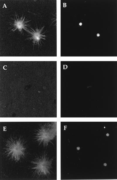
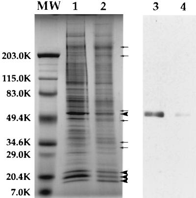
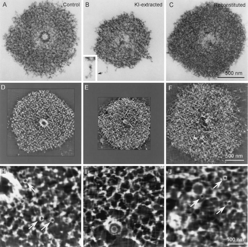
Animal cells contain a single centrosome that nucleates and organizes a polarized array of microtubules which functions in many cellular processes. In most cells the centrosome is composed of two centrioles surrounded by an ill-defined "cloud" of pericentriolar material. Recently, gamma-tubulin-containing 25-nm diameter ring structures have been identified as likely microtubule nucleation sites within the pericentriolar material of isolated centrosomes. Here we demonstrate that when Spisula centrosomes are extracted with 1.0 M KI they lose their microtubule nucleation potential and appear by three-dimensional electron microscopy as a complex lattice, built from 12- to 15-nm thick elementary fiber(s), that lack centrioles and 25-nm rings. Importantly, when these remnants are incubated in extracts prepared from Spisula oocytes they recover their 25-nm rings, gamma-tubulin, and microtubule nucleation potential. This recovery process occurs in the absence of microtubules, divalent cations, and nucleotides. Thus, in animals the centrosome is structurally organized around a KI-insoluble filament-based "centromatrix" that serves as a scaffold to which those proteins required for microtubule nucleation bind, either directly or indirectly, in a divalent cation and nucleotide independent manner.


Figures

Figure 1
Figure 1
Fluorescence micrographs of asters formed by centrosomes and centrosome remnants after indirect immunofluorescent labeling for both tubulin (A, C, and E) and γ-tubulin (B, D, and F). The MNP (A) and γ-tubulin (B) present in centrosomes are removed by treatment with 1.0 M KI (C and D). The MNP recovers (E) and γ-tubulin returns (F) when KI-insoluble centrosome remnants are incubated in oocyte extracts.
Figure 2
Figure 2
SDS/PAGE and immunoblot analyses of centrosomes and KICRs. Some of the proteins present in isolated centrosome fractions (lane 1) are removed by KI treatment (lane 2). Three proteins of ≈20 kDa, which copurify with centrosomes (lane 1), along with a protein of ≈50 kDa are the most abundant proteins in KICR fractions (large arrowheads, lane 2). Additionally, at least six proteins are enriched in the KICR fraction compared with control centrosomes (small arrows, lane 2). Immunoblots of centrosomes (lane 3) and KICRs (lane 4) with affinity-purified polyclonal anti-γ-tubulin antibody reveals that γ-tubulin that is present in isolated centrosome fractions (lane 3), is removed by KI treatment and KICR fractions (lane 4) contain negligible γ-tubulin.
Figure 3
Figure 3
IVEM analyses of control (A, D, and H), KI-extracted (B, E, and I), and reconstituted (C, F, and J) Spisula centrosomes. A–C are electron micrographs of 0.25-μm-thick sections cut through the centriole-containing region of the centrosome. D–F represent single 3-nm-thick slices through the tomographic volume computed from centrosomes similar to those in A–C. H–J are higher magnification images of the centrosome matrix pictured in D–F. Note that the 25-nm-diameter ring-shaped structures seen in control (arrows in H) and reconstituted (arrows in J) centrosomes are absent from the KI-extracted centrosome (I). Note also that the matrix of the KI-extracted centrosome is comprised of an interconnected latticework of 12- to 15-nm-diameter filaments (see D, E, H, and I) that can be resolved in the raw IVEM images at the peripheral edges of the matrix (B Inset).

References

    1. Kellogg D R, Moritz M, Alberts B M. Annu Rev Biochem. 1994;63:639–674. - PubMed
    1. Lee C M, Ferguson M, Chen L B. J Cell Biol. 1989;109:2045–2055. - PMC - PubMed
    1. Soltys B J, Gupta R S. Biochem Cell Biol. 1992;70:1174–1186. - PubMed
    1. Schroer T A, Sheetz M P. Annu Rev Physiol. 1991;53:629–652. - PubMed
    1. Sakai H. Cell Struct Funct. 1994;19:57–62. - PubMed

Publication types

Substances

LinkOut - more resources