. 1998 Aug 4;95(16):9360-5.
doi: 10.1073/pnas.95.16.9360.

Detection of subtle phenotypes: the case of the cell adhesion molecule csA in Dictyostelium

Affiliations

Detection of subtle phenotypes: the case of the cell adhesion molecule csA in Dictyostelium

E Ponte et al. Proc Natl Acad Sci U S A. .

Abstract

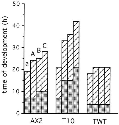
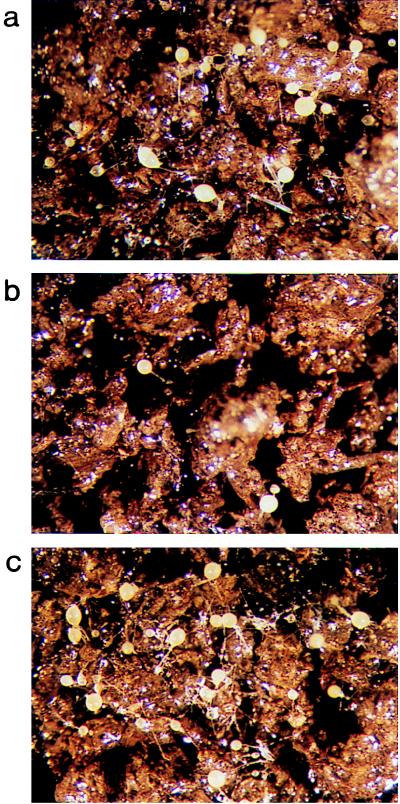
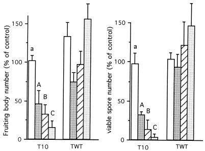
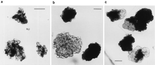
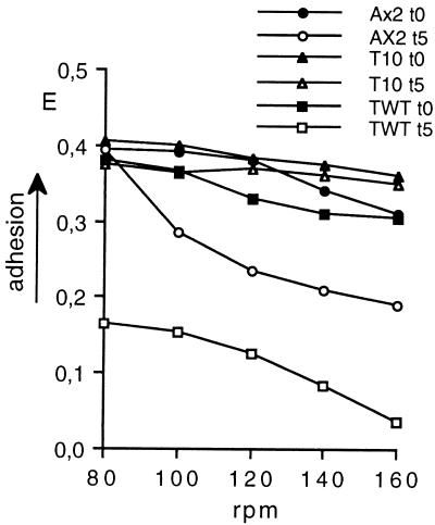
Dictyostelium amoebae aggregate into a multicellular organism by cAMP-driven chemotaxis and cell-cell adhesion. Cell adhesion is mediated by an EDTA-sensitive and an EDTA-resistant adhesion system. The latter is developmentally regulated and triggered by homophilic interactions of the membrane glycoprotein csA; on disruption of the encoding gene, EDTA-resistant contacts fail to form. Nevertheless, csA-null cells under usual laboratory conditions aggregate normally and complete development. By using experimental conditions that reproduce more closely the habitat of Dictyostelium amoebae, evidence is provided that csA is required for development and that its expression confers a selective advantage to populations of wild-type cells over csA-null mutants. The latter display reduced cell-cell adhesion, increased adhesiveness to the substratum, and slower motility, which lead to their sorting out from aggregating wild-type cells. It is proposed that the experimental conditions commonly used in the laboratory are not stringent enough to assess the developmental role of csA and other proteins. The assay described can be used to detect subtle phenotypes, to reexamine the developmental role of apparently nonessential genes, and to test the validity of recent models on emergence and maintenance of apparent genetic redundancy.


Figures

Figure 1
Figure 1
Timing of development of AX2, T10, and TWT cells on agar or soil plates. At the beginning of starvation, cells of the indicated strain were resuspended at 1 × 10⁸/ml in Sörensen phosphate buffer, were plated on agar or soil plates, and were incubated at 23°C. Development was monitored at regular intervals with a stereomicroscope. Time elapsed from beginning of starvation for appearance in the colonies of the first compact aggregates (dotted columns) or fruiting bodies (dashed columns) is indicated in the ordinate. a, agar; A, B, and C, soil plates with increasing degree of moisture, prepared as described in Material and Methods. Average times of three (a) and eight (A, B, and C) experiments are shown. The SDs did not exceed ±1 h for both compact aggregate and fruiting body formation in all cases.
Figure 2
Figure 2
Fruiting bodies formed on soil plate C by AX2, T10, and TWT cells. (a) Wild-type AX2, (b) csA-deletion mutant T10, and (c) csA constitutive expresser TWT. See Fig. 1 for experimental details.
Figure 3
Figure 3
Efficiency of T10 and TWT cells in fruiting body and spore formation on soil. At the beginning of starvation, AX2, T10, or TWT cells were resuspended at 1 × 10⁸/ml in Sörensen phosphate buffer, and a total of 2.5 × 10⁷ cells per colony were plated on agar (a) or soil plates (A, B, and C) and were incubated at 23°C for 48 h. (Left) The number of fruiting bodies per colony formed by T10 or TWT was scored and expressed as percentage of AX2 fruiting bodies formed on each plate. (Right) The number of viable spores in each colony was determined after treating the soil particles with SDS as described in Material and Methods and was expressed as a percentage of the internal AX2 control. Mean values of three (a) and eight (A, B, and C) experiments (Left) or two duplicate experiments (Right) are shown. Vertical bars, SDs.
Figure 4
Figure 4
Types of cell aggregates formed in co-aggregation experiments. AX2 cells, expressing the lac-Z reporter gene (AX2/β-gal), were mixed in binary combinations with T10 or TWT cells in a proportion of 1:1 at a final concentration of 1 × 10⁷ cells/ml and were shaken for 30 min at 150 rpm on a gyratory shaker. The aggregates were fixed and stained to detect AX2 cells in the aggregates (black areas). (a) “Mixed” aggregates formed when AX2/β-gal cells were incubated with T10 cells at the beginning of starvation; (b) “strain-specific” and (c) “partially mixed” aggregates formed when AX2/β-gal cells were incubated with TWT cells at the beginning of starvation as well as with T10 cells at aggregation stage. See Table 2 for quantitative data. (Bars = 0.2 mm.)
Figure 5
Figure 5
Cell adhesion to polystyrene dishes. Growth-phase (t0) or aggregation-competent (t5) cells of AX2, T10, or TWT were washed once and were resuspended at 2.5 × 10⁶ cells/ml in ice-cold Sörensen phosphate buffer. Aliquots of 20-μl cell suspension were spotted at the periphery of 60-mm in diameter polystyrene dishes. After a 10-min incubation at 23°C, 10 ml of Sörensen phosphate buffer were pipetted gently at the center of each dish, and dishes were rotated for 1 min at the speed indicated in the abscissa. Unbound cells were aspirated with the buffer, and bound cells were determined as described under Material and Methods.

Similar articles

Cited by

References

    1. De Lozanne A, Spudich J A. Science. 1987;236:1086–1091. - PubMed
    1. Loomis W. Microbiol Rev. 1996;60:135–150. - PMC - PubMed
    1. Gerisch G, Noegel A A, Schleicher M. Annu Rev Physiol. 1991;53:607–628. - PubMed
    1. Witke W, Schleicher M, Noegel A A. Cell. 1992;68:53–62. - PubMed
    1. Müller K, Gerisch G. Nature (London) 1978;274:445–449. - PubMed

Publication types

Substances

LinkOut - more resources