. 1998 Aug 4;95(16):9436-41.
doi: 10.1073/pnas.95.16.9436.

A very large protein with diverse functional motifs is deficient in rjs (runty, jerky, sterile) mice

Affiliations

A very large protein with diverse functional motifs is deficient in rjs (runty, jerky, sterile) mice

A L Lehman et al. Proc Natl Acad Sci U S A. .

Abstract

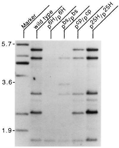
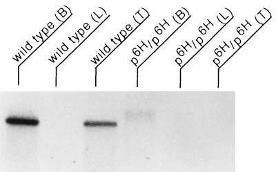
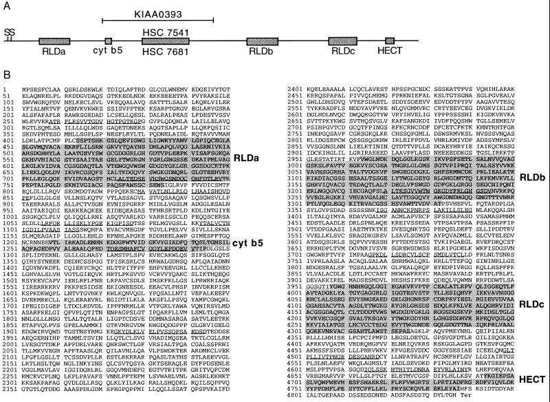
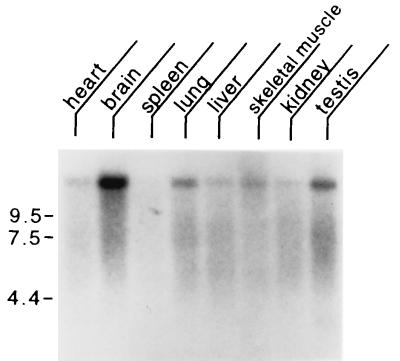
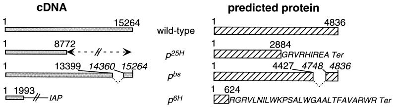
Three radiation-induced alleles of the mouse p locus, p6H, p25H, and pbs, cause defects in growth, coordination, fertility, and maternal behavior in addition to p gene-related hypopigmentation. These alleles are associated with disruption of the p gene plus an adjacent gene involved in the disorders listed. We have identified this adjacent gene, previously named rjs (runty jerky sterile), by positional cloning. The rjs cDNA is very large, covering 15,264 nucleotides. The predicted rjs-encoded protein (4,836 amino acids) contains several sequence motifs, including three RCC1 repeats, a structural motif in common with cytochrome b5, and a HECT domain in common with E6-AP ubiquitin ligase. On the basis of sequence homology and conserved synteny, the rjs gene is the single mouse homolog of a previously described five- or six-member human gene family. This family is represented by at least two genes, HSC7541 and KIAA0393, from human chromosome 15q11-q13. HSC7541 and KIAA0393 lie close to, or within, a region commonly deleted in most Prader-Willi syndrome patients. Previous work has suggested that the multiple phenotypes in rjs mice might be due to a common neuroendocrine defect. In addition to this proposed mode of action, alternative functions of the rjs gene are evaluated in light of its known protein homologies.


Figures

Figure 1
Figure 1
Southern blot analysis of genomic DNA from wild type, p6H/p6H, pbs/pbs, pcp/pcp, and p25H/p25H homozygous mice digested with HindIII and hybridized with the BS2c probe (corresponding to nucleotides 12661–14606 of the final rjs cDNA contig that includes part of RLDc and all of the HECT domain). Marker lane contains size markers with position in kb indicated at left. Note deletion of all bands except lowest molecular weight band in p6H/p6H DNA.
Figure 2
Figure 2
Northern blot of total RNA isolated from wild-type and p6H/p6H tissues (B, brain; L, liver; and T, testis) and hybridized with BS2c probe (corresponding to nucleotides 12661–14606 of the final rjs cDNA contig, which includes part of RLDc and all of the HECT domain). Markers (not shown) indicate this to be a very large transcript, ≥15 kb. Note the expression of the rjs gene in brain and testis of wild type and the absence of transcript in p6H/p6H total RNA. The faint high molecular weight band in p6H/p6H brain total RNA probably represents nonspecific hybridization since RT-PCR from p6H/p6H mRNA was negative with primers after nucleotide 1993.
Figure 3
Figure 3
(A) Graphic representation of the rjs nucleotide sequence and homologies. The solid line represents the rjs nucleotide sequence, with boxed regions indicating significant homologies. S denotes two possible methionine start codons. RLD indicates RCC1-like domains, and cyt b5 indicates homology to cytochrome b5. The HECT domain is found in proteins with homology to E6-AP in their carboxyl terminus. HSC 7541 indicates highly conserved homology (83% identity) to human chromosome 15 sequences related to MN7, and HSC 7681 indicates highly conserved homology (84% identity) to human chromosome 16 sequences related to MN7. KIAA0393 indicates high homology (85% identity) to a human cDNA that maps to chromosome 15, human homologs of the rjs gene (see text). (B) Predicted amino acid sequence encoded by the rjs gene, with regions of homology shaded and listed at right. Underlined amino acids are predicted to be transmembrane domains by the TM Pred program (19) accessible via http://www.isrec.isb-sib.ch/software/TMPRED_form.html.
Figure 4
Figure 4
Mouse multiple tissue Northern blot. Each lane contains 2 μg of poly(A)+ RNA from the tissues indicated hybridized with a probe from the rjs gene (nucleotides 10180–11640). Marker sizes are indicated at left in kilobases. Control hybridization with β-actin (not shown) confirmed the integrity and equal loading of all RNAs.
Figure 5
Figure 5
(Left) Graphical representation of cDNAs encoded by rjs alleles. (Right) Their corresponding proteins. The p25H inversion (indicated by double arrowhead) results in the loss of rjs cDNA sequences after nucleotide 8,772; the predicted p25H rjs peptide ends at amino acid number 2,884, followed by 9 novel amino acids before termination. The pbs deletion results in an internal loss of 960 nucleotides of rjs cDNA (13400–14359, inclusive); the predicted pbs rjs peptide lacks an internal 320 amino acids (4428–4747, inclusive). The p6H deletion results in a loss of rjs cDNA sequences after nucleotide 1,993, with the addition of sequences from an intracisternal A particle (IAP) element; the predicted p6H rjs peptide ends at amino acid 624, followed by 27 novel amino acids encoded by an out-of-reading-frame IAP element before termination.
Figure 6
Figure 6
Light micrographs of sections from mouse mammary tissue (A and B), ovary (C and D), and testis (E and F) taken from wild-type (A, C, and E), p25H/p25H (B), and p6H/p6H (D and F) mice. Mammary tissue was taken from mice 1–2 months of age within hours of giving birth. Ovaries and testis were taken from adult mice 2–6 months of age. Letters on photos indicate important structures. Mammary tissue: D, duct; A, alveoli; and AT, adipose tissue. Ovary: SE, surface epithelium; M, medulla; C, cortex; PF, primary follicle; SF, secondary follicle; and O, oocytes. Testis: G, spermatogonium; I, interstitial cell; P, primary spermatocyte; S, Sertoli cell; and ST, spermatids; and arrow indicates developing spermatozoa. [Approximate final magnifications: ×50 (A and B), ×30 (C and D), and ×45 (E and F).]

References

    1. Silvers W K. The Coat Colors of Mice. New York: Springer; 1979.
    1. Lyon M F, King T R, Gondo Y, Gardner J M, Nakatsu Y, Eicher E M, Brilliant M H. Proc Natl Acad Sci USA. 1992;89:6968–6972. - PMC - PubMed
    1. Rinchik E M, Carpenter D A, Handel M A. Proc Natl Acad Sci USA. 1995;92:6394–6398. - PMC - PubMed
    1. Johnson D R, Hunt D M. J Reprod Fertil. 1975;42:51–58. - PubMed
    1. Hunt D M, Johnson D R. J Embryol Exp Morphol. 1971;26:111–121. - PubMed

Publication types

Associated data