. 1998 Aug 4;95(16):9459-64.
doi: 10.1073/pnas.95.16.9459.

Intestinal intraepithelial lymphocytes include precursors committed to the T cell receptor alpha beta lineage

Affiliations

Intestinal intraepithelial lymphocytes include precursors committed to the T cell receptor alpha beta lineage

S T Page et al. Proc Natl Acad Sci U S A. .

Abstract

The majority of T cells develop in the thymus and exhibit well characterized phenotypic changes associated with their maturation. Previous analysis of intestinal intraepithelial lymphocytes (IEL) from nude mice and a variety of experimentally manipulated models led to the view that at least a portion of these cells represent a distinct T cell population that matures extrathymically. The IEL that are postulated to mature within the intestine include both T cell receptor (TCR) alpha beta- and gamma delta-bearing subpopulations. They can be distinguished from conventional thymically derived T cells in that they express an unusual coreceptor, a CD8alpha homodimer. In addition, they can utilize the Fc receptor gamma-chain in place of the CD3-associated zeta-chain for TCR signaling and their maturation depends on the interleukin 2 receptor beta-chain. Moreover, TCRalpha beta+CD8alpha alpha+ IEL are not subject to conventional thymic selection processes. To determine whether CD3(-)CD8alpha alpha+ IEL represent precursors of T cells developing extrathymically, we examined IEL from knockout mice lacking the recombination activating gene-1 (rag-1), CD3epsilon, or both Lck and Fyn, in which thymic T cell development is arrested. CD3(-)CD8alpha alpha+CD16(+) IEL from all three mutant strains, as well as from nude mice, included cells that express pre-TCRalpha transcripts, a marker of T cell commitment. These IEL from lck-/-fyn-/- animals exhibited TCR beta-gene rearrangement. However, CD3(-)CD8alpha alpha+CD16(+) IEL from epsilon-deficient mice had not undergone Dbeta-Jbeta joining, despite normal rearrangement at the TCRbeta locus in thymocytes from these animals. These results revealed another distinction between thymocytes and IEL, and suggested an unexpectedly early role for CD3epsilon in IEL maturation.


Figures

Figure 1
Figure 1
Some CD3CD8αα+ IEL from ζ−/− mice express cytoplasmic TCR β-chains. IEL were isolated from the indicated mice and stained for three-color flow cytometry by using anti-CD8α-TC, anti-B220-PE, or anti-TCRβ-PE, and anti-CD16-FITC. (a) IEL from 4- to 8-week-old mice were analyzed. Data are shown for lymphocytes pregated on CD8α expression and are presented on a logarithmic scale. cTCRβ and sTCRβ represent cytoplasmic and surface TCRβ expression, respectively. The percentage of CD8α+ cells falling within each rectilinear gate is shown. (b) The data shown are for 6-week-old ɛ−/− and ζ−/− mice. Similar results were seen for 4- to 16-week-old ζ-deficient mice analyzed in at least four independent experiments.
Figure 2
Figure 2
Models of lineage relationships for developing TCRαβ+CD8αα+ IEL. (a) This model is based on published studies (2, 15, 25, 29) and the data for surface and cytoplasmic staining shown in Fig. 1. Arrows indicate postulated developmental transitions. Cell-size relationships are depicted relative to wild-type IEL. (b) This model, where the surface phenotypes are as depicted in a, also incorporates the data for pre-Tα expression and TCRβ gene rearrangement presented in Figs. 4 and 5.
Figure 3
Figure 3
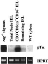
Pre-Tα transcripts are expressed in CD3CD8αα+CD16+ IEL from nude mice. RNA was extracted from sorted CD3CD8αα+CD16+ and CD3CD8α IEL isolated from nude mice, and from thymocytes from rag-1−/− mice and splenocytes from wild-type C57BL/6 mice. RT-PCR was performed by using pre-Tα-specific or hypoxanthine phosphoribosyltransferase (HPRT)-specific primers. Pre-Tα PCR products were detected by Southern blot analysis with an oligonucleotide probe specific for the pre-Tα transmembrane region. HPRT reaction products were visualized by ethidium bromide staining.
Figure 4
Figure 4
Pre-Tα transcripts are expressed in CD3CD8αα+CD16+ IEL from mice exhibiting a block in T cell development. RNA was extracted from sorted CD3CD8αα+CD16+ and CD3CD8α IEL isolated from pools of six 5- to 10-week-old mice of the indicated genotypes. Thymocytes from the same animals and splenocytes from wild-type C57BL/6 mice served as positive and negative controls, respectively. RT-PCR and detection of the amplification products were performed as for Fig. 3.
Figure 5
Figure 5
TCRβ gene rearrangement is detected in CD3 IEL from lck−/−fyn−/− but not ɛ−/− mice. DNA was purified from the indicated sorted cell type and amplified in parallel reactions by using a 5′ Dβ2- or Vβ11-specific primer paired with a 3′ primer immediately downstream of Jβ2.6. PCR products were detected by Southern blot analysis using a Jβ2-specific oligonucleotide probe.

Similar articles

Cited by

References

    1. Lefrancois L. J Immunol. 1991;147:1746–1751. - PubMed
    1. Guy-Grand D, Cerf-Bensussan N, Malissen B, Malassis-Seris M, Broittet C, Vassalli P. J Exp Med. 1991;173:471–481. - PMC - PubMed
    1. Malissen M, Gillet A, Rocha B, Trucy J, Vivier E, Boyer C, Kontgen F, Brun N, Mazza G, Spanopoulou E, Guy-Grand D, Malissen B. EMBO J. 1993;12:4347–4355. - PMC - PubMed
    1. Liu C, Ueda R, She J, Sancho J, Wang B, Weddell G, Loring J, Kurahara C, Dudley E, Hayday A, Terhorst C, Huang M. EMBO J. 1993;12:4863–4875. - PMC - PubMed
    1. Ohno H, Aoe T, Taki S, Kitamura D, Ishida Y, Rajewsky K, Saito T. EMBO J. 1993;12:4357–4366. - PMC - PubMed

Publication types

Substances

LinkOut - more resources