. 1998 Aug 4;95(16):9518-23.
doi: 10.1073/pnas.95.16.9518.

3'-Azido-3'-deoxythymidine drug resistance mutations in HIV-1 reverse transcriptase can induce long range conformational changes

Affiliations

3'-Azido-3'-deoxythymidine drug resistance mutations in HIV-1 reverse transcriptase can induce long range conformational changes

J Ren et al. Proc Natl Acad Sci U S A. .

Abstract

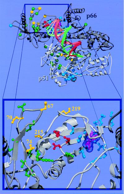
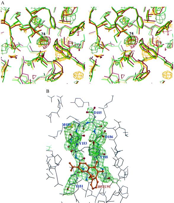
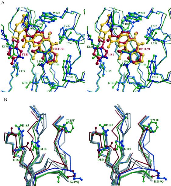
HIV reverse transcriptase (RT) is one of the main targets for the action of anti-AIDS drugs. Many of these drugs [e.g., 3'-azido-3'-deoxythymidine (AZT) and 2',3'-dideoxyinosine (ddI)] are analogues of the nucleoside substrates used by the HIV RT. One of the main problems in anti-HIV therapy is the selection of a mutant virus with reduced drug sensitivity. Drug resistance in HIV is generated for nucleoside analogue inhibitors by mutations in HIV RT. However, most of these mutations are situated some distance from the polymerase active site, giving rise to questions concerning the mechanism of resistance. To understand the possible structural bases for this, the crystal structures of AZT- and ddI-resistant RTs have been determined. For the ddI-resistant RT with a mutation at residue 74, no significant conformational changes were observed for the p66 subunit. In contrast, for the AZT-resistant RT (RTMC) bearing four mutations, two of these (at 215 and 219) give rise to a conformational change that propagates to the active site aspartate residues. Thus, these drug resistance mutations produce an effect at the RT polymerase site mediated simply by the protein. It is likely that such long-range effects could represent a common mechanism for generating drug resistance in other systems.


Figures

Figure 1
Figure 1
Overall structure and drug resistance mutation sites of the RT heterodimer. (Top) The p66 subunit is drawn in dark gray and p51 in light gray. NI resistance mutation sites (26) are shown as green spheres, with RTMC and L74V sites highlighted in yellow. In the p51 subunit, residues 215 and 219 are disordered; their positions are not shown. NNI resistance mutation sites (27) are shown as blue spheres. The three polymerase active site aspartate residues and the bound NNI are shown in red and magenta, respectively. Double-stranded DNA (shown as a spiral ladder with the template strand in green and the primer in red) was modeled into our RT-nevirapine structure (6) from the Cα and phosphate coordinates of the RT-DNA-Fab complex (5) by superimposing the p66 palm domain of the two structures. (Bottom) A close-up view of the polymerase active site and the drug resistance mutation sites in the p66 subunit. The coloring scheme is the same as in the top panel; however, the side chains for mutated residues are shown in ball-and-stick representation and the van der Waals surface for the bound NNI (nevirapine) is shown semitransparent.
Figure 2
Figure 2
Electron density maps. (a) A stereodiagram of  Fo-Fc  maps contoured at −2.5σ showing the negative electron density at residue 74 of the p51 subunit, indicating the Leu to Val substitution. The largest negative feature in the map was close to the inhibitor, which had been omitted from the model. The electron density and models for RTL74V-1, RTL74V-2, and RTL74V-3 are colored as red, yellow, and green, respectively. (b) An omit  Fo-Fc  map for the RTMC-1 data set contoured at 2.5σ showing the electron density for residues 180–189. The map was calculated from a model obtained by further refinement after removing these residues.
Figure 3
Figure 3
The NNI binding site and polymerase active site. (a) A stereodiagram showing the superposition of the NNI binding site in RTMC and wild-type RT. The protein backbone is shown by thin sticks. The NNIs (thick bonds) and side chains that have contacts with the NNIs are shown as ball-and-stick representations. The RTMC is colored in green with residue 181 and the bound 1051U91 highlighted in red. The wild-type RT is colored in blue with residue 181 and bound 1051U91 highlighted in yellow. (b) A stereodiagram of the superposition of the active sites in RTMC (green), the wild type unliganded (red), and six NNI-bound RT structures (blue for RT-1051U91, gray for others) showing the structural changes at the active site in RTMC caused by 215 and 219 mutations. The Cα trace and side chains for residues 110, 185, 186, 215, and 219 are shown for RTMC, wild-type unliganded RT, and RT-1051U91; the Cα traces only are shown for RT-Cl-TIBO, RT-BHAP, RT-nevirapine, RT-MKC-442, and RT-α-APA. In the p51 subunit, residues 215 and 219 are disordered whereas residues 67 and 70 do not show significant rearrangement from the wild-type p51.

Similar articles

Cited by

References

    1. Balter M. Science. 1996;274:1988–1989. - PubMed
    1. Larder B A, Kemp S D. Science. 1989;246:1155–1158. - PubMed
    1. St. Clair M H, Martin J L, Tudor-Williams G, Bach M C, Vavro C L, King D M, Kellam P, Kemp S D, Larder B A. Science. 1991;253:1557–1559. - PubMed
    1. Kohlstaedt L A, Wang J, Friedman J M, Rice P A, Steitz T A. Science. 1992;256:1783–1790. - PubMed
    1. Jacobo-Molina A, Ding J P, Nanni R G, Clark A D J, Lu X, Tantillo C, Williams R L, Kamer G, Ferris A L, Clark P, et al. Proc Natl Acad Sci USA. 1993;90:6320–6324. - PMC - PubMed

Publication types

MeSH terms

Associated data

LinkOut - more resources