. 1998 Aug 4;95(16):9524-9.
doi: 10.1073/pnas.95.16.9524.

A mouse model of severe von Willebrand disease: defects in hemostasis and thrombosis

Affiliations

A mouse model of severe von Willebrand disease: defects in hemostasis and thrombosis

C Denis et al. Proc Natl Acad Sci U S A. .

Abstract

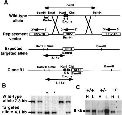
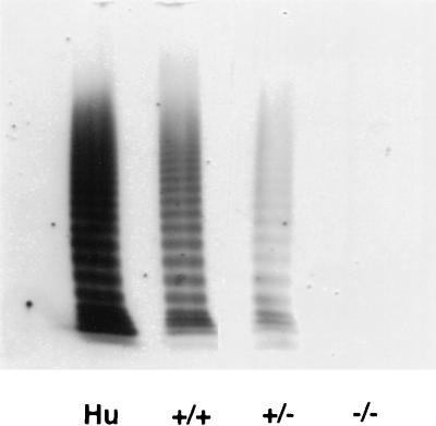
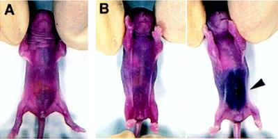
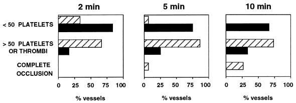
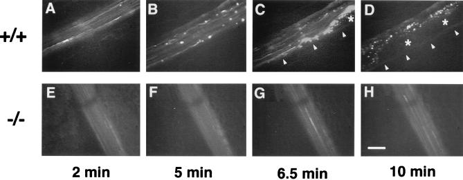
von Willebrand factor (vWf) deficiency causes severe von Willebrand disease in humans. We generated a mouse model for this disease by using gene targeting. vWf-deficient mice appeared normal at birth; they were viable and fertile. Neither vWf nor vWf propolypeptide (von Willebrand antigen II) were detectable in plasma, platelets, or endothelial cells of the homozygous mutant mice. The mutant mice exhibited defects in hemostasis with a highly prolonged bleeding time and spontaneous bleeding events in approximately 10% of neonates. As in the human disease, the factor VIII level in these mice was reduced strongly as a result of the lack of protection provided by vWf. Defective thrombosis in mutant mice was also evident in an in vivo model of vascular injury. In this model, the exteriorized mesentery was superfused with ferric chloride and the accumulation of fluorescently labeled platelets was observed by intravital microscopy. We conclude that these mice very closely mimic severe human von Willebrand disease and will be very useful for investigating the role of vWf in normal physiology and in disease models.


Figures

Figure 1
Figure 1
Targeting of the vWf gene by homologous recombination. (A) The wild-type vWf locus is shown on the top. To make the replacement vector, a 800-bp KpnI–ClaI fragment including exons 4 and 5 of vWf was deleted and replaced with a 1.7-kb neomycin gene cassette driven by a PGK promoter. The 5′ flanking probe used for screening ES cell clones and genotyping mice by Southern blot analysis is indicated. The probe detects a 7.3-kb BamHI fragment in the wild-type allele and a 3.1-kb BamHI fragment in the expected targeted allele. The bottom line shows the targeted clone actually obtained with the retained KpnI–ClaI sites and the neo insertion in intron 5 of vWf. In this clone (clone 91), the probe recognizes a 4.1-kb BamHI fragment. (B) Southern blot analysis of genomic DNA isolated from tail biopsies of one litter resulting from heterozygous crossing. DNA was digested with BamHI, electrophoresed, and probed. Fragments recognized from wild-type and targeted alleles are indicated. Two mice are homozygous for the mutation (asterisks). (C) Northern blot analysis of vWf RNA: Total RNA was isolated from lung (L) and heart (H) of wild-type, heterozygous, and mutant mice. The vWf probe used was a 1-kb fragment from exon 28 of murine vWf cDNA. Several transcripts are present in the −/− mice. The bands indicated by asterisks also hybridized with a neo probe.
Figure 2
Figure 2
Immunolocalization of vWf in platelets and tissue sections. Blood smears (A, B, E, and F), heart sections (C and D), or lung sections (G and H) were prepared from wild-type (+/+) and mutant (−/−) mice and stained by immunofluorescence with a polyclonal antibody to mature vWf (A–D) or to the propeptide (E–H). Bright granular staining characteristic of platelet α-granules (A and E), and Weibel–Palade bodies (C and G), stronger than the autofluorescent background of tissue, is observed only in the samples from the wild-type animals (arrowheads). (Bar = 10 μm.)
Figure 3
Figure 3
Analysis of vWf multimers in plasma. Human plasma (Hu) or mouse plasma from wild-type (+/+), heterozygous (±), or homozygous mutant (−/−) mice was layered on an SDS-1% agarose gel. vWf multimers were separated by discontinuous SDS/agarose gel electrophoresis and detected by incubation with 125I-labeled polyclonal anti-human vWf antibody. Autoradiograph of the gel is shown.
Figure 4
Figure 4
Spontaneous intra-abdominal bleeding in vWf-deficient 1-day-old pups. (A) Wild-type pup. (B) Two vWf-deficient littermates. The pup on the right suffered a massive abdominal bleed visible through the skin (arrowhead).
Figure 5
Figure 5
Platelet-vessel wall interactions after ferric chloride-induced injury. Wild-type (striated columns, n = 15) or vWf-deficient mice (black columns, n = 12) were injected with fluorescently labeled platelets of matching genotype, and their mesenteries were exposed. Arterioles (60–100 μm in diameter) were selected, and a vascular injury was provoked by superfusion with ferric chloride. Any platelet interaction with the vessel wall, either brief or stable, was counted. The presence of platelet thrombi also was recorded. For each vessel studied, the total number of interactive platelets during the first 2 min, 5 min, or during the entire 10 min of filming was recorded and was classified into categories, as indicated on the left.
Figure 6
Figure 6
In vivo thrombosis model in arterioles after ferric chloride-induced injury. An arteriole from a wild-type (+/+) or a vWf-deficient (−/−) mouse was injured by ferric chloride superfusion and photographed at four time points after injury. A progression in the quantity of platelets interacting with the vessel wall is visible in the wild-type arteriole (A–D), leading to complete occlusion and blood stasis in D. Arrowheads in C–D show edge of vessel above which is the forming thrombus. Asterisks indicate the center of a thrombus containing unlabeled and bleached platelets. Almost no platelet interactions are visible in the vWf-deficient arteriole (E–H). (Bar = 50 μm.)

References

    1. Ewenstein B M. Annu Rev Med. 1997;48:525–542. - PubMed
    1. Andrews R K, Lopez J A, Berndt M C. Int J Biochem Cell Biol. 1997;29:91–105. - PubMed
    1. Weiss H J, Sussman I I, Hoyer L W. J Clin Invest. 1977;60:390–404. - PMC - PubMed
    1. Bowie E J, Owen C A. In: Coagulation and Bleeding Disorders. Zimmerman T S, Ruggeri Z M, editors. New York: Dekker; 1989. pp. 305–324.
    1. Wu Q Y, Drouet L, Carrier J L, Rothchild C, Berard M, Rouault C, Caen J-P, Meyer D. Arteriosclerosis. 1987;7:47–54. - PubMed

Publication types

Substances